

Research Article | DOI: https://doi.org/10.31579/2834-5126/133
Vaping As One of The Causes of Death from Complicated Pneumonia in A Teenager
- Ermachenko M.F. 1,2*
- Ivanov R.A. 1
- Sergeeva L.I. 1
- Pakelchuk A.L. 1
- Emelyanova I.I. 1
- Lunenok A.A. 2,4
- Babushkina M.A. 1
- Rodina Yu.R. 1,2
- Goruda I.S. 1,2
- Vanteeva I.A. 1,2
- Bautina Yu.A. 1,2
- Sergeeva S.A. 1,2
- Kokorin V.A. 1,3
1Regional State Budgetary Healthcare Institution "Bratsk Children's City Hospital", Russia, Irkutsk Region, Bratsk.
2Irkutsk State Medical University, Russia, Irkutsk.
3Regional State Budgetary Healthcare Institution "Bratsk District Hospital", Russia, Irkutsk Region, Bratsk.
4Regional State Autonomous Healthcare Institution "City Hospital No. 1", Russia, Irkutsk Region, Bratsk.
*Corresponding Author: Mikhail Еrmасhепkо., Regional State Budgetary Healthcare Institution
Citation: Ermachenko M.F., Ivanov R.A., Sergeeva L.I., Pakelchuk A.L., Emelyanova I.I., et al, (2026), Continuity of Care Versus Episodic Care, Clinical Trials and Clinical Research,5(2); DOI:10.31579/2834-5126/133
Copyright: © 2026, Mikhail Еrmасhепkо. This is an open access article distributed under the creative commons’ attribution license, which permits unrestricted use, distribution, and reproduction in any medium, provided the original work is properly cited.
Received: 06 January 2026 | Accepted: 20 January 2026 | Published: 02 March 2026
Keywords: pediatric; blood; abdominal; ultrasound
Abstract
Upon admission, the patient denied smoking regular or electronic cigarettes. Upon admission, the patient's condition was assessed as severe, due to acute respiratory failure (tachypnea 26-28 bpm, SpO2 93-94%), fever up to 40°C, and severe pneumonia. Auscultation revealed weakened breath sounds, crackling and dry wheezing on both sides. Tachycardia was noted at 128-132 bpm. She was hospitalized in the pediatric department with a diagnosis of acute respiratory viral infection. Chest X-ray revealed infiltrative changes, mainly on the right.
Introduction
The article describes a clinical case of lung injury associated with e-cigarette smoking, called EVALI (E -cigarette and Vaping use-Associated Lung Injury) brief review Literature. Due to the proliferation and popularization of e-cigarettes, lung damage associated with their use is becoming increasingly common. The term EVALI emerged in 2019 during the largest outbreak of the disease in the United States, and criteria for verifying the diagnosis were developed at the same time [9, 10]. The use of electronic cigarettes and vapes in the world has given rise to a new disease - EVALI , which stands for " electronic cigarette " of vaping product use associated lung injury » - lung damage associated with the use of electronic cigarettes or vapes [1,3]. The danger of EVALI is that this disease is difficult to diagnose, since to date there is not a single special study that could reliably help confirm or refute the diagnosis [4]. The new "fashionable" disease that has spread in the United States has also affected Russian schoolchildren, who continue to vape despite all the bans. Currently, 40% of schoolchildren in Russia periodically or regularly use e-cigarettes and vape [1,5]. Vaping liquid still contains nicotine, but it also contains oils, which ensure proper evaporation. The vapor in e-cigarettes is a cloud of white, aromatic smoke, created by adding glycerin, an oil solution of vitamin E, propylene glycol, and other additives to the liquid. These substances themselves are harmless to the body. However, if oil droplets enter the lungs, this can cause serious problems. The oil clogs the small bronchioles, preventing oxygen from reaching the lungs. Although we breathe air that is far from clean, it doesn't contain enough oil to turn into small droplets in the lungs. If this happens, the body may perceive them as a foreign object and initiate an immune response. The result can be inflammation and fluid accumulation in the lungs, leading to the development of so-called "lipoid pneumonia," which develops with regular inhalation of the oily compounds present in vape liquid. This is a potentially life-threatening condition [8,9]. Vaping dries out lung tissue and the mucous membranes of the respiratory tract, making it easier for infections (bacterial, viral, or fungal) to penetrate. Gas exchange is disrupted, leading to oxygen deficiency (hypoxia) [6,7]. EVALI is considered a diagnosis of exclusion because there is no specific test or marker for its diagnosis. Physicians primarily exclude community-acquired pneumonia and acute eosinophilic pneumonia: laboratory testing includes a complete blood count with differential for inflammatory markers ( C-reactive protein , erythrocyte sedimentation rate ), liver transaminase levels , and urine toxicology , including the presence of THC. Physicians traditionally prescribe CT angiography or chest CT, and bronchoscopy with bronchoalveolar lavage [2]. Chest radiographs of patients with EVALI typically show bilateral diffuse, turbid, or consolidated opacities, but patients show negative tests for respiratory infectious diseases. Lung biopsy is not required for diagnosis but is performed according to clinical indications [10]. Among other things, it is necessary to exclude coronavirus infection, the symptoms of which are similar to EVALI [5]. As of 2020, there were no consensus guidelines for the treatment of EVALI [6]. Systemic corticosteroids are the mainstay of therapy; their effectiveness is likely related to attenuation of the inflammatory response. A course of treatment with methylprednisolone equivalent typically lasts 5–10 days, depending on the clinical condition [2]. In some cases, patients were prescribed bronchoalveolar lavage [6]. Because the signs and symptoms of the disease overlap with community-acquired pneumonia and other infectious diseases, empirical antibiotics or steroids are also recommended, depending on the clinical context [11]. Statistics: The average age of EVALI patients was 24 years, 62% of patients were aged 18 to 34 years, and another 16% were under 18 years old. The average age of deceased patients was 49.5 years (minimum 15, maximum 85 years). No reliable mortality statistics for adolescent vaping users have been found in the available literature [9, 10, 11]. Purpose of the study. To demonstrate the complexity of diagnosis and treatment challenges in vape-induced lung disease associated with COVID-19, complicated by bacterial destruction of the lungs and sepsis in a 15-year-old girl. Materials and Methods. Patient B., 15, was admitted to the pediatric department of the Bratsk Children's City Hospital (a level 2 medical facility) on September 29, 2025, with complaints of cough, fever up to 39.6°C, left ear pain, nosebleeds, and difficulty breathing. The illness developed over the course of a week, and outpatient treatment was ineffective. Upon admission, the patient denied smoking regular or electronic cigarettes. Upon admission, the patient's condition was assessed as severe, due to acute respiratory failure (tachypnea 26-28 bpm, SpO2 93-94%), fever up to 40°C, and severe pneumonia. Auscultation revealed weakened breath sounds, crackling and dry wheezing on both sides. Tachycardia was noted at 128-132 bpm. She was hospitalized in the pediatric department with a diagnosis of acute respiratory viral infection. Chest X-ray revealed infiltrative changes, mainly on the right. Complete blood count (CBC): anemia ( Hb 86 g/l), leukocytosis 12 x 10x9/l (without left shift), thrombocytosis 611 x 10x9/l. Coagulogram from September 30, 2025, showed an increase in fibrinogen to 6.58 g/l. MSCT of the lungs from September 30, 2025: bilateral polysegmental destructive pneumonia, the total lesion volume is 35-40% (CT 2). Ultrasound of the abdominal cavity revealed splenomegaly. Nasopharyngeal swabs for influenza, acute respiratory viral infections and sputum culture for microflora were negative. Positive results of ELISA for mycoplasma and PCR for COVID-19 (from September 29, 2025) were revealed. On October 1, 2025, due to persistent hyperthermia, signs of intoxication, and respiratory failure, the child was transferred to the intensive care unit with a diagnosis of community-acquired bilateral polysegmental infiltrative-destructive pneumonia, severe (S 1,2,3,6 of the right lung; S 1 of the left lung with massive infiltration and large cavities of decay) of viral-bacterial etiology (total lesion volume 35-40%). DN grade 1-2 severity. The patient's condition was severe upon admission . Respiratory support with humidified oxygen via nasal cannula was added to the treatment regimen, and antibacterial therapy was changed to meronem at a dose of 60 mg/kg/day and vancomycin at 40 mg/kg/day. Immunostimulating therapy with intravenous human immunoglobulin and nebulized mucolytic therapy ( ambroxol ) were prescribed. To treat the hemic component of hypoxia, a replacement transfusion of red blood cells was administered . Enoxaparin sodium at a dose of 1 mg/kg was prescribed to prevent thrombosis. Despite the ongoing therapy, the patient's condition remained severe, with a persistent need for oxygen, hyperthermia, unproductive cough, shortness of breath with retraction of the compliant areas of the chest, auscultation - mosaic areas of weakened breathing in the lungs on both sides, respiratory rate 22-23 per min, SpO2 - 95-96%. Laboratory signs of acute inflammation persisted: leukocytosis up to 20 x 10 9 /l, CRP up to 245 mg/l, left shift in the leukocyte formula, D-dimer up to 6000 ng /l. A control MSCT scan of the lungs performed on October 3, 2025, showed an increase in the lesion area to 50% with foci of destruction on the right. By October 5, 2025, the child's condition showed positive dynamics: her body temperature had returned to normal, the child no longer required respiratory support, her appetite had improved, and she was actively coughing up large, dense pieces of purulent sputum. She subjectively noted an improvement in her well-being. On October 6, 2025, she was transferred to the infectious diseases department, where by evening another episode of hyperthermia up to 39°C was noted. A control CT scan of the lungs showed negative dynamics, the lesion volume had increased to 65%, pleural effusion was present on the right and left sides, and foci of destruction were enlarged. Following a telemedicine consultation, a decision was made to transport the patient to the regional center, the Irkutsk Children's Ivano-Matreninskaya Hospital (a third-level medical care facility) for further treatment, where the patient stayed from October 7, 2025, to October 18, 2025. Intensive therapy was administered with the assistance of leading regional medical specialists. Further medical history revealed that the girl had been using smoking mixtures, such as vaping, for approximately one year. Due to lung damage from vaping products, the addition of viral and bacterial flora, and complications affecting organs and body systems, treatment was ineffective, and a fatal outcome was recorded on October 18, 2025.
Results and discussion
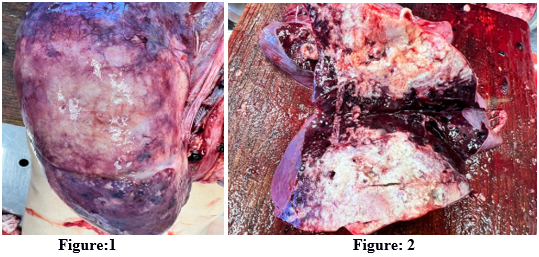
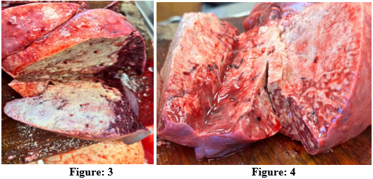
Figures 1, 2, 3, 4: show a macroscopic preparation of the lungs.
Due to the fact that a pathological report was not received from the medical institution where the patient died, it was decided to turn to the opinion of an independent pathological expert ( Krasnoyarsk ) to describe the pathological picture of the lungs .
Description of the lung macropreparation by an independent pathological expert:
1. General appearance (Fig. 1 – external view, pleura)
Color and surface:
- The lungs are significantly enlarged in volume and have an uneven, variegated, mosaic color.
- Large areas of the surface have a dark red, bluish-purple color, which indicates pronounced venous congestion, hyperemia and hemorrhage into the parenchyma.
- Against the background of dark red areas, extensive, irregularly shaped, whitish-gray or yellowish-gray spots are visible. These zones correspond to massive inflammatory infiltration (consolidation) and destructive changes.
- Pleura: The visceral pleura is dull and has lost its luster. Thin, diffuse or focal whitish fibrin deposits are visible on the surface, indicating fibrinous pleurisy.
Consistency:
- Lungs of a dense consistency, with the absence of normal crepitation on palpation, which is a sign of extensive consolidation and edema
2. Sectional view (Figure. 2, 3, 4)
Consolidation and destruction:
- On section, the lung demonstrates massive, almost total consolidation of lung tissue.
- Color on section: a predominantly dirty gray, yellowish-gray, or whitish color indicates massive alveolar infiltration with fibrin, leukocytes, and exudate (bacterial pneumonia, gray hepatization phase ). Yellowish hues in these areas are caused not only by purulent exudate, but also by the presence of lipid components that could have been inhaled with the vape aerosol.
- Variegation: areas of dark red, almost black color remain (especially along the periphery of consolidated zones), which is a sign of severe congestion, hemorrhagic edema and hemorrhages.
- Consistency on section: The tissue is dense, homogeneous, the structural pattern of the lung is completely erased.
- Foci of destruction: Fig. 2 shows small, necrotic foci or areas where the tissue appears loose, heterogeneous, with signs of destruction, which corresponds to the diagnosis of “destructive pneumonia” and abscess formation .
- Fluid release: when pressure is applied to the tissue, a foamy, cloudy, pinkish-yellowish fluid is released in abundance, which indicates severe edema, inflammatory exudate and the presence of lipid components.
Bronchi and vessels:
- The lumens of the bronchi are narrowed due to swelling of the walls and compression by the surrounding tissue.
- The bronchial mucosa is hyperemic and edematous.
- The section shows that the vessels (especially arteries) are full of blood (stagnation).
Macroscopic picture of the lungs in severe acute injury associated with vaping, complicated by destructive pneumonia:
The lungs are significantly enlarged in volume and weight, dense, with a marked decrease in airiness and crepitations. The visceral pleura is dull, covered with diffuse fibrinous deposits. On the surface and in cross-section, the lungs have a sharply mottled, mosaic appearance.
On section: The lung tissue exhibits a pattern of massive, almost total consolidation, predominantly dirty gray or yellowish-gray in color, indicating bacterial infiltration, purulent exudate, and the gray hepatization phase . Yellowish hues in the consolidated areas, as well as a "greasy" sensation upon tactile contact, indicate the contribution of components of vaping liquids, such as lipid-containing additives (e.g., vitamin E acetate) dissolved in propylene glycol and vegetable glycerin, to the development of primary acute lung parenchyma injury. These changes alternate with extensive zones of dark red and purple color (signs of severe congestion, hemorrhagic edema, and hemorrhage). The structural pattern of the lung is completely erased. Pressure leads to the profuse release of a foamy, turbid, pinkish-yellowish fluid. Foci of destruction of the pulmonary parenchyma are noted in the form of loose, necrotic areas, which confirms the diagnosis of destructive pneumonia. This macroscopic picture reflects the most severe acute toxic and inflammatory lung injury associated with vaping (EVALI-like syndrome), which was superimposed by secondary bacterial destructive pneumonia, leading to a fatal outcome in a teenager.
Conclusion
Doctors need to more thoroughly and persistently collect smoking histories from adolescents. The dangers of vaping, especially among adolescents, are underestimated. Due to their toxicity and the rapid development of lung damage symptoms, these smoking products are far more dangerous than regular tobacco products. Lung damage from the products contained in smoking mixtures leads to damage to the lung parenchyma, resulting in weakened immunity and the development of viral and bacterial infections, especially in lungs compromised by vaping. To protect the health and lives of children, adolescents, and adults, a state-level ban on the production and sale of vapes is needed.
References
- (2020). Vaper's disease (EVALI): what is its danger and how to avoid it [Electronic resource] Federal Budgetary Institution of Health
View at Publisher | View at Google Scholar - Vapers' disease [Electronic resource] 10th City Clinical Hospital.
View at Publisher | View at Google Scholar - Vaper's Disease [Electronic resource] runi.rf : encyclopedia.
View at Publisher | View at Google Scholar - Diagnosis EVALI, or Vaper's Disease [Electronic resource] Rospotrebnadzor.
View at Publisher | View at Google Scholar - Summary of cases of lung diseases associated with the use of electronic cigarettes (Electronic nicotine delivery systems) and vaping products (EVALI) [Electronic resource] Ministry of Health of the Russian Federation.
View at Publisher | View at Google Scholar - Shilov V. V. N. M. Lutova , A. A. Krolevets (2020). E-cigarette smoking-associated lung injury (EVALI) [Electronic resource]. Bulletin of SMSU. -. - No. 3. - P. 30-34.
View at Publisher | View at Google Scholar - Huaa M. P. Talbotb. (2016). Potential health effects of electronic cigarettes: A systematic review of case reports [Текст] : [англ.] Preventive Medicine Reports. — Vol. 4. — P. 169–178.
View at Publisher | View at Google Scholar - McCauley L. C. Markin, D. Hosmer. (2012). An Unexpected Consequence of Electronic Cigarette Use [Текст] : [англ.] Postgraduate Education Corner Pulmonary and Critical Care Pearls. — Vol. 141, no. 4. — P. 1110–1113.
View at Publisher | View at Google Scholar - (2020). Outbreak of Lung Injury Associated with the Use of E-Cigarette, or Vaping, Products [Электронный ресурс] : [англ.] / Office on Smoking and Health, National Center for Chronic Disease Prevention and Health Promotion CDC.
View at Publisher | View at Google Scholar - Schier J. G. J. G. Meiman, J. Layden. (2019). Severe Pulmonary Disease Associated with Electronic-Cigarette–Product Use — Interim Guidance [Текст] : [англ.] MMWR. — CDC,. — Vol. 68, no. 36 (13 September). — P. 787–790.
View at Publisher | View at Google Scholar - Siegel D. A. T. C. Jatlaoui, E. H. Koumans (2019). Update: Interim Guidance for Health Care Providers Evaluating and Caring for Patients with Suspected E-cigarette, or Vaping, Product Use Associated Lung Injury — United States, October 2019 [Текст] : [англ.] MMWR. — CDC. — Vol. 68, no. 41— P. 919–927.
View at Publisher | View at Google Scholar
