

Review Article | DOI: https://doi.org/10.31579/2834-8486/002
Utilization of EEG for Monitoring in Subarachnoid hemorrhage Recovery
- Kevin Pierre 1
- Abeer Dagra 2
- Mohammad Reza Hosseini Siyanaki 3
- Yusuf Mehkri 2
- Samuel Woodford 2
- Rami Hatem 2
- Brandon Lucke-Wold 3*
1 University of Florida Department of Radiology, Gainesville, FL, 32603
2 University of Florida College of Medicine, Gainesville, FL, 32603
3 University of Florida Department of Neurosurgery, Gainesville, FL, 32603
*Corresponding Author: Brandon Lucke-Wold, University of Florida Department of Neurosurgery, Gainesville, FL, 32603
Citation: Pierre K., Dagra A., Mohammad R. H. Siyanaki, Mehkri Y., Woodford S. et al. (2022). Utilization of EEG for Monitoring in Subarachnoid hemorrhage Recovery. Biomedical and Clinical Research. 1(1); DOI:10.31579/2834-8486/002
Copyright: © 2022 Brandon Lucke-Wold, This is an open access article distributed under the Creative Commons Attribution License, which permits unrestricted use, distribution, and reproduction in any medium, provided the original work is properly cited.
Received: 12 September 2022 | Accepted: 16 September 2022 | Published: 27 September 2022
Keywords: EEG; subarachnoid hemorrhage; delayed cerebral ischemia; morbidity
Abstract
Seizures and delayed cerebral ischemia following subarachnoid hemorrhage are associated with significant morbidity and mortality. In this article, we briefly review subarachnoid hemorrhage, its complications, and the current literature on information gained from EEG monitoring in subarachnoid hemorrhage. We review when EEG should be used implemented in the multi-modal monitoring of patients with subarachnoid hemorrhage. Finally, we discuss the recent advances and future directions in the field.
Introduction
Subarachnoid hemorrhage (SAH) results in constriction of the microcirculation, disruption of the blood brain barrier, neuronal, and endothelial cell death [1] (Figure 1) SAH is associated with high mortality and morbidity [2] 23Percentage of patients with SAH develop seizures within the first 48 hours following SAH [3] Delayed seizures may also occur 6 weeks following SAH. The development of these seizures are associated with poor prognosis [4] While convulsive episodes are clinically evident, many non-convulsive seizure occurrences (NCSE) are missed without EEG monitoring [5,6] In fact, in a study of 233 patients with SAH and coma or neurological deterioration of uncertain etiology, 8% were found to have NCSE through EEG [7].

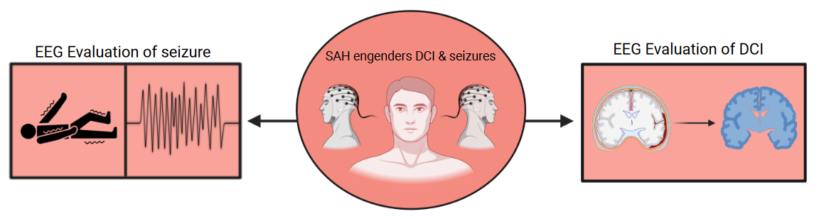
Similarly, DCI (delayed cerebral ischemia), which occurs in approximately 30 Percentage of all patients 4-14 days post-SAH, can also result in long-term functional limitations in patients with SAH.[ 8, 9, 10-12] DCI occurs when there is a new neurological deficit or cerebral infarction on neuroimaging or autopsy following SAH.11 While vasospasms are thought to be responsible for these ischemic lesions, other factors can also contribute, such as microembolism from impaired endothelial function or hypercoagulopathy, inflammation, and impaired autoregulatory vasodilatation in small, intraparenchymal arteries leading to spreading ischemia secondary to abberant hemodynamic response to cortical spreading depolarization [5, 8, 9, 11, 13, 14] Considering that patients with SAH, particularly those at high risk of developing seizures and DCI are often comatose, sedated, or paralyzed, these conditions have been have been historically difficult to diagnose. In this paper, we review the use of EEG as an adjunct in diagnosing DCI and seizures that develop in patients with subarachnoid hemorrhage. (Figure 2)
Current clinical practice and review of the evidence
Continuous EEG (cEEG) monitoring is often used in the neurointensive care unit to identify and direct the management of nonconvulsive seizures, particularly those that follow convulsive status epilepticus[6, 13, 15-17] Additionally, the management of pharmaceutical coma for the treatment of elevated intracranial pressure is guided by cEEG [6, 17] Finding new or worsening brain ischemia in high-risk patients, notably those with subarachnoid hemorrhage, is a growing application for cEEG [13, 15]. In general, transcranial duplex sonography consistently shows the constriction of major intracranial arteries, which has long been thought to be the only factor in DCI [8, 18]. Therefore, EEG may assess the effects of other hypothesized processes in DCI that cannot be measured or detected with duplex sonography.
Quantitative continuous EEG (qEEG) continually monitors brain activity to detect changes in real time, enabling earlier and more precise identification of DCI symptoms [5, 6, 8, 10, 13, 15-17, 19] In EEG, the areas of cerebral ischemia are characterized by focal slowing with prominent delta foci with decreased alpha/delta wave ratio (ADR), which can be used to identify delayed cerebral ischemia early and before symptomatic manifestations [19, 20] An ADR of less than 50 Percentage is highly sensitive and specific for SAH [17] Furthermore, patients with vasospasm often have a lower relative alpha variability (RAV). A study with 32 patients showed that most patients have changes in RAV 2 days prior to abnormalities detected on TCD.10 Other studies confirmed these findings by showing that EEG allows for earlier detection of vasospasm compared to transcranial doppler (TCD) sonography and neuroimaging (magnetic resonance imaging and computed tomography) by up to 7 hours to 1.9 days.[8, 21]. A prospective study with a blinded reviewer who made 59 predictions found that the use of EEG improved prediction of clinical deterioration and improvement by 27 Percentage and 42 Percentage, respectively [22] Lastly, periodic and rhythmic patterns and epileptiform discharges indicate that patients are at risk for DCI [23].
EEG also detects seizures following SAH. Most patients with nonconvulsive seizures (NCS) develop nonconvulsive status epilepticus (NCSE), which is associated with significant morbidity and mortality [5, 16, 24] Per the American Clinical Neurophysiology Society’s Standardized Critical Care EEG Terminology, an electrographic seizure occurs when there are epileptiform discharges that average more than 2.5Hz and occur for more than 10 seconds or with a pattern of evolution occurring at least 10 seconds. The same findings over a 10-minute period or at least 20 Percentage of a 60-minute period are representative of electrographic status epilepticus. If a patient does not meet these exact criteria, a seizure may still be present if there is spike-wave activity, periodic discharges, or rhythmic delta activity with synchronous neurological exam findings or improvement with antiepileptics [25]. In general, studies have shown that EEG correlates with DCI and are of important value in detecting DCI in seizures following subarachnoid hemorrhage [12].

Practice recommendations
Detection of vasospasm and DCI relies on routine clinical neurological examinations, TCD measurements and confirmatory imaging. The gold standard diagnostic technique is catheter angiography, though CT and MR angiography can also be of use. Although individual administration of an EEG displays high specificity in detecting vasospasm, the sensitivity of the assessment is relatively low. Therefore, cEEG should be considered in the appropriate clinical situations in addition to a multimodal approach involving clinical examinations, TCD measurements, and clinical imaging [26]
cEEG is recommended after SAH in patients who do not have neurological improvement or have neurological deterioration following SAH to help exclude NCS, NCSE, and potentially detect DCI earlier. Patients who experience a convulsive witnessed seizure are at high risk for further seizures, including subclinical seizures and therefore should undergo cEEG if their neurological examination is abnormal. The duration of monitoring depends on the indication. If evaluating for NCS, patients should be evaluated for at least 1-2 days. If DCI is suspected, monitoring over several days to weeks may be appropriate [8, 9, 10, 11, 27]
When available, quantitative EEG, which transforms raw EEG data to quantitative, quickly interpretable data on the patient’s display, should be considered. It serves as an additional tool available to the neurointensivist for earlier detection of acute brain events like seizures, ischemia, increasing intracranial pressure, decreasing cerebral perfusion pressure, hemorrhage, and systemic abnormalities affecting the brain [6, 13, 28 29]
Advances and Future Directions
Recent studies have shown that EEG results may also have prognostic value. A study by Lissak et. Al showed that new or worsening epileptiform abnormalities on cEEG following SAH are associated with sustained functional outcome impairments [30] Similarly, a study by Gollwitzer et al. showed that patients with alpha power decrease on qEEG had poor functional outcome 6 months after SAH [15] Furthermore, the presence of NCSE, periodic discharges, abnormal sleep architecture and reactivity are associated with poor neurological outcomes (Modified Rankin Scale > 4) [31] Similarly, higher seizure burden is associated with worse 3 month functional and cognitive outcomes, and worse disability and mortality [24]
The current literature includes relatively few studies evaluating EEG monitoring in subarachnoid hemorrhage [12] We look forward to future studies and clinical trials investigating the use of cEEG and qEEG data, implementation of bedside qEEG, effectiveness in training neurointensivists to interpret bedside qEEG, and further technological developments to account for sedation level and minimize artifacts. A recent study demonstrated that treatment with antiepileptics in patients with subarachnoid hemorrhage with cEEG abnormalities did not have improved functional outcomes [32]. Further studies investigating the use of EEG in guiding decision to start and choice of anticonvulsant treatment and in evaluating subsequent treatment response will be of value.
Early clinical studies have suggested that intracortical EEG may be superior to scalp cEEG in detecting ischemia using ADR [20, 33] Further studies on this topic, particularly regarding the feasibility and outcomes, will be beneficial.
Conclusion
In conclusion, cEEG allows for non-invasive, real time detection of seizures and DCI in patients who suffer from subarachnoid hemorrhage who have unreliable neurological examinations secondary to sedation or comatose state. Future studies investigating the clinical prognostic value of cEEG and bedside implementation, clinical training of neurointensivists for use of qEEG, and value and feasibility of cortical EEG in this patient population will be of value.
References
- Welty, T. E, Horner, T. G. (1990). Pathophysiology and treatment of subarachnoid hemorrhage. Clin Pharm. 9 (1), 35-39.
View at Publisher | View at Google Scholar - Chen, S, Xu, P, Fang, Y, Lenahan, C. (2020). The Updated Role of the Blood Brain Barrier in Subarachnoid Hemorrhage: From Basic and Clinical Studies. Curr Neuropharmacol. 18 (12), 1266-1278.
View at Publisher | View at Google Scholar - Ciurea, A. V, Palade, C, Voinescu, D, Nica, D. A. (2013). Subarachnoid hemorrhage and cerebral vasospasm - literature review. J Med Life. 6 (2), 120-125.
View at Publisher | View at Google Scholar - Peeyush Kumar, T, McBride, D. W, Dash, P. K, Matsumura, K, Rubi, A, et al. (2019). Endothelial Cell Dysfunction and Injury in Subarachnoid Hemorrhage. Mol Neurobiol. 56 (3), 1992-2006.
View at Publisher | View at Google Scholar - Chen, S, Feng, H, Sherchan, P, Klebe, D, Zhao, G, et al. Controversies and evolving new mechanisms in subarachnoid hemorrhage. Progress in Neurobiology. 115, 64-91.
View at Publisher | View at Google Scholar - Santos, G. A, Petersen, N, Zamani, A. A, Du, R, LaRose, S, et al. (2016). Pathophysiologic differences in cerebral autoregulation after subarachnoid hemorrhage. Neurology. 86 (21), 1950.
View at Publisher | View at Google Scholar - Lantigua, H, Ortega-Gutierrez, S, Schmidt, J. M, Lee, K, Badjatia, N, et al. (2015). Subarachnoid hemorrhage: who dies, and why? Crit Care. 19 (1), 309.
View at Publisher | View at Google Scholar - Raper, D. M, Starke, R. M, Komotar, R. J, Allan, R, Connolly, E. S. (2013). Seizures after aneurysmal subarachnoid hemorrhage: a systematic review of outcomes. World Neurosurg. 79 (5-6), 682-690.
View at Publisher | View at Google Scholar - Butzkueven, H, Evans, A. H, Pitman, A, Leopold, C, Jolley, D. J, et al. (2000). Onset seizures independently predict poor outcome after subarachnoid hemorrhage. Neurology. 55 (9), 1315-1320.
View at Publisher | View at Google Scholar - Kondziella, D, Friberg, C. K, Wellwood, I, Reiffurth, C, Fabricius, M, et al. (2015). Continuous EEG monitoring in aneurysmal subarachnoid hemorrhage: a systematic review. Neurocrit Care. 22 (3), 450-461.
View at Publisher | View at Google Scholar - Friedman, D, Claassen, J, Hirsch, L. J. (2009). Continuous electroencephalogram monitoring in the intensive care unit. Anesth Analg. 109 (2), 506-523.
View at Publisher | View at Google Scholar - Dennis, L. J, Claassen, J, Hirsch, L. J, Emerson, R. G, Connolly, E. S, et al. (2002). Nonconvulsive status epilepticus after subarachnoid hemorrhage. Neurosurgery. 51 (5), 1136-1143.
View at Publisher | View at Google Scholar - Gollwitzer, S, Groemer, T, Rampp, S, Hagge, M, Olmes, D, et al. (2015). Early prediction of delayed cerebral ischemia in subarachnoid hemorrhage based on quantitative EEG: A prospective study in adults. Clin Neurophysiol. 126 (8), 1514-1523.
View at Publisher | View at Google Scholar - Ohkuma, H, Manabe, H, Tanaka, M, Suzuki, S. (2000). Impact of cerebral microcirculatory changes on cerebral blood flow during cerebral vasospasm after aneurysmal subarachnoid hemorrhage. Stroke. 31 (7), 1621-1627.
View at Publisher | View at Google Scholar - Budohoski, K. P, Guilfoyle, M, Helmy, A, Huuskonen, T, Czosnyka, M, et al. (2014). The pathophysiology and treatment of delayed cerebral ischaemia following subarachnoid haemorrhage. J Neurol Neurosurg Psychiatry. 85 (12), 1343-1353.
View at Publisher | View at Google Scholar - Claassen, J, Hirsch, L. J, Kreiter, K. T, Du, E. Y, Connolly, E. S, et al. (2014). Quantitative continuous EEG for detecting delayed cerebral ischemia in patients with poor-grade subarachnoid hemorrhage. Clin Neurophysiol. 115 (12), 2699-2710.
View at Publisher | View at Google Scholar - Vergouwen, M. D, Vermeulen, M, van Gijn, J, Rinkel, G. J, Wijdicks, E. F, et al. (2010). Definition of delayed cerebral ischemia after aneurysmal subarachnoid hemorrhage as an outcome event in clinical trials and observational studies: proposal of a multidisciplinary research group. Stroke. 41 (10), 2391-2395.
View at Publisher | View at Google Scholar - Kondziella, D, Friberg, C. K, Wellwood, I, Reiffurth, C, Fabricius, M, et al. (2015). Continuous EEG Monitoring in Aneurysmal Subarachnoid Hemorrhage: A Systematic Review. Neurocritical Care. 22 (3), 450-461.
View at Publisher | View at Google Scholar - Rots, M. L, van Putten, M. J, Hoedemaekers, C. W, Horn, J. (2016). Continuous EEG Monitoring for Early Detection of Delayed Cerebral Ischemia in Subarachnoid Hemorrhage: A Pilot Study. Neurocrit Care. 24 (2), 207-216.
View at Publisher | View at Google Scholar - Brown, R. J, Kumar, A, Dhar, R, Sampson, T. R, Diringer, M. N. (2013). The relationship between delayed infarcts and angiographic vasospasm after aneurysmal subarachnoid hemorrhage. Neurosurgery. 72 (5), 702-707.
View at Publisher | View at Google Scholar - Dreier, J. P, Major, S, Manning, A, Woitzik, J, Drenckhahn, C, et al. (2009). Cortical spreading ischaemia is a novel process involved in ischaemic damage in patients with aneurysmal subarachnoid haemorrhage. Brain. 132 (Pt 7), 1866-1881.
View at Publisher | View at Google Scholar - Lauritzen, M, Dreier, J. P, Fabricius, M, Hartings, J. A, Graf, R, et al. (2011). Clinical relevance of cortical spreading depression in neurological disorders: migraine, malignant stroke, subarachnoid and intracranial hemorrhage, and traumatic brain injury. J Cereb Blood Flow Metab. 31 (1), 17-35.
View at Publisher | View at Google Scholar - Dreier, J. P, Körner, K, Ebert, N, Görner, A, Rubin, I, et al. (1998). Nitric oxide scavenging by hemoglobin or nitric oxide synthase inhibition by N-nitro-L-arginine induces cortical spreading ischemia when K+ is increased in the subarachnoid space. J Cereb Blood Flow Metab. 18 (9), 978-990.
View at Publisher | View at Google Scholar - Dreier, J. P, Major, S, Manning, A, Woitzik, J, Drenckhahn, C, et al. (2009). Cortical spreading ischaemia is a novel process involved in ischaemic damage in patients with aneurysmal subarachnoid haemorrhage. Brain. 132 (7), 1866-1881.
View at Publisher | View at Google Scholar - Shin, H. K, Dunn, A. K, Jones, P. B, Boas, D. A, Moskowitz, M. A, et al. (2006). Vasoconstrictive neurovascular coupling during focal ischemic depolarizations. J Cereb Blood Flow Metab. 26 (8), 1018-1030.
View at Publisher | View at Google Scholar - Gollwitzer, S, Müller, T. M, Hopfengärtner, R, Rampp, S, Merkel, J, et al. (2019). Quantitative EEG After Subarachnoid Hemorrhage Predicts Long-Term Functional Outcome. J Clin Neurophysiol. 36 (1), 25-31.
View at Publisher | View at Google Scholar - Claassen, J, Mayer, S. A, Kowalski, R. G, Emerson, R. G, Hirsch, L. J. (2004). Detection of electrographic seizures with continuous EEG monitoring in critically ill patients. Neurology. 62 (10), 1743-1748.
View at Publisher | View at Google Scholar - Vespa, P. M, Nuwer, M. R, Juhász, C, Alexander, M, Nenov, V, et al. (1997). Early detection of vasospasm after acute subarachnoid hemorrhage using continuous EEG ICU monitoring. Electroencephalogr Clin Neurophysiol. 103 (6), 607-615.
View at Publisher | View at Google Scholar - Carrera, E, Schmidt, J. M, Oddo, M, Ostapkovich, N, Claassen, J, et al. (2009). Transcranial Doppler ultrasound in the acute phase of aneurysmal subarachnoid hemorrhage. Cerebrovasc Dis. 27 (6), 579-584.
View at Publisher | View at Google Scholar - Claassen, J, Mayer, S. A, Hirsch, L. J. (2005). Continuous EEG monitoring in patients with subarachnoid hemorrhage. J Clin Neurophysiol. 22 (2), 92-98.
View at Publisher | View at Google Scholar - Stuart, R. M, Waziri, A, Weintraub, D, Schmidt, M. J, Fernandez, L, et al. (2010). Intracortical EEG for the detection of vasospasm in patients with poor-grade subarachnoid hemorrhage. Neurocrit Care. 13 (3), 355-358.
View at Publisher | View at Google Scholar - Guo, Y, Fang, S, Wang, J, Wang, C, Zhao, J, Gai, Y. (2020). Continuous EEG detection of DCI and seizures following aSAH: a systematic review. Br J Neurosurg. 34 (5), 543-548.
View at Publisher | View at Google Scholar - Rathakrishnan, R, Gotman, J, Dubeau, F, Angle, M. (2011). Using continuous electroencephalography in the management of delayed cerebral ischemia following subarachnoid hemorrhage. Neurocrit Care. 14 (2), 152-161.
View at Publisher | View at Google Scholar - Rosenthal, E. S, Biswal, S, Zafar, S. F, O'Connor, K. L, Bechek, S, et al. (2018). Continuous electroencephalography predicts delayed cerebral ischemia after subarachnoid hemorrhage: A prospective study of diagnostic accuracy. Ann Neurol. 83 (5), 958-969.
View at Publisher | View at Google Scholar - De Marchis, G. M, Pugin, D, Meyers, E, Velasquez, A, Suwatcharangkoon, S, et al. (2016). Seizure burden in subarachnoid hemorrhage associated with functional and cognitive outcome. Neurology. 86 (3), 253-260.
View at Publisher | View at Google Scholar - Hirsch, L. J, Fong, M. W. K, Leitinger, M, LaRoche, S. M, Beniczky, S, et al. (2021). American Clinical Neurophysiology Society's Standardized Critical Care EEG Terminology: 2021 Version. J Clin Neurophysiol. 38 (1), 1-29.
View at Publisher | View at Google Scholar - Scherschinski, L, Catapano, J. S, Karahalios, K, Koester, S. W, Benner, D, et al. (2022). Electroencephalography for detection of vasospasm and delayed cerebral ischemia in aneurysmal subarachnoid hemorrhage: a retrospective analysis and systematic review. Neurosurg Focus. 52 (3), E3.
View at Publisher | View at Google Scholar - Diringer, M. N, Bleck, T. P, Claude Hemphill, J., Menon, D, Shutter, L, Vespa, P, et al. (2011). Critical care management of patients following aneurysmal subarachnoid hemorrhage: recommendations from the Neurocritical Care Society's Multidisciplinary Consensus Conference. Neurocrit Care. 15 (2), 211-240.
View at Publisher | View at Google Scholar - Claassen, J, Velazquez, A, Meyers, E, Witsch, J, Falo, M. C, et al. (2016). Bedside quantitative electroencephalography improves assessment of consciousness in comatose subarachnoid hemorrhage patients. Ann Neurol. 80 (4), 541-553.
View at Publisher | View at Google Scholar - Kurtz, P, Hanafy, K. A, Claassen, J. (2009). Continuous EEG monitoring: is it ready for prime time? Current Opinion in Critical Care. 15 (2).
View at Publisher | View at Google Scholar - Lissak, I. A, Locascio, J. J, Zafar, S. F, Schleicher, R. L, Patel, A. B, et al. (2021). Electroencephalography, Hospital Complications, and Longitudinal Outcomes After Subarachnoid Hemorrhage. Neurocrit Care. 35 (2), 397-408.
View at Publisher | View at Google Scholar - Claassen, J, Hirsch, L. J, Frontera, J. A, Fernandez, A, Schmidt, M, et al. (2006). Prognostic significance of continuous EEG monitoring in patients with poor-grade subarachnoid hemorrhage. Neurocrit Care. 4 (2), 103-112.
View at Publisher | View at Google Scholar - Zafar, S. F, Rosenthal, E. S, Postma, E. N, Sanches, P, Ayub, M. A, et al. (2022). Antiseizure Medication Treatment and Outcomes in Patients with Subarachnoid Hemorrhage Undergoing Continuous EEG Monitoring. Neurocrit Care. 36 (3), 857-867.
View at Publisher | View at Google Scholar - Foreman B, Albers D, Schmidt J. M, Falo C. M, Velasquez A, et al. (2018). Intracortical electrophysiological correlates of blood flow after severe SAH: A multimodality monitoring study. J Cereb Blood Flow Metab. 8 (3), 506-517.
View at Publisher | View at Google Scholar
