

Review Article | DOI: https://doi.org/10.31579/ 2834-5118/005
Unmasking Hypercalcaemia – A challenge
- K Suresh 1*
Family Physician, Public Health Consultant and Hon. Professor of Practice KSRDPRU Gadag, RGIPH and IIHMR Bengaluru.
*Corresponding Author: K Suresh, Family Physician, Public Health Consultant and Hon. Professor of Practice KSRDPRU Gadag, RGIPH and IIHMR Bengaluru.
Citation: K Suresh. (2022). Unmasking Hypercalcaemia – A challenge. Journal of Surgical Case Reports and Images 1(1); DOI: 10.31579/ 2834-5118/005
Copyright: © 2022, K Suresh, this is an open access article distributed under the Creative Commons Attribution License, which permits unrestricted use, distribution, and reproduction in any medium, provided the original work is properly cited.
Received: 07 September 2022 | Accepted: 15 September 2022 | Published: 23 September 2022
Keywords: hypercalcaemia; hyperparathyroidism; hyperparathyroidism screening; diagnosis; hypercalcemia; hypophosphatemia; Vit. D intoxication; Malignancies
Abstract
Hypercalcaemia is not a disease but just a biomarker of a total serum calcium concentration > 10.4 mg/dL (> 2.60 mmol/L) or ionized serum calcium > 5.2 mg/dL (> 1.30 mmol/L). Principal causes include hyperparathyroidism, vitamin D toxicity, and cancer. It appears that Calcium and Phosphorous (Ca/P) ratio is a valuable tool for the diagnosis of PHPT and is of superior value compared to serum Ca alone.
Incidentally detected hypercalcemia is most likely caused by primary hyperparathyroidism. In contrast, hypercalcemia in the patient with a history of cancer presents in a wide range of clinical settings and may be severe enough to warrant hospitalization and can be fatal. Hypercalcemia of malignancy is mostly mediated by tumoral production of parathyroid hormone–related protein or by cytokines activating osteoclast degradation of bone. Indiscriminate and rampant use of Vit D supplementation especially in diabetes management is leading to an increase in Vitamin D intoxication in the last decade in India.
Clinical features include polyuria, constipation, muscle weakness, confusion, and coma.
Diagnosis is by measuring serum ionized calcium and parathyroid hormone concentrations.
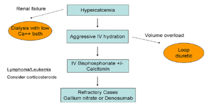
Treatment to increase calcium excretion and reduce bone resorption of calcium involves saline, sodium diuresis, and drugs such as zoledronate.
I present here 2 cases of hypercalcaemia seen in 2021 and quote some more cases from published articles to infer that hypercalcaemia is biomarker of hyperparathyroidism, Cancers, and Vitamin D toxicity, with major morbidity, serious complications, and deaths.
Physicians and Laboratories in developing countries including India need to develop a highly reliable index of suspicion (Ca/P Ratio?) because PHT escape diagnosis due to the subnormal calcium and its dependence on albumin levels and lack of PTH level assessment facilities.
Material & Methods: I, the author a family physician and with a standing experience in Public Health for 54 years, have used 2 cases of hypercalcaemia seen in 2021 and some other published cases, along with a few retrospective studies in India and abroad. Have reviewed the available literature globally of various conditions that lead to hypercalcaemia and current recommendations for the management of conditions with elevated serum calcium levels.
Introduction
Hypercalcaemia is not a disease, but a disorder characterized by laboratory test results showing an elevation of calcium levels in the peripheral blood. It is a common metabolic abnormality seen in both inpatient and outpatient settings. Hypercalcemia is a common metabolic abnormality seen in both inpatient and outpatient settings.
In plasma, calcium exists in 3 different forms: (1) 50% as ionized or the biologically active form, (2) 45% bound to plasma proteins (mainly albumin), and (3) 5% complexed to phosphate and citrate. Almost all our body's calcium is stored in your bones. A very small amount -- about 1% -- is in our blood as Ionized (Free) calcium in our blood (not attached to proteins), that helps in muscle contraction, nerve signalling, and blood clotting. Approximately, 40% to 45% of the serum calcium is attached to albumin, and serum calcium levels may fluctuate based on the serum albumin levels. Therefore, ionized, or free calcium levels should be measured when hypercalcemia is suspected. Hypercalcemia is categorized based on serum calcium levels, as i) Mild if calcium levels are 10.0–12 mg/dL (2.5–3 mmol/L) OR ionized calcium 5.25–8 mg/dL (1.31–2 mmol/L), ii) Moderate when total calcium levels is 12–14 mg/dL (3–3.5 mmol/L) and hypercalcaemic crisis when the total calcium level is > 14 mg/dL (> 3.5 mmol/L) [1, 2].
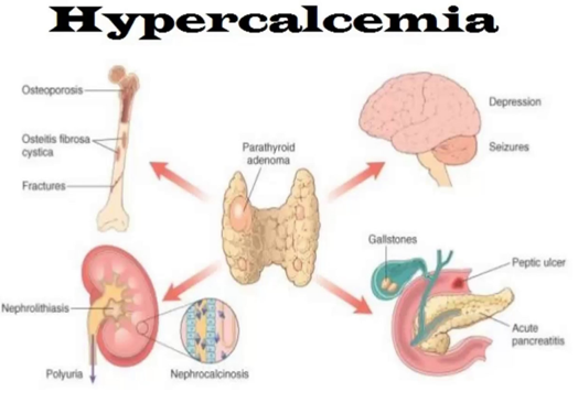
Hypercalcemia is caused by a) overactive parathyroid glands (hyperparathyroidism) that can stem from a small, benign tumour or enlargement of one or more of the four parathyroid glands b) Some types of Cancers and c) Some medicines like Thiazide diuretics, Teriparatide, Oestrogens, Tamoxifen and Lithium d) Over supplementation of Calcium- main forms of calcium supplements are carbonate, citrate, gluconate, and lactate. Some calcium supplements are combined with vitamins and other minerals.
Foods high in calcium include cheese, cottage cheese, yogurt, pudding, and ice cream. Too much calcium in your blood can weaken bones, create kidney stones, and interfere with how heart and brain work. Clinical manifestations include fatigability, muscle weakness, depression, anorexia, nausea, and constipation.
Clinicians should suspect if a routine Calcium level shows abnormal level and seek a PTH test. The confirmation of diagnosis in small settings depend solely on measuring parathyroid hormones in blood /serum. The test would cost between INR 700-1500 depending upon the city. Clinical laboratories generally measure the total serum calcium level. Changes in the albumin level causes misleading increases or decreases, respectively, in the total serum calcium level. It is valuable to measure the serum level of ionized calcium. Alternately there are formulas to calculate the serum ionized calcium level or to “correct” the total calcium level (add 0.8 mg per DL to the total calcium level for every 1.0 g per DL of serum albumin below the level of 3.5 g per DL).
Differential Diagnosis of Hypercalcaemia:
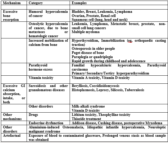
The differential diagnosis of hypercalcemia consists of A) Parathyroid hormone (PTH)-mediated hypercalcemia and B) non-PTH-mediated hypercalcemia. Causes of PTH-mediated hypercalcemia include primary hyperparathyroidism, familial hypocalciuric hypercalcemia, and ectopic PTH secretion. Causes of non-PTH-mediated hypercalcemia comprise vitamin D-mediated hypercalcemia (vitamin D intoxication, lymphoma, granulomatous diseases), vitamin A intoxication, hyperthyroidism, drug-induced hypercalcemia (hydrochlorothiazide, theophylline, and lithium), immobilization, milk-alkali syndrome, adrenal insufficiency, and humoral hypercalcemia of malignancy.
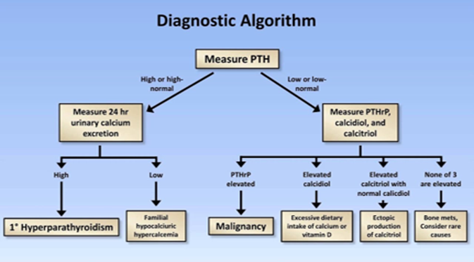
True hypercalcemia occurs through three basic mechanisms: i) enhanced osteoclastic bone resorption (in local osteolytic hypercalcemia, HHM, 1,25(OH)2D-secreting lymphomas, and the rare case of ectopic hyperparathyroidism); ii) enhanced renal tubular reabsorption of calcium (in HHM and ectopic hyperparathyroidism); and iii) enhanced intestinal absorption of calcium (in 1,25(OH)2D-secreting lymphomas and possibly ectopic hyperparathyroidism).
The diagnosis of hypercalcaemia is made when the corrected serum calcium concentration is 2 standard deviations above the mean of values found in people with normal calcium levels, in at least two samples at least one week apart over a period of three months. The presence of high or not adequately suppressed serum parathyroid hormone levels should point the diagnosis towards hypercalcaemia of parathyroid origins.
Mild hypercalcaemia is usually caused by primary hyperparathyroidism, the treatment for which is typically surgery. Persons aged 50 or more with serum calcium levels <0>
ICD 10 Classification:
In International classification (10) the hypercalcaemia is categorised under Disorders of mineral metabolism. E-83- Disorders of Mineral Metabolism, E83.50 - Unspecified disorder of calcium metabolism, E83.51 – Hypocalcaemia.
E 83.52 Disorders of Calcium Metabolism, - Hypercalcaemia, hypocalciuric, familial E83.52, Milk-alkali disease, or syndrome, Burnett's syndrome, Dementia (degenerative primary/ old age/ persisting) due to hypercalcemia with behavioural disturbance.
E83.59 - Other disorders of calcium metabolism [3]
Prevention: Not all hypercalcemia can be prevented but avoiding excess intake of calcium pills and calcium-based antacid tablets is recommended [2].
Management Principles:
When biochemical screening is common, asymptomatic primary hyperparathyroidism is the most likely form of the disease. In countries where vitamin D deficiency is prevalent and biochemical screening is not
a feature of the health care system, symptomatic disease with skeletal abnormalities is likely to predominate. When PTH levels are part of the evaluation for low bone mass, the normo-calcaemic variant is seen. Guidelines for surgical removal of hyperfunctioning parathyroid tissue apply to all three clinical forms of the disease.
Case Reports:
- Cases I saw in 2021:
- Shova Pradhan, daughter of A - 66-year-old Kamala Pradhan (names changed) called me in early December 2021 from Gurung Basti Kalimpong, West Bengal complaining that her mother had been complaining of pruritus, muscle weakness, lack of appetite and weight lost for 3 months. Kamala was a known diabetic of a decade under my treatment in Delhi with FBS-182, Hb1Ac (9,2) Vitamin D3 (25 Hydroxy) levels of 12.4 ng/ml in March 2018 and was advised to take oral antidiabetics, diet regulation and walking for 2-3 Kms a day and D-rise a Vitamin D supplement before once a week for 4 weeks, then once a fortnight for 4 times and once every month regularly thereafter. I moved to Bengaluru in April 2018, and they moved to Kalimpong in 2019, but our telephonic consultations continued once in 4-6 months. The history revealed that She continued to take D-Rise at weekly interval for almost 3 years and due to Covid 19 Pandemic walking was almost stopped. A blood tests in December 2021 in a local laboratory showed Hb1Ac 8.2, FBS 160, Creatinine 0.83 mg/dl and serum Calcium level 20 mg/dl and Vitamin D 150ng/ml.
We stopped the Vitamin D supplementation and advised over hydration with frequent fluids intake and diuretics and the recent report of 8 April the Calcium levels has come down to 10mg/dl and Vitamin D levels to 50ng/ml.
- A rural 45 year’s old women from Chikkaballapur 100 kms away from Bengaluru had reported on 4th May 2021 with symptoms vaginal bleeding, back pain, urine leakage & pelvic pain for 2 months to a gynaecologic oncology service at a cancer hospital & research centre, Bangalore. She had consulted a family doctor first who referred her to the Cancer hospital. On admission her Pulse was 72/min, BP 85/135 mm hg, Temperature 98.200 F, SPO2 97% (mandated in Covid 19 Pandemic) & Weight 44kgs.
INVESTIGATIONS: A battery of tests like blood sugar, cholesterol, LFT, THYROID and Parathyroid hormones & serological RA factors were normal, Serum Calcium was high at 120mg/dl & abnormal & Hb% was low (8dl/mg). After an evaluation for abnormal vagina bleeding, cervical biopsies that demonstrated invasive adenocarcinoma of the cervix (Stage IV B CA Cervix) the diagnosis was confirmed. Since it was in an advanced stage, a palliative management including radiation therapy for control of bleeding & pain, and systemic chemotherapy for disseminated disease was opted and was given IV fluids and with diuretic for hypercalciuria. After 22 days of treatment, she got discharged along with medications prescribed Inj. Amikacin 100mg IV OD, Inj. Pantoprazole 40mg IV OD, DNS which was given by the local doctor. However, there was no progress and she died on 17th June 2021.
- Multiple myeloma with hypercalcaemic encephalopathy, AIIMS Jodhpur 2019:
An 84-year-old female presented to the emergency department of AIMS Jodhpur, in October 2019 with complaints of constipation, vomiting, decreased appetite, and excessive urination for the last 10 days and altered sensorium for the last 2 days. There was a history of generalised bone pain for last 3 months for which she was prescribed calcium. Except Bell’s palsy 5 months back with complete recovery, past and family history was unremarkable. On examination, the patient was drowsy but arousable. She was dehydrated, and blood pressure was 146/90 mm Hg. Her systemic examination including neurological did not reveal any localisation.
Investigations revealed anaemia (Hb—6.5 g/dl), deranged renal function (urea—118 mg/dl, creatinine 5.62 mg/dl), increased total protein (9.27 g/dl) and globulin (7.34 g/dl), low albumin (1.93 g/dl), and hypercalcemia (total calcium-15.07 mg/dl). Random plasma glucose (131 mg/dl) and serum electrolytes (sodium/potassium—141/3.92 meq/L) were in the normal range. Serum PTH levels and serum ammonia levels were normal (39.4 mmol/L, normal 17–90 mmol/L).
Non-contrast computerised tomography of brain showed age-related changes in the bilateral cerebral hemisphere with multiple lytic lesions of varying sizes noticed in the cranial vault and base of the skull. The patient was diagnosed as hypercalcaemic encephalopathy with suspicion of multiple myeloma.
To confirm the diagnosis of multiple myeloma, serum protein electrophoresis was performed, which showed M protein (in the beta region) 4.9 g/dL with increased beta 2 microglobulin (9284 ng/ml, normal 609–2366 ng/ml). Bone marrow aspiration showed 60% plasma cells. The final diagnosis of multiple myeloma with hypercalcaemic encephalopathy was made.
The patient was treated with iv. fluids (0.9% normal saline, 200 ml/hr with the rate adjusted to maintain urine output 100–150 ml/hr). with loop diuretics and calcitonin. The next day, zolendronic acid (at modified dose according to GFR) was added. On day 9 of hospitalisation, the patient was put on dexamethasone, lenalidomide, and bortezomib. At the end of 2nd week patient’s sensorium was improved, with normalization of serum calcium and renal function [5].
- Hypercalcaemia with Breast Cancer:
- A 60-year-old woman with a history of breast cancer presents with confusion and dehydration. She had postural hypotension and low central venous pressure on examination of the jugular veins. The serum calcium level is 18.0 mg per decilitre (4.5 mmol per litre), Phosphorus level was 5.0 mg per decilitre (1.6 mmol per litre), the blood urea nitrogen level is 80.0 mg per decilitre (28.6 mmol per litre), the serum creatinine level is 2.0 mg per decilitres (177 μmol per litre), and the albumin level is 3.3 g per decilitres.
- A 48-year-old woman was given a diagnosis of infiltrating ductal breast carcinoma (T2N1M0; grade I; positive for oestrogen receptors, progesterone receptors and human epidermal receptor-2 [HER2] status; with 3/31 positive nodes). left lumpectomy with axillary node dissection followed by chemotherapy and radiotherapy was the treatment given. Thirteen years later, she was given a diagnosis of breast cancer recurrence. The patient had hypercalcemia on multiple occasions, with corrected calcium levels ranging from 2.72 to 2.85 (normal 2.1–2.6) mmol/L, dating back to the time of diagnosis of her breast cancer. She did not have symptoms directly attributable to hypercalcemia apart from one episode of nephrolithiasis 11 years after diagnosis. Hypercalcemia was again noted two years later, with a serum corrected calcium level of 2.95 mmol/L [7].
- Vitamin D intoxication:
- A 53-year-old male patient, with history diabetes mellitus, hypertension, non-dialysis chronic renal failure and smoking was admitted to a hospital in September 2011 to investigate worsening of the renal function, pruritus, muscle weakness, lack of appetite and weight lost. Table 1 displays the results of the biochemical tests before hospitalization
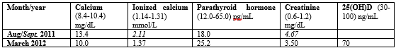
The treatment included hyperhydration; administration of furosemide, corticosteroid, and iron; and replacing erythropoietin by methoxy polyethylene glycol-epoetin beta due to suspected skin allergy. Intranasal and subcutaneous calcitonin was also introduced [8].
ii) A 70-year-old female, known hypertensive for thirty-five years and diabetic for seven years underwent total knee replacement (TKR) for osteoarthritis left knee in December 2010. For perioperative glycaemic control, multiple subcutaneous injections of insulin were advised. Patient later presented with poor glycaemic control, decreased appetite, and constipation for last 1 month with history of episodes of transient loss of consciousness for 15 days and recurrent vomiting. Biochemical work-up showed hypercalcemia (Serum calcium 12.4 mg/dL). Sr. albumin, ALP, Sr. phosphorus and PTH levels were normal, thus suggesting PTH independent hypercalcemia. A check of vitamin D levels were 2016 ng/mL, thus confirming vitamin D toxicity. Retrospective analysis of treatment history revealed patient receiving 4 injections of Architol (6 Lac units IM injections) prior to presentation. Work-up for malignancy was negative, brain imaging and EEG were normal. Holter was suggestive of intermittent CHB. Patient was given hydration, injection calcitonin 100 I.U. subcutaneously, injection pamidronate 60 mg infusion, with serum calcium levels normalizing, with relief in constipation, vomiting and behavioural improvement. However, persistence of rhythm disturbances led to permanent pacemaker placement. (Vitamin D toxicity presenting as hypercalcemia and complete heart block [9].
5. A Case of Constipation due to hypercalcemia secondary to TB in Dharwad, Karnataka:
A 75-year-old monk was admitted to SDM College of Medical Sciences and Hospital, Sattur, Dharwad-580009, Karnataka, India in 2014 with complaints of constipation for the past 3-4 months who was on Laxatives and intermittent enema with partial benefit. He had mild anorexia and no significant weight loss.
On examination he was well built and moderately nourished elderly male with borderline hypertension. Systemic examination revealed mild right sided pleural effusion and other systems were unremarkable.
Investigations revealed normal hemogram, normal thyroid function studies, mild renal dysfunction, normal urinalysis, and moderate Hypercalcemia.
He was treated with hydration for hypercalcemia but was not getting optimized & renal failure was worsening. The intact parathyroid hormone (PTH) was low suggestive of non-PTH related hypercalcemia. His Vitamin D3 {1, 25 (OH) 2} level was available after a week which was very much elevated.
HRCT Chest showed multiple nodular lesions along the broncho-vascular bundles, in both upper lobes, right middle lobe and anterior segment right lower lobe. Fibrotic streaks were also noted in bilateral apical region. Few calcified bilateral hilar lymph nodes noted
A provisional diagnosis of sarcoidosis was made, and he was put on prednisolone (0.5 mg / Kg / day). After 4 days of starting prednisolone, he started having high grade fever with chills and cough with expectoration. His sputum samples were positive for acid fast bacilli and hence was put on Anti-Tuberculous Therapy (ATT). After about 15 days of starting steroids and 1 week of ATT his fever subsided, sensorium improved. His hypercalcemia and renal dysfunction normalized. His constipation got relieved.
Steroids were withdrawn completely at the end of 4 weeks. He completed the full course of ATT for 6 months and at the completion of ATT he was free from constipation, renal functions remained normal and there was no hypercalcemia.
Hypercalcemia is seen in ~ 25
Discussions:
Many organs are involved in the regulation of calcium. Chief among these are the parathyroid glands and, when calcium levels drop, the parathyroid glands increase secretion of parathyroid hormone (PTH). Vitamin D, which is partially regulated through PTH, also plays an important role in the regulation of calcium. The first step of vitamin D metabolism occurs at the skin- ultraviolet light catalyses the production of Vitamin D3, (cholecalciferol), from 7-dehydrocholesterol. Cholecalciferol is then hydroxylated at the 25 positions by the liver to form 25-hydroxycholecalciferol (called calcifediol). Calcifediol is then hydroxylated at the 1 position in the kidney to form 1,25-dihydroxycholecalciferol (calcitriol). This final step is regulated by PTH, and calcitriol is the active form of vitamin D. Calcitriol increases serum calcium by causing increased calcium absorption in the intestines, increased calcium reabsorption in the kidneys, and stimulation of osteoblasts to reabsorb calcium from bone
Primary Hyperparathyroidism:
Primary hyperparathyroidism (PHPT), the most common cause of hypercalcemia, is often identified in postmenopausal women with hypercalcemia and parathyroid hormone (PTH) levels that are either frankly elevated or inappropriately normal. PHPT can present as adenoma (85%), hyperplasia (14%) or carcinoma (1%) with 1% of the adult population and incidence increasing to 2
Conclusions:
Hypercalcaemia is a biomarker for detecting hyperparathyroidism currently with major morbidity and serious complications. Assessing ionized, calcium levels is the method of choice when hypercalcemia is suspected but costly and rarely available in smaller towns. If biochemical screening becomes a national norm, asymptomatic primary hyperparathyroidism will be the most likely form of hypercalcaemia to be unmasked. In countries where vitamin D deficiency is prevalent and biochemical screening is not a feature of the health care system, symptomatic disease with skeletal abnormalities is likely to predominate.
Physicians and Laboratory science in developing countries including India need to develop a high index of suspicion because PHT escapes diagnosis due to the subnormal calcium and PTH levels assessment facilities. If PTH levels are part of the evaluation for low bone mass, the normo-calcaemic variant is seen. In such situation Ca/P ratio is the best indicator for detecting Normocalcaemic hyperparathyroidism.
Ca/P ratio is a valuable tool for the diagnosis and screening of PHPT in combination with serum Ca levels and is of superior value compared to serum Ca alone, especially in normo-calcaemic hyperparathyroidism cases (NCPHT). Ca/P is simple, inexpensive, and easily accessible worldwide, especially in laboratory/medical settings relying on limited resources.
Surgical removal of hyperfunctioning parathyroid tissue apply to all three clinical forms of the disease.
Not all hypercalcemia can be prevented but avoiding excess intake of calcium pills and calcium-based antacid tablets is recommended.
References
- Zhu N, Zhang D, Wang W, et al. (2020) A Novel Coronavirus from Patients with Pneumonia in China, 2019. N Engl J Med. 382(8):727-733.
View at Publisher | View at Google Scholar
