

Case report | DOI: https://doi.org/10.31579/2834-8761/052
Treatment of seborrheic keratosis with non-ablative laser
1 First and Second Degree Specialist in General Surgery and Aesthetic Medicine, Assistant Professor, Assistant Researcher and Consulting Professor.
2 Specialist in Laser Medicine and phototherapy in dermoaesthetic pathologies and Researcher in the development of different dermatological and vascular lesions with the use of lasers.
3 Lic.M In general genetic engineering and main in viruses and bacteria
*Corresponding Author: Pedro Rolando López Rodríguez., First and Second Degree Specialist in General Surgery and Aesthetic Medicine, Assistant Professor, Assistant Researcher and Consulting Professor.
Citation: Pedro Rolando López Rodríguez, Dr.M. Lázaro Pérez Rodríguez, Lic.M Mercedes Gil García, (2024), Thromboembolic Risk and Testosterone Replacement Therapy: Debunking Myths and Clarifying Evidence with Recent Systematic Reviews and Meta-Analyses, 3(3): 10.31579/2834-8761/052
Copyright: © 2024, Pedro Rolando López Rodríguez. This is an open access article distributed under the Creative Commons Attribution License, which permits unrestricted use, distribution, and reproduction in any medium, provided the original work is properly cited.
Received: 03 May 2024 | Accepted: 18 May 2024 | Published: 20 June 2024
Keywords: seborrheic keratosis, non-ablative laser
Abstract
Introduction. Seborrheic keratosis is a common non-cancerous (benign) skin neoplasm. It can develop as a single tumor or in a group. Its treatment with non-ablative laser is excellent. Objective. To describe the clinical characteristics of a patient with a large seborrheic keratosis lesion and to be able to treat it with non-ablative laser. Clinical Case: An observational study was carried out on a 56-year-old SRC patient, with a dermatological diagnosis of a seborrheic keratosis lesion, who was treated with non-ablative laser with excellence, without leaving a scar, with the lesion falling off after three weeks and without performing surgery. Conclusions: Seborrheic keratosis is a benign dermatological lesion and its treatment with non-ablative laser is effective, it leaves no scar without the use of surgery.
Introduction
Seborrheic keratoses are common, noncancerous (benign) skin growths. People tend to get more of them as they get older. 1 Seborrheic keratoses are usually brown, black, or light brown in color. The growths (lesions) have a waxy, scaly, and slightly raised appearance. 2 They appear progressively, usually on the face, neck, chest, or back. Seborrheic keratoses are harmless and not contagious. Seborrheic keratoses are usually circular or oval growths that range in color from light tan to black. They may develop as a single tumor or in a group 3. Seborrheic keratoses (SK) are easily recognized through a clinical and dermoscopic approach, however, some lesions behave in a way that simulates different skin conditions that lack typical clinical and dermoscopic criteria 4. Seborrheic keratoses (SK) are easily recognized through a clinical and dermoscopic approach, however, some lesions behave in a way that simulates different skin conditions that lack typical clinical and dermoscopic criteria 5. Seborrheic keratoses are very common benign tumors, especially in the elderly. It is not uncommon for them to coexist with other dermatological pathologies, including skin lesions of mycosis fungoides. We present a 41-year-old patient with mycosis fungoides and multiple seborrheic keratoses, predominantly located on the mycosis fungoides plaques. The histopathological findings of one of these lesions showed infiltration of the seborrheic keratosis by atypical T lymphocytes . Seborrheic keratoses are absolutely benign lesions, so they are removed due to the symptoms that cause itching, bleeding due to irritation or rubbing with clothing and because they are aesthetically bothersome. Lesions can be removed without risk of scarring using laser therapy 6.
Method
An observational study was carried out on a case diagnosed and treated with seborrheic keratosis (SK) on the right side of the face. The patient's clinical history was reviewed in detail, and an adequate application of the clinical method was carried out, which allowed collecting the necessary data for the interrogation and physical examination. Diagnostic and therapeutic means were used, among which were: They find dermatological evidence, analysis and the use of the XEO machine, Genesis laser handpiece with a programming of: Fluence 300 j/cm 2, Pulse duration 6 ms., Repetition frequency 00 Hz and Spot size 3 mm.
ClinicalCase
A 56-year-old white male patient attended the International Medical Technology Center in Waas, Madrid, due to a sensation of itching on the right side of the face caused by a large lesion that has increased over time.
Physical examination revealed a large, warty, pigmented, blackish lesion in the right temporofrontal region, approximately 2-3 cm in diameter, irregular, with well-defined edges, painless, firm, and somewhat mobile for 3-4 years.
A dermatological study was performed, confirming a benign lesion diagnosed as a Seborrheic Keratosis lesion.
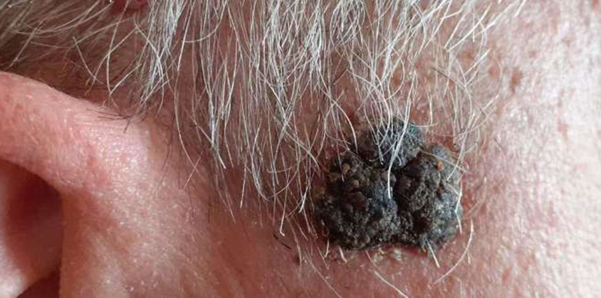
Figure 1: Seborrheic Keratosis lesion of the patient.
This lesion was treated with a 1064 nm Neodymium YAG Laser. XEO, with a programming of 300 j/cm2, 6 milliseconds, 00 Hz and 3 mm. The point does not require anesthesia and is painless and is performed on an outpatient basis and you can return to work without days of rest.The results are obtained when the lesion falls off in three to four weeks without leaving a scar or recurrence. No need for surgery.The treatment performed by conventional surgery requires general anesthesia and the use of an operating room.The surgical action is performed in two ways: 1.- We remove the lesion in block and the skin is closed spontaneously. This process takes about two months, called healing by secondary intention, frequently causing wet cures. 2.- We remove the lesion and close the skin with the flap interposition technique, leaving as sequelae a significant scar in the aesthetics for the patient and a high cost and loss of days of activity for the patient and the family, in addition to the discomfort caused by the complications that may arise.The real advantages that we offer in the treatment of these lesions with the application of the non-ablative laser in our Center are evident.
Discussion
Seborrheic keratoses are benign skin tumors that occur very frequently and most people will develop them in very variable numbers (from a few to hundreds) during their lives. They most frequently affect people over 30 years of age. These tumors can appear in any location, although they have not been described in mucous membranes. When located on the trunk and are numerous, they may present a ‘Christmas tree’ pattern, with their longest axis parallel to the skin folds or Blaschko lines 7. SK have not been associated with malignancy; they are considered banal lesions that usually constitute an aesthetic problem and that occasionally cause mild symptoms, such as itching or irritation 8. There are several histological forms of seborrheic keratosis (acanthotic, reticulated, irritated, macular, among others). The clonal subtype is characterized by well-demarcated or larger basaloid cell nests within a seborrheic keratosis 9. Seborrheic keratoses do not require treatment and in patients with a high number of lesions, their excision may be impractical. If they cause discomfort due to irritation, rubbing, or cosmetic problems, they can be treated with cryotherapy, electrocoagulation, laser therapy, or excised.10 Melanomas that clinically mimic other skin lesions, particularly seborrheic keratosis, may delay diagnosis and timely treatment. Differentiating between melanocytic and non-melanocytic lesions by dermoscopy allows for early detection of melanomas and prevents the misdiagnosis of SK instead of melanocytic tumor(9). Dermoscopy has been shown to improve diagnostic accuracy among dermatologists and to improve skin cancer detection even among non-dermatologists. For this reason, the use of this tool should be encouraged as an additional and complementary technique, since it significantly improves the sensitivity for the detection of melanocytic lesions.2 However, suspicious lesions should be biopsied, to achieve not only a diagnosis, but also histological microstaging of the lesion 11, 12. Seborrheic keratosis appears on the skin and is distinguished by a diagnostic triad: hyperkeratosis (thickening of keratin in the skin), acanthosis (thickening of the epidermis) and papillomatosis, which is the formation of invaginations of the epidermis in the dermis. The diagnosis based on morphological findings was seborrheic keratosis. This tends to have multiple and varied presentations that occur, above all, in areas of the body with skin 13. The use of IPL can also be effective in the treatment of seborrheic keratoses, since these are superficial and discretely pigmented lesions14. For this purpose, filters must be used that allow the selection of short λ (530nm), which act on the epidermis and are safe in the treatment of superficial lesions. The mechanism of action is probably related to the absorption of radiation by epidermal melanin and subsequent thermal diffusion to the surrounding cells; This causes epidermolysis that increases the regeneration of keratinocytes with the elimination of necrotic ones through microcrusts, thus eliminating these keratinocyte lesions14. There are no published studies, but the British Skin Laser Study Group presented at the meeting of the British Medical Laser Association (2011) a series of cases of seborrheic keratosis treated successfully and with good cosmetic results using a session of IPL 14, 15,16,17.
Conclusions
Seborrheic keratosis is a benign dermatological lesion and its treatment with non-ablative laser is effective. The lesion falls off spontaneously within 3-4 weeks, leaves no scar, does not require anesthesia, does not require surgery, is performed on an outpatient basis and the patient can continue with their normal activity.
Conflict of interest.
The authors declare that they have no conflict of interest.
References
- Melgosa Ramos FJ, Aguado Vásquez A, Mateus Puchades A. Queratosis seborreica irritadas múltiples, una causa infrecuente de intertrigo.Med Faml Imag.2022; Vol 48 (5): 39-40 DOI: ((doi.org/10.1016/j.semerg.2022.01.005
View at Publisher | View at Google Scholar - Oliveira A, Cardosa J, Zalande k I, Solitary angiokeratoma with meyerson phenomenon . J Am Acad Dermatol.2017; 76, pp 516-518.
View at Publisher | View at Google Scholar - Massco M, Manfreda V, Diluvio L, Dattola A, Bianchi L, Campione E. Análisis dermatoscópico de 72 queratosis seborreicas atípicas. Actas Dermosifiogr. 2019; Vol 110 (5): 366-371.
View at Publisher | View at Google Scholar - Kirilovsky P, Peralta Montes S, Diaz D, Sanchez MP, Mariarch P, Eidilstein D. Queratosis seborreica: variante clonal .Reporte de un caso. Rev Argent Dermatol . 2020; Vol 101 (2): https: //www.dermatolog.org.ar
View at Publisher | View at Google Scholar - Pinilla Martin B, Tercedor Sánchez J, Queratosis seborreica: ¿solo en adultos ‘Piel.2020; Vol 35(3): 139-140 https: //10.1016/j.piel.2019.05.008
View at Publisher | View at Google Scholar - Sturgeon de la Serna V, Guiangualano E, Celeste Pizarro M, Vannelli M, Garcia S, Rossi V. Queratosis folicular invertida.Dermatol Arg. 2024; vol 30 (1): https://www.dermatolarg.org.ar
View at Publisher | View at Google Scholar - Perez Dieste JM, Castro Viejo Bolivar M, Santos C. Queratosis seborreica en margen palpebral. Arch Socied Esp Oftalmol.2020; Vol 95(11): https://doi.org/10.1016/j.oftal.2020.05.007
View at Publisher | View at Google Scholar - Monteagudo B, Mosquera Martinez MT, Figueroa O, Suarez Magdalena O. Queratosis seborreica en la concha auricular. Piel J. 2018; Vol 33(10): 665-667 https://doi.org/10.1016/j.piel(2018.01.014
View at Publisher | View at Google Scholar - Sanchez MP, Mariarch P, Eidilstein D, Kirilovsky P, Peralta Montes S, Diaz D.Queratosis seborreica variante clonal. Reporte de un caso. Rev Arg Dermatol.2020; Vol 101 (2): https://www.clinicapiel.com.ar
View at Publisher | View at Google Scholar - Mayorga Rios RM, Sierra Silva G, Torres Delgadillo R, Hernández Torres MM. Paraqueratosis granular. Dermatol Rev Mex.2023; Vol 67 (3): 410-414 https://doi.org/10.24245/dom/bmv.v6713
View at Publisher | View at Google Scholar - Arciniegas Grisoles V, Jimenez Oliveros C, González Alvares T, Andres Flores H. Melanoma y queratosis seborreica: a propósito de dos casos de difícil diagnóstico. Rev Cienc Biolg.2021; Vol 10 (4):281-287 https://doi.org/10.32997/rcb-2021-3672
View at Publisher | View at Google Scholar - Goldsmith JF, Montaser Kouhsari L, Tahan SR. Queratosis seborreica de patron clonal: riesgo de recurrencia y progresión. Am J Surg Pathol.2022; Vol 46 (12):1642-1649(Pub Med )
View at Publisher | View at Google Scholar - Gómez Londoño M, Echevarria Restrapo LM, Gutierrez Sanmartin JM, Meriño Correa SC, Patricia Henao C, Sanin Ramirez D. Seborrheic keratoses in the recto-uterine pouch:case report. Rev Ginecol Obst Mex.2021; Vol 87(12): https: //doi.org/10.24245/gom.V87i1d.3265
View at Publisher | View at Google Scholar - González Rodríguez AJ, Lorente Gual R. Indicaciones actuales y nuevas aplicaciones de los sistemas de luz pulsada intensa. Actas Dermosifiliog.2015; Vol 16(5): 350-364 https://doi.org/10.1016/jad.2014.10.004
View at Publisher | View at Google Scholar - Uriarte Ruiz K, Amador Rogero ME, Vega Memije ME, Tounssaint Caire S, Torres Barragan G, Hernandez Vera M. Quiz queratosis seborreica.Dermatol Cosmet Med Quirg.2022; Vol 19(4): 415-417 https://reseahgate.net/publication/360001091
View at Publisher | View at Google Scholar - Barthelmanns Butsch F, Lang BM, Stege H, Grobmann B, Scchepler H, Grabbe S. Queratosis seborreica. J. Dtsch Dermatol Ges. 2023; Vol 23(3): 265-277 (Pub Med) https://doi.org/10.1111/ddg.14984
View at Publisher | View at Google Scholar - Sun MD, Halpern AC. Advance in the etiology , detectión and clínical management of seborrehic keratoses. Dermatology.2022; 238(2):205-217 (Pub Med) https: //doi.org/10.1159/000517070
View at Publisher | View at Google Scholar
