 Clinic
Case Report | DOI: https://doi.org/10.31579/2834-8761/111
Successful Medical Management of Pituitary Apoplexy Presenting with Severe Neuro‑Ophthalmic Deficits
1UCLA Olive View Medical Center, Department of Internal Medicine
2UCLA Olive View Medical Center, Department of Endocrinology
*Corresponding Author: Ahmed N, Internal Medicine Department Hepatology and Gastroenterology unit, Faculty of Medicine, Mansoura University.
Citation: Rebekah C. Smith, Nasser Mikhail, (2026), Successful Medical Management of Pituitary Apoplexy Presenting with Severe Neuro‑Ophthalmic Deficits, Clinical Endocrinology and Metabolism, 5(3); Doi:10.31579/2834-8761/111
Copyright: © 2026, Rebekah Cossette Smith. This is an open access article distributed under the Creative Commons Attribution License, which permits unrestricted use, distribution, and reproduction in any medium, provided the original work is properly cited.
Received: 08 May 2026 | Accepted: 19 May 2026 | Published: 26 May 2026
Keywords: hypothyroidism; hemodynamically; neurosurgery
Abstract
Pituitary apoplexy is an acute neuroendocrine syndrome caused by hemorrhage or infarction of the pituitary gland, usually within an unrecognized pituitary adenoma, and may present with headache, ophthalmoplegia, visual disturbance, and hypopituitarism. We report a 71-year-old man with atrial fibrillation, hypertension, and type 2 diabetes mellitus who developed progressive diplopia and ptosis after elective parotidectomy. Initial postoperative imaging identified a hemorrhagic 2.6 cm sellar/suprasellar mass with extension into the right cavernous sinus, consistent with pituitary apoplexy, with evolving cranial nerve III and later VI palsies. Despite severe ocular motor deficits, he remained hemodynamically stable without optic neuropathy or major visual field compromise. Multidisciplinary management included Intensive Care Unit (ICU) monitoring, stress-dose hydrocortisone, serial ophthalmologic and endocrine assessment, levothyroxine initiation for central hypothyroidism, interval imaging, and delayed resumption of anticoagulation. Neurosurgery deferred urgent decompression in favor of conservative management with close follow-up and offered elective decompression. The patient improved clinically, with near-complete resolution of ptosis and marked recovery of cranial nerve III and VI palsies by 2 months, accompanied by radiographic regression of the hemorrhagic sellar mass without surgical correction. This case highlights how pituitary apoplexy may not always represent a neurosurgical emergency. Future studies should investigate whether stable patients with more severe neuro-ophthalmic deficits may achieve favorable outcomes with multidisciplinary conservative management.
Introduction
Pituitary apoplexy is a rare and potentially life-threatening clinical syndrome caused by acute hemorrhage and/or infarction of the pituitary gland, most often within a previously undiagnosed pituitary macroadenoma [1]. Clinical presentation is widely variable depending on whether there is pituitary infarction causing subacute, gradual symptoms; or rapid hemorrhage producing acute, more severe symptoms involving compression of cavernous sinus structures [1]. Most common signs include sudden headache, ophthalmoplegia from cranial nerve III, VI, and IV palsies, visual deficits from optic nerve compression, and endocrine dysfunction from pituitary injury[1]. It is exceptionally rare, with one study identifying a prevalence of about 6 people for every 100,000 within Oxfordshire, UK[2]. At its original discovery, pituitary apoplexy was treated as a neurosurgical emergency [3], but contemporary evidence suggests that outcomes after conservative management may be comparable to surgery[4]. Roughly 30 – 40% of cases of pituitary apoplexy have an identified precipitating cause, ranging widely from pituitary tumors, surgical procedures, pregnancy and pregnancy complications, medications including hormone therapies, arterial hypertension, diabetes mellitus, coagulopathies, shock, and head trauma 1.
This case illustrates successful management of pituitary apoplexy presenting with headache, cranial nerve III and VI palsies including complete ptosis with prompt glucocorticoid intravenous therapy and thyroid replacement therapy.
Case Report
A 71-year-old man with atrial fibrillation (on rivaroxaban, held preoperatively), hypertension, and type 2 diabetes mellitus underwent elective right superficial parotidectomy for pleomorphic adenoma at an academic teaching hospital in Los Angeles County. The procedure was complicated by transection of branches of the right facial nerve with immediate intraoperative repair. On postoperative day one he demonstrated slight right buccal weakness and developed a headache and endorsed having diplopia since awakening from surgery. He subsequently developed right eyelid swelling, ptosis, and diplopia. He otherwise did not have facial paralysis. A code stroke was activated for complete ptosis and worsening extraocular movement deficits in adduction, elevation, and depression consistent with a right cranial nerve III palsy. A CT scan of the head revealed a suprasellar mass with extension into the right cavernous sinus. Further studies included cerebral perfusion studies, angiography and venography of the head and neck, dedicated orbit and sella CT, and MRI brain, Sella, and internal auditory canal. These were grossly normal except for a small subacute infarct in the right cingulate cortex which did not correlate with ocular symptoms. MRI confirmed a hemorrhagic mass of the right sella and suprasellar cistern suspicious for a pituitary hemorrhage with mass effect measuring 1.9 x 3.1 x 2.0 cm (Figure 1). Reassuringly, the patient remained hemodynamically stable throughout the hospital course. Additionally, endocrine function was stable based on blood markers including FSH of 5.1 mlU/mL (normal range 1.5 – 12.4 mlU/mL), LH of 1.8 mlU/mL (normal range 1.7 – 8.6 mlU/mL), prolactin of 6.3 ng/mL (normal range 4.0 – 15.2 ng/mL), TSH of 0.24 ulU/mL (normal range 0.27 – 4.20 ulU/mL), and initial free T4 of 1.12 ng/dL (normal range 0.92 – 1.68 ng/dL). However, four days after symptom onset, the repeat free T4 dropped to 0.86. A multidisciplinary team was involved by the 4th postoperative day including ophthalmology, neurology, neurological surgery, and endocrinology. The patient was upgraded to the Intensive Care Unit (ICU) for once every hour neurological checks and close blood pressure management for high concern for hemorrhagic expansion. An AM cortisol on the 4th postoperative day returned low at 3.2 mcg/dL (normal range 4.8 – 19.5 mcg/dL) concerning for adrenal insufficiency however other labs were notable for a sodium of 138 (normal range 135 – 145 mmol/L), blood glucose of 111 mg/dL (normal range 65 – 99 mg/dL), with normal blood pressures. Stress dose steroids were started at a regimen of hydrocortisone 50mg intravenous (IV) push every 6 hours. A subsequent ophthalmology evaluation indicated gaze restriction in elevation, depression, adduction as seen before in addition to an abduction deficit concerning for a new CN VI palsy. Reassuringly, there was no evidence of papilledema or optic nerve damage based on Optical Coherence Tomography (OCT) testing of both optic nerves and a dilated fundus exam. Given the overall reassuring ophthalmic evaluation and continued hemodynamic stability, the patient was cleared by neurosurgery surgery from requiring expedient surgical intervention by the 6th postoperative day and outpatient follow up for evaluation of elective surgical intervention was scheduled. Daily assessment of serum free t4 initially trended from 1.12 to 0.86 ng/dL, along with a reduction in TSH from 0.24 to 0.16 ulU/mL suggestive of new-onset central hypothyroidism so Levothyroxine 100 mcg IV daily in the morning was initiated. Additionally, stress steroids were continued for the concern of adrenal insufficiency as well as for the anti-inflammatory benefit of steroids to relieve edema within the Cavernous Sinus from acute hemorrhage. Given the overall clinical stability, the patient was resumed on anticoagulation for atrial fibrillation with a continuous heparin drip for quick reversal if necessary. The patient remained in the ICU for a total of four days before downgrading to the medical/surgical unit on post operative week one, hemodynamically stable on daily levothyroxine 100 mcg / daily, a steroid taper, and a heparin drip. The original ENT surgical team continued to follow the patient daily for post operative surgical management of the right parotidectomy. On post operative day eight the patient was transitioned from a heparin drop to his home oral anticoagulation regimen. Steroids were tapered at a regimen of hydrocortisone 50 mg IV every 6 hours for the first 72 hours, followed by 50mg IV every 8 hours for the next 48 hours, followed by 50 mg IV every 12 hours for the next 48 hours, followed by 25 mg orally twice a day for the next 48 hours, followed by maintenance dose of 20 mg orally daily for one month. Additionally, the patient was transitioned from IV levothyroxine 100 mcg daily after three days to maintenance once daily levothyroxine 125 mcg orally. By post operative week two the patient was discharged from the hospital. A week later the patient consulted with neurosurgery outpatient for elective resection of the sellar mass. He opted to continue radiographic monitoring without surgical intervention. Two months after the original presentation, the patient experienced a near total resolution of ptosis with marked improvement in cranial nerve III and VI palsies, and no field deficits by confrontational visual field exam. The patient continued to require oral levothyroxine 125 mcg supplementation for residual central hypothyroidism. Interval MRI imaging showed regression in size of the original Sellar hemorrhagic mass, shrinking from 1.9 x 3.1 x 2.0 cm to 1.8 x 2.3 x 1.4 cm (Figures 1 and 2) in just a little over a month. The patient continues to be followed by ophthalmology, endocrinology, and neurosurgery outpatient.
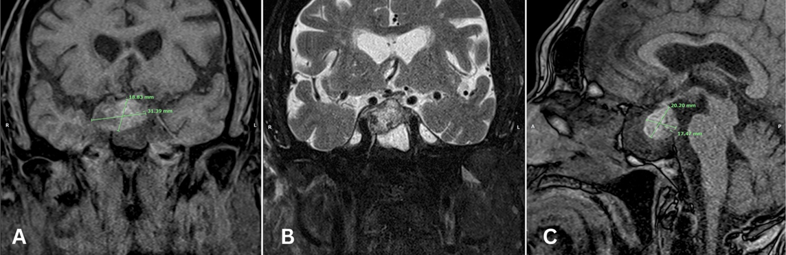
Figure 1: Original imaging of pituitary macroadenoma with associated hemorrhage measuring 1.9 x 3.1 x 2.0 cm; showing T1 Coronal (A), T2 Coronal (B), and Sagittal views (C).
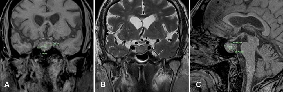
Figure 2: Subsequent imaging 1 month later with reduced size of hemorrhagic mass 1.8 x 2.3 x 1.4 cm; showing T1 Coronal (A), T2 Coronal (B), and Sagittal views (C).
Discussion
This case highlights the importance of multidisciplinary evaluation in determining appropriate management of acute pituitary apoplexy. Since the earliest publications on pituitary apoplexy management, providers have gradually shifted focus from initially recommending prompt neurosurgical decompression [3] to considering a conservative approach involving only medical management with aggressive glucocorticoid and hormonal replacement as spontaneous regression is common[1,4,5]. Currently, this decision point remains controversial especially given the wide variability in how pituitary apoplexy presents clinically [1]. The available academic literature is limited, often revealing conflicting results between studies and demonstrating a bias to pursue surgery in patients with more severe symptoms[4,6]. Also, though some patients are initially treated conservatively, they may later elect for surgical management for varying long-term complications [6]. The 2025 Pituitary Society practice guideline recommends considering surgery for most patients with visual symptoms or somnolence caused by pituitary apoplexy yet encourages an interdisciplinary discussion to account for the unique features in patients with incidentally discovered pituitary adenomas including functional status, age, co-morbidities, and severity of symptoms [7]. A prospective, multicenter study followed 97 patients with similar presentations and discovered that between the 67 patients who underwent surgery, and the 30 who were managed medically there were similar 3- and 6- month outcomes in visual acuity, cranial nerve palsies, and hormonal derangements [4]. The case report we present offers an example of a positive 2-month outcome in medically managed pituitary apoplexy which originally presented with severe features including pronounced oculomotor restriction of both CN III and VI, complete ptosis, and somnolence caused by mass effect. Our case additionally highlights the potential importance of serial endocrine monitoring in patients with pituitary apoplexy when surgical intervention is deferred. Although the patient’s free T4 level was initially within normal limits, it subsequently declined four days after symptom onset, consistent with delayed-onset central hypothyroidism. To our knowledge, delayed development of central hypothyroidism in pituitary apoplexy is not well described. While current guidelines recommend serial assessment of thyroid function for up to 8 weeks following surgical intervention to evaluate the need for thyroid hormone replacement [8], our findings suggest that close interval monitoring may also be warranted in conservatively managed patients during the first 7 days after presentation at the minimum. Additionally, it exemplifies how surgery and anticoagulation can be precipitants of pituitary apoplexy, two recognized risk factors based on existing literature[1]. It is important to continue investigating how medical management alone compares to surgical intervention in this disease entity, accounting for both short term and long-term data.
Conclusion
Pituitary apoplexy may not universally require emergent neurosurgical intervention. In hemodynamically stable patients, even the presence of severe or progressive visual compromise may be managed conservatively with close neurologic monitoring acutely, aggressive hormonal replacement, and serial imaging. Our single case of pituitary apoplexy demonstrates safe deferral of neurosurgical intervention using high dose steroid therapy with near total resolution of complete ptosis, and significant improvement in both cranial nerve III and VI palsies as well as a return to baseline mental status. Interval radiographic imaging showed regression in the size of the hemorrhagic mass, corresponding to the observed clinical improvement.
References
- Iglesias P. Pituitary apoplexy: an updated review. J Clin Med. 2024;13(9). doi:10.3390/jcm13092508
View at Publisher | View at Google Scholar - Fernandez A, Karavitaki N, Wass JAH. Prevalence of pituitary adenomas: a community-based, cross-sectional study in Banbury (Oxfordshire, UK). Clin Endocrinol (Oxf). 2010;72(3):377-382. doi:10.1111/j.1365-2265.2009.03667.x
View at Publisher | View at Google Scholar - Laws ER, Ebersold MJ. Pituitary apoplexy--an endocrine emergency. World J Surg. 1982;6(6):686-688. doi:10.1007/BF01655359
View at Publisher | View at Google Scholar - Mamelak AN, Little AS, Gardner PA, et al. A prospective, multicenter, observational study of surgical vs nonsurgical management for pituitary apoplexy. J Clin Endocrinol Metab. 2024;109(2):e711-e725. doi:10.1210/clinem/dgad541
View at Publisher | View at Google Scholar - Boyke AE, Michel M, Mamelak AN. Pituitary Apoplexy: a re-appraisal of risk factors and best management strategies in the COVID-19 era. Pituitary. 2024;27(6):898-908. doi:10.1007/s11102-024-01420-0
View at Publisher | View at Google Scholar - Donegan D, Erickson D. Revisiting Pituitary Apoplexy. J Endocr Soc. 2022;6(9):bvac113. doi:10.1210/jendso/bvac113
View at Publisher | View at Google Scholar - Fleseriu M, Gurnell M, McCormack A, et al. Pituitary incidentaloma: a Pituitary Society international consensus guideline statement. Nat Rev Endocrinol. 2025;21(10):638-655. doi:10.1038/s41574-025-01134-8
View at Publisher | View at Google Scholar - Rajasekaran S, Vanderpump M, Baldeweg S, et al. UK guidelines for the management of pituitary apoplexy. Clin Endocrinol (Oxf). 2011;74(1):9-20. doi:10.1111/j.1365-2265.2010.03913.x
View at Publisher | View at Google Scholar