

Review Article | DOI: https://doi.org/10.31579/2835-7957/158
Successful Management of Infectious Keratoconjunctivitis in Sheep
*Corresponding Author: Luciana de Barros Correia Fontes, Federal University of Pernambuco (Clinic Hospital – HC-UFPE/Ebserh).
Citation: Natinael D. Kalacho, (2026), Successful Management of Infectious Keratoconjunctivitis in Sheep, Clinical Reviews and Case Reports, 5(2); DOI:10.31579/2835-7957/158
Copyright: © 2026, Natinael Dawit Kalacho. This is an open-access article distributed under the terms of the Creative Commons Attribution License, which permits unrestricted use, distribution, and reproduction in any medium, provided the original author and source are credited.
Received: 27 February 2026 | Accepted: 12 March 2026 | Published: 19 March 2026
Keywords: infectiouskeratoconjuctivitis; sheep; treatment; wolaita sodo
Abstract
Infectious keratoconjunctivitis is a highly contagious ocular inflammation frequently reported in domestic sheep and goats. In present case study about seven local bred sheep were brought to Wolaita Sodo town veterinary clinic with history of ocular discharges, opacity of cornea and reduced feed intake. The clinical investigation reveals that the sheep were dull, with conjuctival hyperemia, corneal opacity and presence of ulceration on the corneal surface. Based on history and presented clinical signs the present condition was tentatively diagnosed as ovine infectious keratoconjunctivitis (OIKC). Parenteral short acting Oxytetracycline administration (10%) for three cosequetive days was found to be effective in treating pink eye in small ruminants.
Introduction
Infectious keratoconjunctivitis (IKC), also known as pink eye is disease of cattle, sheep and goats, and is characterized by blepharospasm, conjunctivitis, lacrimation, and varying degrees of corneal opacity and ulceration [1]. In sheep the disease is known to be ovine infectious keratoconjunctivitis (OIKC). It affects sheep of all sex and ages, and more prominent in ewes than lambs and often occurs as a flock level outbreak [2]. Clinically the disease can be recognized by progressive cloudiness of eye, severe congestion of ocular mucous membrane with inflammation of cornea; ocular discharges vary from serous to purulent which usually bilateral and temporary to permanent blindness [3]. In the early stages, the appearance of the disease is in the form of unilateral or bilateral conjunctivitis as well as hyperemia in the scleral veins. In untreated cases, temporary blindness and corneal opacification can be seen as a result of mucopurulent conjunctivitis and corneal ulceration. The affected animals affected by IKC usually recover spontaneously, but keratoconjunctivitis may also progress to staphyloma and perforation of the cornea when no treatment is applied, causing irreversible eye lesions and, consequently, permanent blindness [4]. Several bacterial species namely Listeria monocytogenes, Moraxella ovis, Mycoplasma conjunctivae, Chlamydophila pecorum, C. abortus, Coxiella burnetii, etc., were isolated from sheep affected with keratoconjunctivitis which were supposed to be the causative agents of infectious keratoconjuctivitis in small ruminants [2, 5]. However, mycoplasma conjunctivae and chlymyda are the most common infectious causes of keratoconjunctivitis in small ruminants [2]. There are a lot of contributing factors involved with the disease infectious keratoconjunctivitis. These include environmental factors like bright UV sunlight, conditions in the paddock like long stalky grass, mechanical irritation (awns from plants such as foxtail grasses), dust and overhead hay feeders. Nutritional deficiencies also play a role with vitamin A, and the minerals copper and selenium [6]. and the highest incidence of disease is in late summer and early fall, correlating with the increase in fly populations, plant growth, pollen production, and an abundance of UV light. In addition, close contact of sheep and goats when trough feeding enables rapid spread of infection [7]. The pathogenesis of the disease is influenced by many factors, such as season, mechanical irritation, host immune response, eyelid pigmentation, and concurrent presence of pathogenic bacteria. Young animals are more susceptible to disease as older animals typically develop surface immunity following exposure as lambs. There is usually very low to no mortality reported associated with IKC however, the morbidity rate can reach as high as 80%. Losses of productions are compounded by the cost of keratoconjunctivitis for producers in terms of incurring additional labour and treatment costs [1]. Presumptive diagnosis of pinkeye is usually sufficient based on ocular sign and systemic infection. However, microbial culture, cytological evaluation and PCR could be done to confirm the disease. Bacterial culture and susceptibility testing is advised before any treatment is carried out because antibiotic susceptibility may vary depending on different geographical regions [8]. Effective treatment of IKC can be done by use of a specific antimicrobial therapy along with proper management approach. Early treatment of affected animals is important, first for a successful outcome for the affected individual animal and then to stop the shedding of the bacteria, decreasing the risk of transmission to other animals [6]. Furthermore, knowledge of the incidence of keratoconjunctivitis, the microorganisms involved and the predisposing and environmental factors are important for correct treatment and prophylaxis [9]. The Management practices that reduce the risk factors associated with IKC are the most effective tools in decreasing the incidence of the disease. Fly control is one of the most important factors. Insecticide impregnated ear tags in both ears have been shown to decrease the spread of disease [3].
- Case history
Seven (three female and four male) sheep were brought to Wolaita Sodo town veterinary clinic with history of ocular discharges, opacity of cornea and reduced feed intake. According to the owner complaint all of the sheep reside in the same house were affected three days before their presentation to clinic. The sheep were managed extensively and kept in house having poor hygiene and ventilation.
- Physical examination
Clinical examination findings revealed that the sheep were dull, and the examination of eye the animals found to have conjuctival hyperemia, corneal opacity and presence of ulceration on the corneal surface in some of sheep affected and lacrimation, photophobia and discomfort seen on the rest of sheep examined. Regardless of these signs the body temperature, respiratory rate and heart beat of all examined animals were in normal range and also rumination, urination and defecation was normal. Based on history and presented clinical signs condition was tentatively diagnosed as ovine infectious keratoconjunctivitis.
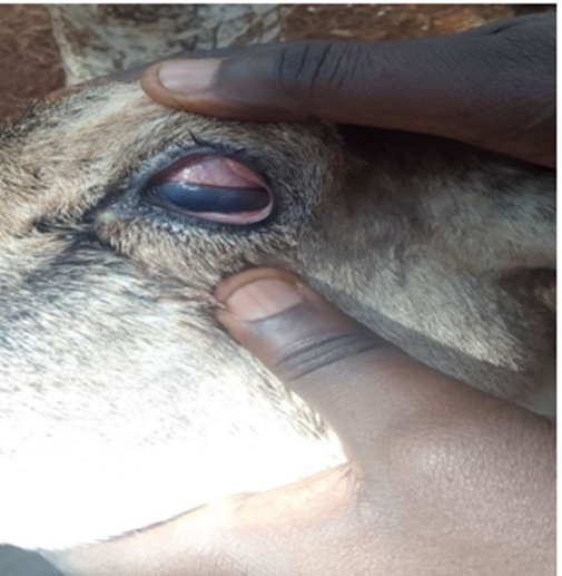
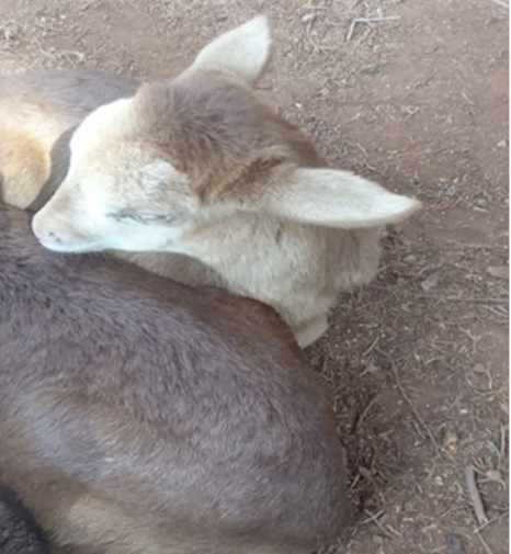
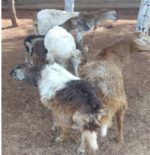
Figure 1: Image showing the status of examined flock of sheep affected of IKC; A. affected flock; B. Congested mucous membrane and cloudy eyeball C. Lambs with matted eyes with discharge
- Treatment
Based on the tentative diagnosis made, short acting Oxytetracycline 10% 2ml for lambs and 5ml for adults I.M Sid. was given for three consecutive days. The prognosis for this case was good because all sheep were treated and followed promptly and effectively. Up on follow-up undertaken after 10 days of post treatment, all affected animals were responded well towards the treatment and recovered.
- Result and discussion
Keratoconjunctivitis in sheep is a painful disease. It may cause temporary, or in severe cases permanent blindness. The first signs are hyperaemia, serous lachrymation, increased blinking and blepharospasm [9]. As the disease progresses without treatment, ocular discomfort and visual disturbance may lead to inappetance or inability to locate food. These will then consequently affect the body condition score (BCS) resulting in emaciation as was observed in the present case [8]. The etiology of infectious keratoconjunctivitis involves many predisposing factors such as age, race, daylight, dust irritations which facilitate the colonization of the eye are important in the formation of the disease [1]. In current clinical investigation all the affected sheep were completely recovered within 10 days of starting treatment, so that all the symptoms disappeared along with cleared corneal opacity and the sheep were found active with normal feeding and watering. Based on treatment regimen followed, Parenteral short acting Oxytetracycline administration (10%) for three cosequetive days was found to be effective in treating pink eye in small ruminants. Similar treatment regimens were followed by [8]. Articles suggested that for treatment of undifferentiated pinkeye in small ruminant’s broad spectrum parenteral and/or topical tetracycline were effective [10].
1.3. Conclusion and recommendation
Effective treatment of IKC can be done by use of a specific antimicrobial therapy along with proper manage approach. In present clinical case Parenteral short acting Oxytetracycline administration (10%) for three cosequetive days was found to be effective in treating pink eye in small ruminants. Therefore, Early detection, segregation and treatment of infected stock can minimize the disease incidence in animals. Furthermore, reducing the incidence of flies and subsequent spreading of bacteria is important to control diseases in population.
References
- Gülmez Sağlam A, Erkiliç EE, Büyük F, Kirmizigül AH, Gökçe G, et al. (2018). Moraxella ovis and Mycoplasma conjunctivae isolation from an ovine infectious keratoconjunctivitis outbreak and fortified treatment approaches. Kafkas Univ Vet Fak Derg. 24(4):551-556.
View at Publisher | View at Google Scholar - Williams HJ, Duncan JS, Fisher SN,et al. (2019). Ovine infectious keratoconjunctivitis in sheep: the farmer’s perspective. Veterinary Record Open. 6:e000321.
View at Publisher | View at Google Scholar - Xavier Fernández-Aguilar, Luca Rossi, Óscar Cabezón, Andrea Giorgino, Isis Victoriano Llopis, et al. (2017). Veterinary Record.
View at Publisher | View at Google Scholar - López-Olvera JR, Ramírez E, Martínez-Carrasco C, Granados JE. (2024). Wildlife–Livestock Host Community Maintains Simultaneous Epidemiologic Cycles of Mycoplasma conjunctivae in a Mountain Ecosystem. Vet Sci. 11:217.
View at Publisher | View at Google Scholar - Karthik K, Manimaran K, Mahaprabhu R, Shoba K. (2017). Isolation of Moraxella sp. from Cases of Keratoconjunctivitis in an Organized Sheep Farm of India Open Journal of Veterinary Medicine. 7:138-143.
View at Publisher | View at Google Scholar - Seid A. (2019). Review on Infectious Bovine Keratoconjunctivitis and its Economic Impacts in Cattle. Cent Dairy and Vet Sci J. 9(5):555774.
View at Publisher | View at Google Scholar - M.Sh. Rhaymah, B.Y. Rasheed, Hussain KJ. (2013). Comparative Study On The Bacterial Causes Of Ovine Keratoconjunctivitis In Native Breed Sheep And Goats. Assiut Veterinary Medical Journal. 59:137.
View at Publisher | View at Google Scholar - Jesse FFA, Chung ELT, Abba Y, Bitrus AA, Hambali IU, et al. (2017). Clinical management of stage i pinkeye with concurrent pneumonic pasteurellosis in a goat: A case report. Journal of Advanced Veterinary and Animal Research. 4(4):390-393.
View at Publisher | View at Google Scholar - Åkerstedt J, Hofshagen M. (2004). Bacteriological Investigation of Infectious Keratoconjunctivitis in Norwegian Sheep. Acta vet scand. 45(2):19-26.
View at Publisher | View at Google Scholar - CD. K. (1983). 'Pink eye' or 'zere oogjes' or keratoconjunctivitis infectiosa ovis (KIO). Clinical efficacy of a number of antimicrobial therapies. . Vet Q. 5:122-127.
View at Publisher | View at Google Scholar
