

Research Article | DOI: https://doi.org/10.31579/2835-785X/005
Risk Factors, Diagnosis, Pathophysiology and Management of Pulmonary Embolism
- Gudisa Bereda 1*
Department of Pharmacy, Negelle Health Science College, Guji, Ethiopia.
*Corresponding Author: Gudisa Bereda, Department of Pharmacy, Negelle Health Science College, Guji, Ethiopia.
Citation: Gudisa Bereda, (2022) Risk Factors, Diagnosis, Pathophysiology and Management of Pulmonary Embolism. International Journal of Clinical Research and Reports.1(1); DOI:10.31579/2835-785X/005
Copyright: © 2022 Gudisa Bereda, This is an open-access article distributed under the terms of the Creative Commons Attribution License, which permits unrestricted use, distribution, and reproduction in any medium, provided the original author and source are credited.
Received: 07 October 2022 | Accepted: 21 October 2022 | Published: 28 October 2022
Keywords: diagnosis; management; pathophysiology; pulmonary embolism; risk factors
Abstract
Pulmonary embolism is a relatively common acute cardiovascular disorder with high early mortality rates. Prolonged immobility, advanced age, postoperative period, post-infarction period, heart failure, obesity and pregnancy are common risk factors for thromboembolic disease via venous stasis. The pathophysiology and clinical manifestations of pulmonary embolism based upon four main factors such as a) the extent of occlusion of the vascular tree and the size of the emboli; b) the patient’s pre-existing cardiopulmonary condition; c) chemical vasoconstriction due to the secretion of serotonin and thromboxane from platelets that adhere to the embolus, as well as to fibropeptide B, which is a product of fibrinogen breakdown; and d) the reflex vasoconstriction that is likely to occur as a consequence of pulmonary artery dilatation. The cornerstone therapy for pulmonary embolism is prevention of new embolic episodes with anticoagulant treatment or a filter in the inferior vena cava, since it has been resulted that the majority of patients do not die from the embolism that leads to diagnosis, but to the continuing deterioration of their condition due to new emboli. Anticoagulant therapy can be classified into two overlapping phases. The first is treatment of the presenting episode of pulmonary embolism, which takes about three months. The second, which is optional, is extended therapy designed to inhibit new episodes of venous thromboembolism. Standard primary therapy is with subcutaneous low molecular weight heparin, fondaparinux, or unfractionated heparin, or intravenous unfractionated heparin.
Introduction
Pulmonary embolism (PE) is a relatively common acute cardiovascular disorder with high early mortality rates [1]. PE is a dramatic and life-threatening complication of deep venous thrombosis (DVT). Pulmonary embolism (PE) is the third greatest cause of mortality from cardiovascular disease, after myocardial infarction and cerebrovascular stroke [2]. Pulmonary embolism (PE) is an emergency condition in which a fragment of a thrombus travels through the venous system to the lungs via the right ventricle. Due to pulmonary bed obstruction, PE can result in acute right ventricular failure, a life-threatening condition because most patients eventually die within the first hours of presentation, and early diagnosis is of paramount importance [3]. Pulmonary embolism covers a broad variety of clinical conditions, which range from asymptomatic, coincidentally revealed subsegmental thrombus found on chest computed tomographic (CT) scan to pressure-dependent PE complicated by multisystem organ failure and cardiogenic shock. Pulmonary thromboembolism is a serious cardiovascular disease, causing a considerable level of morbidity and mortality. Hemodynamic status, concomitant comorbidities, and dysfunction of the right ventricle are predictors of short-term mortality. Indeed, 30-day mortality rates in patients stratified by the European Society of Cardiology (ESC) classification into high, intermediate-high, and intermediate-low risk groups were resulted to be 22%, 7.7%, and 6.0%, respectively [4]. Pulmonary embolism is a leading cause of death in the general population and its incidence rises with age, and patients aged more than 65 years constitute nearly 60% of these cases. Three professional society statements from the American Heart Association, American College of Chest Physicians, and European Society of Cardiology encapsulate contemporary thinking and principles of risk stratification for PE, primarily based on hemodynamic consequences and right ventricular dysfunction. Patients with acute PE perhaps present on a spectrum from sudden cardiac arrest or death at one extreme to incidental clots without hemodynamic insult or cardiopulmonary dysfunction [5].
Risk factors:
The classical triad of risk factors for the occurrence of thromboembolic disease proposed by Virchow in 1856 such as local injury to the vascular wall, increased coagulability, and circulatory stasis can explain PE. Prolonged immobility, advanced age, postoperative period, post-infarction period, heart failure, obesity and pregnancy are common risk factors for thromboembolic disease via venous stasis. Events such as local trauma, vasculitis and previous thrombosis cause injury to the endothelium of the venous wall. Polycythemia, contraceptive pills, as well as malignant cancers, and particularly adenocarcinomas, are associated with coagulability disorders and an elevated risk of DVT and PE [6, 7]. Major risk factors for pulmonary embolism are idiopathic, primary, and unprovoked involves no apparent cause, old age (>65 years), prolong-haul travel, associated with thrombophilia (eg, factor V Leiden or prothrombin gene mutation), obesity, cigarette smoking, hypertension, metabolic syndrome, air pollution and secondary and provoked includes immobilization, postoperative, trauma, oral contraceptives, pregnancy, postmenopausal hormonal replacement, cancer and acute medical illness (eg, pneumonia, congestive heart failure) [8].
Pathophysiology of pulmonary embolism:
The pathophysiology and clinical manifestations of PE based upon four main factors such as a) the extent of occlusion of the vascular tree and the size of the emboli; b) the patient’s pre-existing cardiopulmonary condition; c) chemical vasoconstriction due to the secretion of serotonin and thromboxane from platelets that adhere to the embolus, as well as to fibropeptide B, which is a product of fibrinogen breakdown; and d) the reflex vasoconstriction that is probably to occur as a consequence of pulmonary artery dilatation [9]. The contribution of reflex and humoral pulmonary vasoconstriction observed in an experimental setting is not considered to be relevant in clinical practice. The acutely developing pulmonary hypertension in PE leads to elevate in RV afterload, morphologically presenting as right ventricular dilation and may ultimately cause right-heart failure. Once pulmonary vascular resistance has risen to a level that the RV is unable to tolerate, PE can result in sudden death through pulseless electrical activity (formerly electromechanical dissociation) or a systole. A less sudden drop in RV cardiac output results in reducing left ventricular (LV) filling, deteriorated diastolic LV function due to RV dilation (ventricular interdependence) and inter ventricular septal bulging. These events can lead to a fall in blood pressure and present as syncope, hypotension or cardiogenic shock. RV overload and reduction in coronary flow secondary to high RV pressure in the presence of massive PE can lead to subendocardial RV ischemia or infarction, with a potential contribution of coronary atherosclerosis. Patients surviving the primary episode of right-heart failure advance compensatory mechanisms via activation of the sympathetic nervous system. Inotropic and chronotropic stimulation, together with the Frank-Starling mechanism, results in the development of pulmonary hypertension crucial for maintaining pulmonary artery flow and, hence, systemic circulation. Together with systemic vasoconstriction, these mechanisms can preserve systemic blood pressure and organ function. It is believed that the RV of a healthy man exposed to acute overload will not generate more than 40 mmHg of mean pulmonary artery pressure [10, 11]. Oxidative stress upholds an essential role in the pathophysiologic mechanism of various diseases. Levels of uric acid (UA), the ultimate oxidation product of purine metabolism, arise in conditions of injured oxidative metabolism, namely cardiovascular disease, idiopathic pulmonary arterial hypertension, chronic thromboembolic pulmonary hypertension, metabolic syndrome, diabetes mellitus and obesity sleep apnea syndrome (OSAS). Hyperuricemia has been associated with elevated mortality and is considered as an independent predictor of death in patients at high risk of cardiovascular disease. High serum UA levels have long been known to indicate poor prognosis in certain acute diseases [12].
Diagnostic procedure:
Pulmonary embolism was diagnosed, based on the presence of positive computerized pulmonary angiography findings, as complete or partial filling defect in the main, lobar, segmental or sub-segmental branches of the pulmonary tree [13]. The clinical diagnosis of pulmonary embolism is difficult, especially when there is coexisting heart or lung disease, and it is notoriously inaccurate when depending on clinical signs alone. About two thirds patients who present with suspected pulmonary embolism do not have these conditions. Very rarely, pulmonary embolism presents in such a dramatic fashion that the diagnosis is intuitively known and treatment will be started, but the usual presentation is often vague and variable in severity, so that further testing is necessary to establish or exclude the diagnosis. Diagnostic evaluation is best carried out by first attempting to identify a provable alternative diagnosis that can explain the patient’s symptoms [14].
Clinical pre-test probability: Diagnosis of pulmonary embolism starts with an assessment of clinical pre-test probability. This is depending on assessment of whether symptoms and signs are typical for pulmonary embolism, if there are risk factors for pulmonary embolism, if pulmonary embolism is considered to be the most probably diagnosis, and if there is evidence of deep vein thrombosis. Clinical pre-test probability (CPTP) assessment is facilitated by use of a clinical prediction rule, of which the Wells score is most broadly used and extensively validated. Although CPTP alone cannot diagnose pulmonary embolism, and generally does not exclude pulmonary embolism, it guides the selection of diagnostic tests (for example, a confirmatory test with high CPTP, an exclusionary test with low CPTP) and perhaps diagnostic in combination with these test results. Every patient for whom pulmonary embolism is primarily considered does not need to be tested for pulmonary embolism; a convincing alternative diagnosis may subsequently be resulted [15].
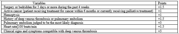
A score of <2>
D-dimer blood testing: D-dimer is formed when cross-linked fibrin is lysed by plasmin, and elevated levels frequently occur with pulmonary embolism. However, because elevations of D-dimer are nonspecific (e.g., elevated by aging, inflammation, cancer), an abnormal result has a low positive predictive value. The value of D-dimer is that a negative result can help to exclude pulmonary embolism. There are a wide variety of D-dimer assays, some of which are not suitable as diagnostic tests for pulmonary embolism because they have such poor operating characteristics (i.e., they are inaccurate). D-dimer assays that have been validated as tests for pulmonary embolism vary in their sensitivity and specificity, partly because of differences in their accuracy and partly because of the cutoff value they use to define normality (i.e., trade-off between sensitivity and specificity) [16].
Computed tomographic pulmonary angiography: Computed tomographic (CT) pulmonary angiography which outlines thrombi in the pulmonary arteries with intravenous contrast medium, has become the primary diagnostic test for pulmonary embolism. Its positive predictive value varies with the extent of pulmonary embolism and CPTP: the positive predictive value is (a) 97% with main or lobar, 68% with segmental, but only 25% with isolated subsegmental pulmonary artery abnormalities, and (b) 96% with high CPTP, 92% with moderate CPTP, but only 58% with low CPTP [17].
Pulmonary angiography: Pulmonary angiography is the criterion standard for the diagnosis of pulmonary embolism, but it is associated with serious side effects (e.g., mortality of about 0.5%), is technically demanding to perform, perhaps difficult to interpret and is costly. It is contraindicated in patients with renal impairment and may not be feasible in the sickest patients. For these reasons, pulmonary angiography is frequently reserved for patients who have had nondiagnostic noninvasive tests for pulmonary embolism when it is considered unsafe to withhold anticoagulation, while performing serial testing to detect evolving proximal deep vein thrombosis, or when it is necessary to establish a diagnosis to manage patients with severe symptoms. Of patients with normal pulmonary angiograms, about 1% has an episode of symptomatic venous thromboembolism during the following 6 months; this is the standard against which the safety of withholding anticoagulant therapy follows [18].
Blood gases: Arterial or venous blood sampling for blood gas analysis is a routine laboratory test. A typical picture of acute PE is a combination of hypoxemia and hypocapnia with respiratory alkalosis as a result of compensatory hyperventilation. Rarely, in severe forms of PE, hypercapnia can be present. Blood gas analyses in PE patients enable more accurate risk stratification of an individual patient, initially in terms of development of organ dysfunction (both current, when establishing the diagnosis, and over time when monitoring the future course of disease and response to therapy). Monitored parameters of organ function initially involve the presence and development of respiratory and renal dysfunction consistent both with the extent of PE (level of ventilation-perfusion mismatch) and its impact on forward cardiac output and potential development of low cardiac output syndrome. However, any blood sampling for blood gas analysis or any cannulation for regular blood drawing should be carefully considered because unnecessary puncture or cannulation before thrombolysis can result not only in inconvenience but also in fatal bleeding [19].
Echocardiography: Echocardiography, which should be available at all hours in any intensive care unit, is recently considered in addition to physical examination the main adjunct method of examination in acute PE. Echocardiography offers a potential for emergency risk stratification depending on evaluation of the hemodynamic impact of the disease on the right-heart chambers while also permitting for comprehensive non-invasive assessment of the patient’s hemodynamic status [20]. Magnetic resonance imaging Magnetic resonance imaging (MRI) offers both morphological and functional information on lung perfusion and right heart function, but its image quality still needs improvement to be comparable with CT. MRI has several attractive advantages, involving the avoidance of nephrotoxic iodinated contrast and ionizing radiation, and excellent sensitivity and specificity for DVT, together with the potential for performing lung perfusion imaging. This technique may eventually allow simultaneous and accurate detection of both DVT and pulmonary embolism. Additional data are needed, however. A disadvantage of MRI compared to CT is the long time (15–30 minutes) needed to perform the examination, which is not suitable for clinically unstable patients [21].
Differential diagnosis of pulmonary embolism
The below following are differential diagnosis of pulmonary embolism: Pneumonia or bronchitis, asthma, exacerbation of chronic obstructive pulmonary disease, myocardial infarction, pulmonary oedema, anxiety-hysteria, aortic dissection, lung cancer, primary pulmonary hypertension, rib fractures, pneumothorax and musculoskeletal pain [22].
Treatment
The cornerstone therapy for PE is inhibition of new embolic episodes with anticoagulant treatment or a filter in the inferior vena cava, since it has been resulted that the majority of patients do not die from the embolism that leads to diagnosis, but to the continuing deterioration of their condition due to new emboli [23]. Several therapeutic implications exist for patients with pulmonary embolism: (1) high-risk patients (who represent about 5% of all symptomatic patients, with about a 15% short-term mortality) should be treated aggressively with thrombolytic medicines or surgical or catheter embolectomy; (2) low-risk patients (most patients with pulmonary embolism), with a short-term mortality of about 1% might benefit from early discharge or even outpatient treatment; (3) intermediaterisk patients (who represent about 30% of all symptomatic patients) should likely be admitted to hospital, with potential benefit of thrombolytic treatment, pending results of ongoing clinical trials. Low-risk and intermediate risk categories are referred to as non-massive pulmonary embolism [24].
Supportive therapy: The patients frequently present with hypoxaemia, which responds to O2 administration, since the main pathophysiological mechanism is V/Q disturbances. Bed rest appears to help via two mechanisms. First, the restriction of movement decreases the likelihood of thrombus detachment from its peripheral location; second, it lowers O2 consumption (VO2) and therefore the need for accelerated cardiac output [25]. Noradrenaline perhaps used in severe cases, since by initiating peripheral vasospasm it can enhance the pressure in the aorta and the flow to the coronary vessels (ameliorating right heart ischaemia), without affecting right ventricular afterload. Other inotropic drugs (dopamine, dobutamine, isoproterenol and adrenaline) appear to have no place in PE, since they enhance O2 consumption without a corresponding improvement in cardiac output. Fluid administration is contraindicated, as further dilatation of the right ventricle leads to accelerate in myocardial O2 consumption and greater restriction of the left ventricle, because of the displacement of the interventricular septum, and therefore a reduction in cardiac output [26].
Anticoagulant therapy: Anticoagulant therapy can be classified into two overlapping phases. The first is treatment of the presenting episode of pulmonary embolism, which takes about three months. The second, which is optional, is extended therapy designed to prevent new episodes of venous thromboembolism. Standard primary therapy is with subcutaneous low molecular weight heparin, fondaparinux, or unfractionated heparin, or intravenous unfractionated heparin. There is no strong evidence that any one method is superior. Subcutaneous low molecular weight heparin and fondaparinux do not require intravenous infusion or laboratory monitoring, whereas intravenous unfractionated heparin is preferred if there is shock, severe renal impairment (low molecular weight heparin and fondaparinux are renally excreted), thrombolytic therapy is being considered, or it perhaps necessary to reverse anticoagulation rapidly. These treatments should be overlapped with a vitamin K antagonist (such as warfarin) and stopped after a minimum of five days provided the international normalised ratio (INR) has been above 2.0 for at least a day [27, 28]. Heparin is the basic treatment for PE, inhibiting the formation of new thrombi and giving time for the endogenous fibrinolysis to take effect, dissolving older thrombi [29]. Low molecular weight heparin can be continued long term, a treatment technique that is generally preferred in patients with cancer associated pulmonary embolism because of superior efficacy of low molecular weight heparin, difficulty in controlling vitamin K antagonist therapy, and greater compatibility of low molecular weight heparin with chemotherapy and the need for invasive procedures [30]. The new oral anticoagulants rivaroxaban and dabigatran (apixaban is at an earlier stage of assessment in the treatment of venous thromboembolism) are as effective as conventional anticoagulant therapy, do not require laboratory monitoring, and are associated with a lower risk of intracranial bleeding but a higher risk of gastrointestinal bleeding. Dabigatran is preceded by heparin therapy, whereas rivaroxaban does not require primary heparin therapy but requires a higher dose for the first three weeks of treatment [31].
Antiplatelet therapy: Results from two recent placebo controlled randomised trials support that aspirin lowers risk of recurrent venous thromboembolism by about a third in patients with a first unprovoked venous thromboembolism who have completed at least three months of anticoagulant therapy. Therefore, if patients are not candidates for extended anticoagulant therapy, this reduction in recurrent venous thromboembolism can be involved in the overall assessment of benefit to risk for indefinite aspirin therapy [32].
Systemic thrombolysis: Benefits of systemic thrombolysis (ST) accrue from rapid recanalization of the pulmonary artery, with corresponding improvements in pulmonary pressures, RV function, ventilation/perfusion mismatches and overall hemodynamics. Although ST achieves these benefits more rapidly when compared with systemic anticoagulation, physiological benefits of anticoagulation (reduced pulmonary pressures and RV dilatation) are identical within days [33]. ST has the most benefits if administered within 1 to 2 days of embolism, so that the clot is not impervious to thrombolysis (note, guidelines permit for thrombolysis ≤2 weeks after symptom onset) [34].
Catheter-directed fibrinolysis: Local delivery of thrombolytic agent’s catheter-directed fibrinolysis (CDF) has been an area of elevating interest. Navigation of a catheter physically through an obstructive pulmonary artery thrombus creates a channel for drug delivery and accelerates the surface area of thrombus exposed to thrombolytic agent [35].
Embolectomy: In patients with haemodynamic instability, in which thrombolysis has failed or is contraindicated (intracranial haemorrhage, current surgery or trauma); transvenous catheter thrombectomy is performed [36].
Conclusion:
Pulmonary embolism (PE) is the third greatest cause of mortality from cardiovascular disease, after myocardial infarction and cerebrovascular stroke. Prolonged immobility, advanced age, postoperative period, post-infarction period, heart failure, obesity, pregnancy, and other factors, predispose for thromboembolic disease via venous stasis. Pulmonary embolism was diagnosed, based on the presence of positive computerized pulmonary angiography findings, as complete or partial filling defect in the main, lobar, segmental or sub-segmental branches of the pulmonary tree. Anticoagulant therapy can be classified into two overlapping phases. The first is treatment of the presenting episode of pulmonary embolism, which takes about three months.
Abbreviations
aPTT: Activated partial thromboplastin time; CDF: Catheter-directed fibrinolysis; CPTP: Clinical pre-test probability; CT: Computed tomographic; DVT: Deep vein thrombosis; ESC: European Society of Cardiology; INR: International normalized ratio; LV: Left ventricular; LMWH: Low-molecular-weight heparin; MRI: Magnetic resonance imaging; OSAS: Obesity sleep apnea syndrome; PT: Prothrombin time; PE: Pulmonary embolism, RV: Right ventricular; ST: Systemic thrombolysis; tPA: Tissue plasminogen activator; UFH: Unfractionated heparin; UA: Uric acid
Acknowledgments
The author would be grateful to anonymous reviewers by the comments that increase the quality of this manuscript.
Data Sources: Sources searched include Google Scholar, Research Gate, PubMed, NCBI, NDSS, PMID, PMCID, Scopus database, Scielo and Cochrane database. Search terms included: risk factors, diagnosis, pathophysiology and management of pulmonary embolism
Funding
None
Competing interests
The author has no financial or proprietary interest in any of material discussed in this article.
References
- Bartziokas, K.; Kyriakopoulos, C.; Potonos, D.; Exarchos, K.; Gogali, A.; Kostikas, K. (2022) The Diagnostic Role of Uric Acid to Creatinine Ratio for the Identification of Patients with Adverse Pulmonary Embolism Outcomes. Diagnostics, 12, 193.
View at Publisher | View at Google Scholar - Konstantinides, S.V.; Meyer, G.; Becattini, C.; Bueno, H.; Geersing, G.-J.; Harjola, V.-P.; Huisman, M.V.; Humbert, M.; Jennings, C.S.; Jiménez, D.; et al. (2020) ESC Guidelines for the Diagnosis and Management of Acute Pulmonary Embolism Developed in Collaboration with the European Respiratory Society (ERS). Eur. Heart J. 41, 543–603.
View at Publisher | View at Google Scholar - Bartziokas, K.; Papaioannou, A.I.; Haniotou, A.; Nena, E.; Kostikas, K.; Steiropoulos, P. (2021) Serum Uric Acid and Arterial Lactate Levels in Patients with Obstructive Sleep Apnea Syndrome: The Effect of CPAP Treatment. Postgrad. Med. 133, 518–524.
View at Publisher | View at Google Scholar - Rumora, L.; Hlapˇci´c, I.; Popovi´c-Grle, S.; Rako, I.; Rogi´c, D.; Cepelak, I. (2020) Uric Acid and Uric Acid to Creatinine Ratio in the ˇ Assessment of Chronic Obstructive Pulmonary Disease: Potential Biomarkers in Multicomponent Models Comprising IL-1beta. PLoS ONE 15, e0234363.
View at Publisher | View at Google Scholar - Tao, J.; Shen, X.; Li, J.; Cha, E.; Gu, P.P.; Liu, J.; Zhu, W.; He, L.L.; Li, G.Q.; Wang, Z. (2020),Serum Uric Acid to Creatinine Ratio and Metabolic Syndrome in Postmenopausal Chinese Women. Medicine 99, e19959.
View at Publisher | View at Google Scholar - Li, M.; Gu, L.; Yang, J.; Lou, Q. Serum Uric Acid to Creatinine Ratio Correlates with β-Cell Function in Type 2 Diabetes. Diabetes. Metab. Res. Rev. 2018, 34, e3001.
View at Publisher | View at Google Scholar - T, ăpoi, L.; S, alaru, D.L.; Sascău, R.; Stătescu, C. (2021) Uric Acid-An Emergent Risk Marker for Thrombosis? J. Clin. Med. Res. 10, 1951–1953.
View at Publisher | View at Google Scholar - Pugliese, N.R.; Mengozzi, A.; Virdis, A.; Casiglia, E.; Tikhonoff, V.; Cicero, A.F.G.; Ungar, A.; Rivasi, G.; Salvetti, M.; Barbagallo, C.M.; et al. The Importance of Including Uric Acid in the Definition of Metabolic Syndrome When Assessing the Mortality Risk. Clin. Res. Cardiol. 110, 1073–1082.
View at Publisher | View at Google Scholar - Casiglia, E.; Tikhonoff, V.; Virdis, A.; Masi, S.; Barbagallo, C.M.; Bombelli, M.; Bruno, B.; Cicero, A.F.G.; Cirillo, M.; Cirillo, P.; et al. (2021) Serum Uric Acid and Fatal Myocardial Infarction: Detection of Prognostic Cut-off Values: The URRAH (Uric Acid Right for Heart Health) Study. J. Hypertens. 2020, 38, 412–419.
View at Publisher | View at Google Scholar - Zhang, X.; Hu, M.; Wang, X.; Zhang, C.; Chen, W.; Chen, S.; Zhou, J.; Chen, Y.; Lou, L.; Chen, G.; et al. (2020) New Perspective on the Risk Markers for Left Atrial Thrombosis in Patients with Atrial Fibrillation. Eur. J. Prev. Cardiol. 28, 641–647.
View at Publisher | View at Google Scholar - Konstantinides SV, Meyer G, Becattini C et al.; (2020) ESC Scientific Document Group. 2019 ESC Guidelines for the diagnosis and management of acute pulmonary embolism developed in collaboration with the European Respiratory Society (ERS). Eur Heart J 41: 543–603.
View at Publisher | View at Google Scholar - Klok FA, (2020) Huisman MV. How I assess and manage the risk of bleeding in patients treated for venous thromboembolism. Blood 135:724–734.
View at Publisher | View at Google Scholar - Lyman GH, Carrier M, Ay C et al. (2021) American Society of Hematology 2021 guidelines for management of venous thromboembolism: prevention and treatment in patients with cancer. Blood Adv 5:927–974.
View at Publisher | View at Google Scholar - Robertson L, Yeoh SE, Broderick C, Stansby G, Agarwal R. (2018) Effect of testing for cancer on cancer- or venous thromboembolism (VTE)-related mortality and morbidity in people with unprovoked VTE. Cochrane Database Syst Rev 11:Cd010837.
View at Publisher | View at Google Scholar - Elsebaie MAT, van Es N, Langston A, (2019) Buller HR, Gaddh M. Direct oral anticoagulants in patients with venous thromboembolism and thrombophilia: a systematic review and meta-analysis. J Thromb Haemost 17:645–656.
View at Publisher | View at Google Scholar - Miranda S, Park J, Le Gal G et al. (2020) Prevalence of confirmed antiphospholipid syndrome in 18-50 years unselected patients with first unprovoked venous thromboembolism. J Thromb Haemost 18:926–930.
View at Publisher | View at Google Scholar - Pengo V, Denas G, Zoppellaro G et al. (2018) Rivaroxaban vs warfarin in high-risk patients with antiphospholipid syndrome. Blood 132:1365–1371.
View at Publisher | View at Google Scholar - Ordi-Ros J, Saez-Comet L, Perez-Conesa M et al. (2019) Rivaroxaban versus vitamin K antagonist in antiphospholipid syndrome: a randomized noninferiority trial. Ann Intern Med 171:685–694.
View at Publisher | View at Google Scholar - Devreese KMJ, de Groot PG, de Laat B et al. (2020) Guidance from the Scientific and Standardization Committee for lupus anticoagulant/antiphospholipid antibodies of the International Society on Thrombosis and Haemostasis: update of the guidelines for lupus anticoagulant detection and interpretation. J Thromb Haemost 18:2828–2839.
View at Publisher | View at Google Scholar - Devreese KMJ, Ortel TL, Pengo V, de Laat B; (2018) Subcommittee on Lupus Anticoagulant/Antiphospholipid Antibodies. Laboratory criteria for antiphospholipid syndrome: communication from the SSC of the ISTH. J Thromb Haemost 16:809–813.
View at Publisher | View at Google Scholar - Pengo V, Hoxha A, Andreoli L et al. (2021) Trial of Rivaroxaban in AntiPhospholipid Syndrome (TRAPS): two-year outcomes after the study closure. J Thromb Haemost 19:531–535.
View at Publisher | View at Google Scholar - Klok FA, Barco S. (2019) Optimal management of hormonal contraceptives after an episode of venous thromboembolism. Thromb Res 181 Suppl 1:S1–S5.
View at Publisher | View at Google Scholar - Kim NH, Delcroix M, Jais X et al. (2019) Chronic thromboembolic pulmonary hypertension. Eur Respir J 53:1801915.
View at Publisher | View at Google Scholar - Le Gal G, Carrier M, Castellucci LA et al.; ISTH CDE Task Force. Development and implementation of common data elements for venous thromboembolism research: on behalf of SSC Subcommittee on official Communication from the SSC of the ISTH. J Thromb Haemost 19:297–303.
View at Publisher | View at Google Scholar - Claeys M, Claessen G, La Gerche A et al. (2021) Impaired cardiac reserve and abnormal vascular load limit exercise capacity in chronic thromboembolic disease. JACC Cardiovasc Imaging 2019;12:1444–1456.
View at Publisher | View at Google Scholar - Held M, Kolb P, Grun M et al. (2016) Functional characterization of patients with chronic thromboembolic disease. Respiration 91:503–509.
View at Publisher | View at Google Scholar - Boon G, Bogaard HJ, Klok FA. (2020) Essential aspects of the follow-up after acute pulmonary embolism: an illustrated review. Res Pract Thromb Haemost 4: 958–968.
View at Publisher | View at Google Scholar - Murgier M, Bertoletti L, Darmon M, et al.(2019) Frequency and prognostic impact of acute kidney injury in patients with acute pulmonary embolism. Data from the RIETE registry. Int J Cardiol. 291:121-126.
View at Publisher | View at Google Scholar - Cosmai L, Porta C, Foramitti M, Perrone V, Mollica L, Gallieni M, Capasso G: (2021) Preventive strategies for acute kidney injury in cancer patients. Clin Kidney J. 14:70-83.
View at Publisher | View at Google Scholar - Streiff MB et al. (2016) Guidance for the treatment of deep vein thrombosis and pulmonary embolism. J Thromb Thrombolysis 41:32–67.
View at Publisher | View at Google Scholar - Rieded M. (2017) Venous thromboembolic disease. Acute pulmonary embolism 1: pathophysiology, clinical presentation, and diagnosis. Heart 85:229–240.
View at Publisher | View at Google Scholar - Uma, S.; Hirai, T.; Nakagawa, K.; Ohara, K.; Fukuda, N.; Nozawa, T.; Inoue, H. (2014) Hyperuricemia and Transesophageal Echocardiographic Thromboembolic Risk in Patients with Atrial Fibrillation at Clinically Low-Intermediate Risk. Circ. J. 78, 1600–1605.
View at Publisher | View at Google Scholar - Babaoglu, E.; Hasanoglu, H.C.; Senturk, A.; Karalezli, A.; Kilic, H.; Aykun, G.; Oztuna, D. (2014) Importance of Biomarkers in Risk Stratification of Pulmonary Thromboembolism Patients. J. Investig. Med. 62, 328–331.
View at Publisher | View at Google Scholar - Akbar, S.R.; Long, D.M.; Hussain, K.; Alhajhusain, A.; Ahmed, U.S.; Iqbal, H.I.; Ali, A.W.; Leonard, R.; (2015) Dalton, C. Hyperuricemia: An Early Marker for Severity of Illness in Sepsis. Int. J. Nephrol. 2015, 301021.
View at Publisher | View at Google Scholar - Lindman, B.R.; Dávila-Román, V.G.; Mann, D.L.; McNulty, S.; Semigran, M.J.; Lewis, G.D.; de las Fuentes, L.; Joseph, S.M.; Vader, J.; Hernandez, A.F.; et al. (2014) Cardiovascular Phenotype in HFpEF Patients with or without Diabetes: A RELAX Trial Ancillary Study. J. Am. Coll. Cardiol. 64, 541–549.
View at Publisher | View at Google Scholar - Becattini, C.; Agnelli, G.; Lankeit, M.; Masotti, L.; Pruszczyk, P.; Casazza, F.; Vanni, S.; Nitti, C.; Kamphuisen, P.; Vedovati, M.C.; et al. (2016) Acute Pulmonary Embolism: Mortality Prediction by the 2014 European Society of Cardiology Risk Stratification Model. Eur. Respir. J. 48, 780–786.
View at Publisher | View at Google Scholar
