

Research Article | DOI: https://doi.org/10.31579/2834-5126/068
Removal of organic compounds and microorganisms from a pharmaceutical wastewater using Ag-Fe3O4 nanoparticles
- Rukiye Öztekin
- Delia Teresa Sponza *
Dokuz Eylül University, Engineering Faculty, Department of Environmental Engineering, Tınaztepe Campus, 35160 Buca/Izmir.
*Corresponding Author: Delia Teresa Sponza, Dokuz Eylül University, Engineering Faculty, Department of Environmental Engineering, Tınaztepe Campus.
Citation: Rukiye Öztekin, Delia Teresa Sponza, (2024), Removal of organic compounds and microorganisms from a pharmaceutical wastewater using Ag-Fe3O4 nanoparticles, Clinical Trials and Clinical Research. 3(3); DOI:10.31579/2834-5126/068
Copyright: © 2024, Delia Teresa Sponza. This is an open access article distributed under the creative commons’ attribution license, which permits unrestricted use, distribution, and reproduction in any medium, provided the original work is properly cited.
Received: 06 May 2024 | Accepted: 17 June 2024 | Published: 25 June 2024
Keywords: field emission scanning electron microscope (fesem); fourier transform infrared spectrophotometer (ftir); pharmaceutical wastewater; silver loaded-magnetic nanoparticles; x-ray diffraction (xrd) analysis; thermogravimetric analysis (tga)
Abstract
In this study, silver-loaded magnetic nanoparticles (Ag-Fe3O4 NPs) were developed under laboratory conditions to treat the secondary settling effluent coming from the biological aerobic activated sludge proses. This wastewater contained some pharmaceuticals and bacteria at high concentrations which were not treated at the beginning treatment steps. The effect of Ag-Fe3O4 NPs concentrations (0.1, 0.5, 1.0, 1.5 mg/l), contacting time (2, 5, 8, 10 and 15 mins), pH levels (4, 7 and 8), power (2, 4 and 6 W/m2), frequency (2, 5, 8 kHz). Salmonella, Psuedomonas, yeast, fungi, total coliforms, fecal coliforms, heterotrophic bacteria and ibuprophene, oxytetracycline removal yields were detected. The Ag-Fe3O4 NPs has a size of 25 nm and the saturation magnetization was recorded as 49 emu/g. The particle shapes and properties were analised with field emission scanning electron microscope (FESEM) and fourier transform infrared spectrophotometer (FTIR), X-ray diffraction (XRD) and thermogravimetric analysis (TGA). The maximum organism and pollutant yields were around 98% at 0.5 mg/l, after 10 mins at a neutral pH. The recovery of the Ag-Fe3O4 NPs were studied. 89% treatment yields were recorded after 8 months of continous operation. Adsoprtive and photocatalytic studies showed that after a short adsortion time the pollutants and the microorganisms were removed successfully at a power of 2 watt/m2 and at a frequency of 2 kHz. A cost analysis also was performed.
Introduction
Pharmaceuticals are products used in large doses in daily life considered as contaminants of emerging concern. Due to the large amounts of drugs consumed, the hydrogenic sources sufer from contamination processes that give rise to toxicological efects in humans despite its low concentrations (Najafpoor et al., 2007; Hodgson et al., 2017). Many medicines considered as emerging contaminants are constantly detected in groundwater, wastewater treatment plants and water supply. Te inefciency of conventional methods used in water treatment plants to remove the contaminant motivates the development of efective methods to treat efuent contamination (Garcia-Segura et al., 2018). According to the physico-chemical properties of drugs, their degradation products and the characteristics of the soils, these substances can reach the groundwater and contaminate the aquifers or remain retained in the soil, thus afecting the ecosystem and humans through the food chain (Rahmani et al., 2015). Additionally, the portion of medicines not assimilated by the organism, as well as chemical substances administered to animals, usually become part of wastewater. Consequently, diferent ways of removing medicines in waters have been studied (Golbaz et al., 2019).
In recent decades, much attention has been paid to the application of nanoparticles in environmental purposes. This is because nanosized materials have a large surface area-to-mass ratio and high reactivity (Qin et al., 2018; Wu et al., 2019). Thanks to these features, nanoparticles (NPs) have been widely used as catalysts (Zhong and Maye, 2001), adsorbents (Sadegh et al., 2017; Zhou et al., 2018), detectors (Lai et al., 2017), and disinfectants (Shaffiey, S.F. Shaffiey, 2017) in various studies. Due to their non-specific action, NPs as adsorbents are capable of removing a wide variety of contaminants including organics, inorganics, and colloids such as bacterial cells. When making a decrease in microbial contamination is a major goal, one can use some NPs with inherent disinfection capabilities such as magnetic nanoparticles (Zhang et al., 2018). Therefore, the need for adding other disinfectants such as chlorine and ozone, which are associated with adverse side effects, may be eliminated.
Magnetite (Fe3O4) is the strongest magnetic species among the transient metal oxides (Ali et al., 2017). The reactive surface of iron-oxides allows for the adsorption of various impurities from water environment (Daraei et al., 2017). When used as NPs, called Fe3O4 NPs, they release reactive oxygen species (ROS) such as superoxide radicals (O2−), hydroxyl radicals (•OH), hydrogen peroxide (H2O2), and singlet oxygen (1O2) that can decompose proteins and DNA in bacterial cells (Tran et al., 2010). Therefore, Fe3O4 NPs can exert their antibacterial effects through both physical adsorption and chemical disinfection. The magnetic feature of Fe3O4 NPs gives them an excellent separation property in the vicinity of an external magnetic field, which is essential for the removal of small-sized NPs from the effluent.
Silver (Ag) is a noble metal that exhibits a relatively weak oxidation behavior (Sachse et al., 2014). In large sizes, it is a low-reactive metal. However, in nanoscales, its microbicidal characteristic extremely enhances due to the increased specific surface area (Ghaseminezhad and Shojaosadati, 2016). Like Fe3O4 NPs, Ag NPs can exhibit their antibacterial activity through ROS generation (Azócar et al., 2019). In addition, other mechanisms have been mentioned such as the interaction of Ag-NPs with surface structures of bacterial cells and the reaction of released Ag ions with sulfur and phosphorous of cell macromolecules (Ghaseminezhad et al., 2018). Accordingly, Ag has been used successfully in previous studies as a bactericidal agent against Gram-negative bacteria, e.g., coliforms (Davoudi et al., 2012). The easy separation of the magnetite product can facilitate the recovery of invaluable Ag particles. It has been stated that Ag in combination with Fe3O4 NPs can penetrate biofilms more easily than Ag alone does (Ghaseminezhad and Shojaosadati, 2016). Therefore, Fe3O4-Ag NPs can show enhanced characteristics for water and wastewater treatment purposes.
There are several assumptions for the interaction of Ag and Fe3O4NPs. Petrov, Ivantsov (Petrov et al., 2020) modified Fe3O4 nanoparticles with Ag and could detect Ag both around Fe3O4 NPs and as separate particles in the structure of Ag-Fe3O4 NPs. They also showed that Ag ions replaced Fe3+ ions in the Fe3O4 lattice. Therefore, a combination of physical and chemical interactions can play a role in the connection of Ag to Fe3O4 NPs structure.
Various studies in the literature investigated the antibacterial effects of magnetic Fe3O4 NPs alone or in combination with Ag-NPs as shells. For example, (Ghaseminezhad et al., 2018) took advantage of the anti-biofilm activity of the compounds against antibiotic-resistant bacterial species in vitro (Joshi et al., 2014) employed the composite successfully against E. coli using the zone inhibition method on culture plates. However, limited studies are available to compare the effectiveness of Fe3O4 NPs and Ag-Fe3O4 NPs against bacterial cells in environmental applications. Moreover, the efficacy of the nanocomposite in the decrease of organic content of environmental samples would be interesting to determine. As far as we know, there is no similar study in the literature to investigate the application of Fe3O4 NPs and Ag-Fe3O4 NPs for advanced purification and disinfection of WWTP effluents. Therefore, this study aimed to examine the effectiveness of Fe3O4 NPs and Ag-Fe3O4 NPs in the removal of Total Coliforms (TC), Fecal Coliforms (FC), Heterotrophic Bacteria (HB), and Chemical Oxygen Demand (COD) from real WWTP effluents.
Materials and Methods
1.1. Preparation of Ag-Fe3O4 NPs
20 ml of H2O was heated at 80oC and continuously stirred under N2(g) atmosphere. Then, 0.56 g FeCl3.6H2O and 0.2 g FeCl2.4H2O were added. When the solids were dissolved, 2 ml of concentrated ammonia solution were incorporated and the solution was stirred for 10 min. The particles were separated using a permanent magnet and the supernatant was discarded. The solid was washed three times with H2O until the washing liquids were neutral. 0.28 g Fe3O4 NPs were suspended in 20 ml of H2O. After, 5.7 ml of diluted 0.011 g/l AgNO3 solution were added, the mixture was stirred for 5 min and using the magnet were separated and washed several time with H2O. Finally, Ag-Fe3O4 NPs were suspended in 20 ml of H2O again.
1.1.Physicochemical Properties of Ag-Fe3O4 NPs
A Field emission scanning electron microscopy analysis (FESEM) was performed to check the presence of Ag in the samples. In Figure 1a it is shown the 3D image for Fe3O4, while Figure 1b shows the intensity image for Ag-Fe3O4 NPs where the Ag appears as spherical, and shiny due to its high atomic number.
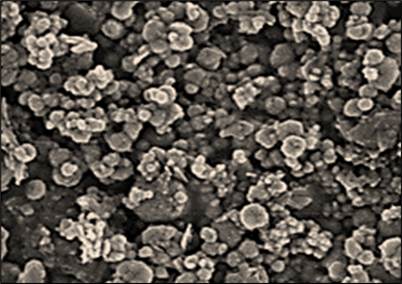
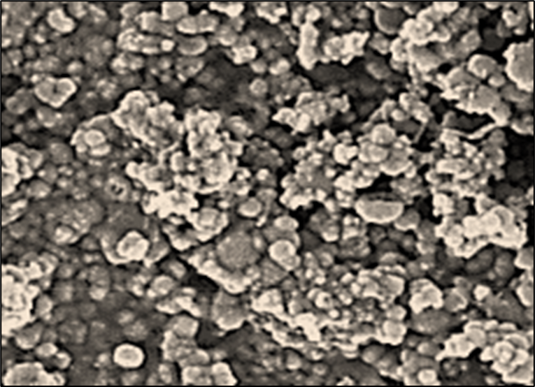
Figure 1: FESEM images of (a) Fe3O4 NPs and (b) Ag-Fe3O4 NPs.
Additionally, energy dispersive X-ray spectroscopy (EDX) graphs for Ag-Fe3O4 NPs are presented in Figure 2. In the latter, the signal corresponding to Ag appears in the plot. A BET test was carried out to determine the contact surface area of the adsorbent, giving a value equal to 116.476 m2/g, and a correlation coefficient for the BET isotherm of 0.999.
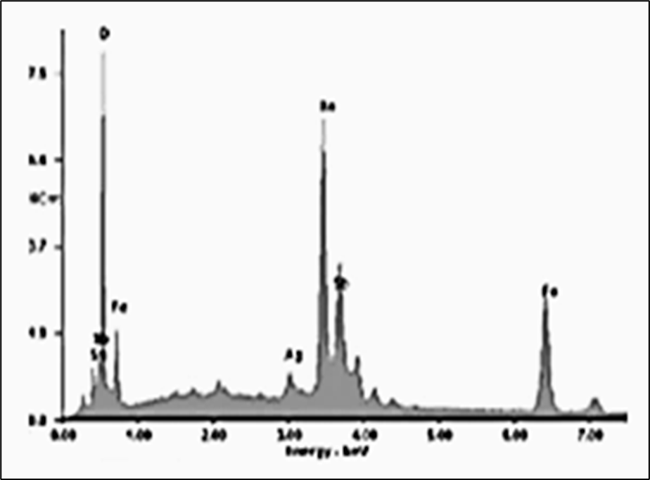
Figure 2: EDX spectra of Ag-Fe3O4 NPs.
- 1. Adsorption Studies
A H2O sample (10 ml) containing pharmaceuticals at concentrations of 0.2 mg/l was placed in a polypropylene tube, 500 µl of Ag-Fe3O4 NPs suspension were added. After shaking 30 min at 21oC, the magnet was placed at the bottom of the tube for 5 min and the adsorbent was separated. The supernatant was analyzed for ibuprophene ana oxytetracycline to determine the maximum removal efciency of the aforementioned chemicals. Figure 3 showed FTIR spectrum for Ag-Fe3O4 NPs after the adsorption process, where the characteristic IB signals are marked (carbonyl group at 1704,12 1/cm stretch frequencies of Csp3-H of isobutyl group at 2951,93 and 2922,15 1/cm; aromatic C=C bond at 1560,83 1/cm; O–H bond at 3100 1/cm).
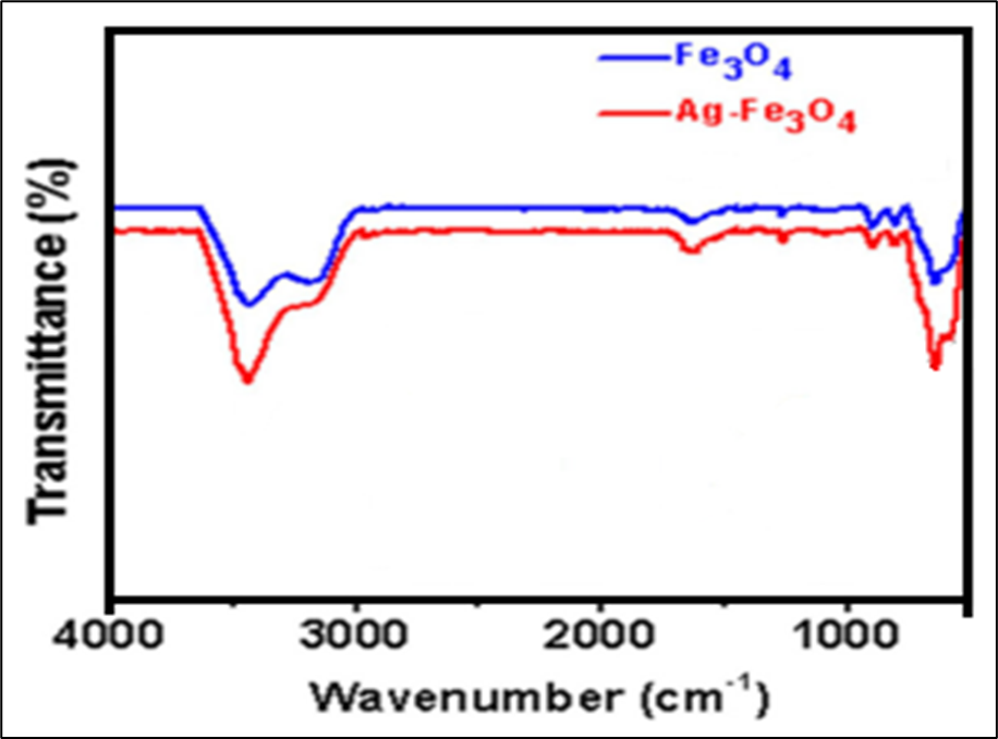
Figure 3: FTIR spectra of Fe3O4 NPs (blue) and Ag-Fe3O4 NPs (red).
1.2. NPs Characteristics
1.1.1.TEM Analysis Results
TEM images were recorded to examine the morphologies of Fe3O4 NPs and Ag-Fe3O4 NPs and determine the particle sizes. TEM images are presented in Figure 4. As can be observed, Fe3O4 NPs (Figure 4a) and Ag-Fe3O4 NPs (Figure 4b) were almost semi-spherical in shape. The TEM image of Ag-Fe3O4 NPs appeared darker than that of Fe3O4 NPs. It is also shown that our method of NPs synthesis could not produce uniform particles in size. The size of magnetic nanoparticles is dependent on various factors including synthesis conditions (e.g. oxygen-free environment) (Wang et al., 2013), pH of the synthesis medium (Park et al., 2010), the ratio of base to iron ions, and the length of the alkyl chain R (Sun et al., 2011). The mean diameter of particles in our study was 41 and 34 nm for Fe3O4 NPs and Ag-Fe3O4 NPs, respectively. The reduced size of particles by adding decorating agents has been also observed in the study by Joshi et al. (2014). They used the same
method of NPs synthesis as in our study and reported a reduced diameter from 26 to 20 nm after decorating Fe3O4 NPs with Ag. It seems that a decrease in the amount of iron-oxide precursor per unit volume of the preparation solution might be a reason for the reduced particle size in Ag-Fe3O4 NPs. However, there are reports in the literature (Tan et al., 2014) showing that the diameter of NPs increases when Ag shells decorated Fe3O4 cores. The procedure of nanoparticle synthesis has a major contribution to the characteristics of the nanocomposites including the particle size. The particle sizes measured in this study conform to the findings of Naqvi et al. (2010) study, reporting the mean size of superparamagnetic iron oxide nanoparticles as 30 nm, and the findings of Ebrahimi et al. (2016) study, producing Ag-Fe3O4 NPs in the range of 23 to 54 nm. The latter study showed that the particle size is dependent on reducing agents used in the synthesis process. Santoyo Salazar et al. (2011) conducted a study on Fe3O4 NPs in the range of 10–40 nm and showed that NPs of smaller than 20 nm had poor Fe3O4 properties.
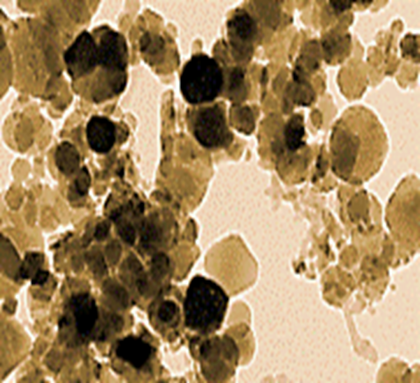
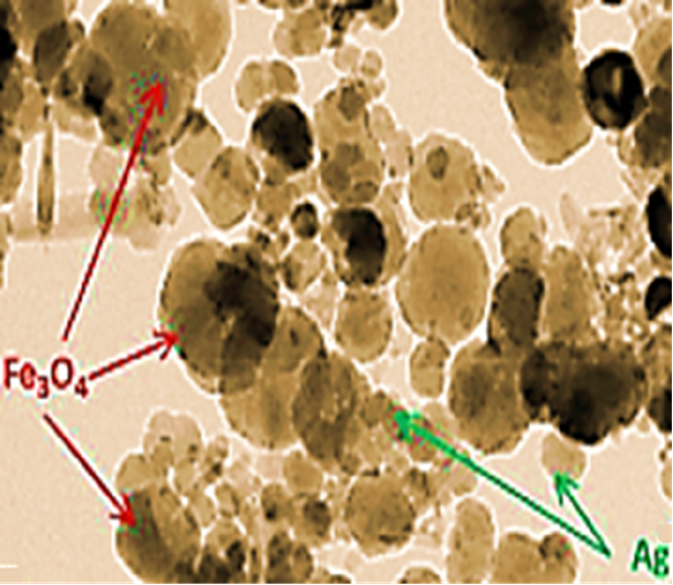
Figure 4: TEM images of (a) Fe3O4 NPs and (b) Ag-Fe3O4 NPs.
1.1.1.XRD Analysis Results
The results of XRD analysis of asprepared nanocomposites (Figure 5). The characterization peaks were observed at 2θ values of 30.12°, 35.51°, 43.15°, 53.23°, 56.82°, and 62.34°, implying pure Fe3O4 in the Fe3O4 NPs structure. These values are in complete agreement with the values reported by
Ebrahimi et al. (2016) and attribute to the indices (220), (311), (400), (422), (511), and (440) for Fe3O4 arises, respectively. The XRD patterns of Ag-Fe3O4 NPs showed the 2θ values of 38.11°, 44.32°, 64.24°, 77.61° and 81.57° corresponding to the (111), (200), (220), (311), and (222) planes of cubic Ag, respectively.
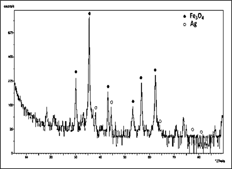
Figure 5: The XRD pattern of Fe3O4NPs and Ag NPs.
1.1.1.VSM Analysis Results
The Vibrating Sample Magnetometer (VSM) analysis was conducted to determine the magnetic properties of NPs (Figure 6). Typical superparamagnetic behavior is observed for Fe3O4 NPs and Ag-Fe3O4 NPs due to the absence of any remanence or coercivity. The analysis showed the saturation magnetization (Ms) values of 61 and 69 emu/g for Fe3O4 NPs and Ag-Fe3O4 NPs, respectively. The increasing Ms. values in the nanocomposite might have been due to the interactions between the NPs that changed the anisotropic energy. There are conflicting results in the literature regarding the VSM analysis of Fe3O4-Ag core-shell structures. Such as, Liu et al. (2008), in line with our study, reported an increase in the Ms. values when Ag was doped on the surface of Fe3O4 NPs, while Li et al. (2015) and Ghaseminezhad and Shojaosadati (2016).
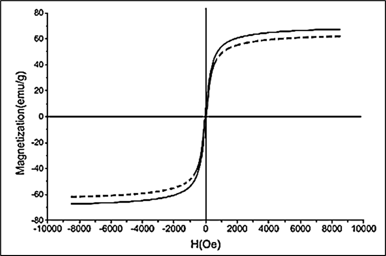
Figure 6: Magnetic hysteresis loops of Fe3O4 NPs (dashed line) and Ag-Fe3O4 NPs at 21oC.
1.3.Effect of pH on ibuprophene and oxytetracycline adsorption on Ag-Fe3O4 NPs
the adsorption of ibuprophene and oxytetracycline on Ag-Fe3O4 NPs were studied within the pH ranges varying between 2.0 and 9.0. The highest degree of adsorption was achieved at pH=7.0 (Table 1). The dependence of adsorption on pH is associated with the point of zero charge (PZC) of the Ag-Fe3O4 NPs and the pKa of the ibuprophene. The PZC is 6.93 for Ag. The ibuprophene is a weak acid (pKa=5.2) and exists as a neutral species por pHpKa41. For pH > 8.0 ibuprophene is deprotonated and Ag surface becomes negatively charged, thus leading to an electrostatic repulsion which reduces the adsorption efciency. For pH values greater than pKa but lower than PZC, electrostatic attraction between anionic ibuprophene and the positively charged surface of Ag NPs improves the adsorption capacity. However, the influence of initial pH of oxytetracycline adsorption on Ag-Fe3O4 NPs was different from the ibupprophene. When the pH was lower than 5.0, the adsorption rate increased with pH increasing. When pH was equal to 5.0, the adsorption rate reached the maximum. The maximum removal yields for ibuprophene and oxytetracycline were 99% with maximum adsorption yields of 2.03 mg/g and 3.04 mg/g for ibuprophene and oxytetracycine, respectively.
pH | Ibuprophene | Oxytetracycline | ||
Removal yields (%) | Adsorption (mg/g) | Removal yields (%) | Adsorption (mg/g) | |
4.0 | 67.00 | 0.99 | 56.00 | 1.34 |
5.0 | 68.00 | 0.99 | 99.00 | 3.04 |
6.0 | 60.00 | 0.76 | 54.00 | 1.03 |
7.0 | 99.00 | 2.03 | 50.00 | 0.99 |
8.0 | 64.00 | 0.97 | 43.00 | 0.76 |
10.0 | 40.00 | 0.56 | 40.00 | 0.65 |
Table 1: Effect of pH on the adsorption yields of 5 mg/l ibuprophene and 5 mg/l oxytetracycline separately in the presence of 3 mg/l Ag-Fe3O4 NPs.
1.4.Effect of Ag-Fe3O4 NPs concentration on the adsorptions of ibuprophene and oxytetracycline
The Ag-Fe3O4 NPs concentrations were increased from 0.1 mg/l up to 6.0 mg/l (Table 2). The ibuprophene and oxytetracycline concentrations were choosen as 3 mg/l at constant concentration. The volume necessary of Ag-Fe3O4 NPs to reach the maximum adsorption effciency is 500 µl. From 900 µl, the percentage of adsorption decreases due to the high volume of adsorbent in the medium and it is not completely removed with the magnet. 93% maximum Ag-Fe3O4 NPs adsorption yield was measured at 45 min contact time, 7 mg in 500 µl of suspension, at 0.1 mg/l Ag-Fe3O4 NPs concentration at 3.0 mg/l ibuprophene and at 3.0 mg/l oxytetracycline, respectively.
Ag-Fe3O4 NPs concentration (mg/l) | Ag-Fe3O4 NPs Adsorption (µl) | Contact Time (min) | Ag-Fe3O4 NPs Adsorption efficiency (%) |
0.1 | 500 | 45 | 93.0 |
0.5 | 531 | 43 | 92.7 |
1.0 | 580 | 30 | 91.2 |
1.5 | 612 | 27 | 90.9 |
2.0 | 654 | 24 | 88.6 |
2.5 | 720 | 10 | 86.5 |
3.0 | 735 | 47 | 83.4 |
3.5 | 756 | 52 | 71.5 |
4.0 | 783 | 65 | 69.8 |
4.5 | 800 | 77 | 63.4 |
5.0 | 841 | 81 | 55.3 |
5.5 | 876 | 88 | 51.7 |
6.0 | 900 | 90 | 48.9 |
Table 2: Effect of Ag-Fe3O4 NPs concentration on the adsorption of 3.0 mg/l ibuprophene and 3.0 mg/l oxytetracycline adsorptions.
1.5. Effect of Contact time on the adsorption of Ag-Fe3O4 NPs to ibuprophene and oxytetracycline
The contact time between Ag-Fe3O4 NPs and the solution containing ibuprophene and oxytetracycline was studied from 1 to 90 min in order to achieve the maximum adsorption efficiency, reached at 45 min. From there, the percentage of adsorption remains constant until 90 min (Table 2).
The proposed method for the adsorption of ibuprophene and oxytetracycline in waters was applied to real water samples partially contaminated with this chemical compound. The concentration of the pollutant in these samples was low or not detected, so they were doped with a known concentrations of ibuprophene (3.0 mg/l) and oxytetracycline (3.0 mg/l) in order to check the viability of the proposed process. Adsorption efficiencies close to 93% were achieved in all cases, thus enabling the method as suitable for its application in real water samples (Table 2).
1.6. Desorption and Recycling Studies of Ag-Fe3O4 NPs
The desorption of the ibuprophene and oxytetracycline from the surface of the NPs, at 1 ml HNO3 solution, at pH=1.0 was used. For that value, interactions between adsorbent and adsorbate are weakened due to the effect of the ionic strength, as stated in the effect of pH on ibuprophene and oxytetracycline adsorption, thus favoring the desorption process. When the ibuprophene and oxytetracycline adsorptions is carried out, after separation from the aqueous solution with the magnet, this solution is decanted. Then, 1 ml HNO3 solution is added and the mixture is sonicated for 3 min. After, the adsorbent is removed with the magnet and the acid solution is quantifed by HPLC, demonstrating that the ibuprophene and oxytetracycline total has been desorbed. Recycling studies were carried out showing that Ag-Fe3O4 NPs can be used during two additional successive adsorption cycles without losing adsorption capacity. In the fourth adsorption cycle, the capacity decreases down to 89.4%.
1.7. Reuse of the Ag-Fe3O4 Nanocomposite
The influence of Ag-Fe3O4 nanocomposite recycling on the degradation rates after one, two and three recycles under UV irradiation were 86%, 86% and 82%, respectively. And the degradation rates of one, two and three recycles under vis light irradiation were 54%, 53% and 50%, respectively. The degradation rate declined slightly with the increasing number of cycles. So Fe3O4-Ag nanocomposite was effective after three recycling times.
Conclusıons
This present study, the proposes a novel simple method was applied for the adsorption of ibuprophene and oxytetracycline in water using Ag-Fe3O4 NPs. The TEM image of Ag-Fe3O4 NPs appeared darker than Fe3O4NPs. Our method of NPs synthesis could not produce uniform particles in size. The XRD patterns of Ag-Fe3O4 NPs showed the 2θ values of 38.11°, 44.32°, 64.24°, 77.61° and 81.57° corresponding to the (111), (200), (220), (311) and (222) planes of cubic Ag, respectively. The Ms values was 61 emu/g for Fe3O4 NPs and 69 emu/g for Ag-Fe3O4 NPs. The increasing Ms values in the nanocomposite might have been due to the interactions between the NPs changed the anisotropic energy.
99% maximum ibuprophene adsorption removal yield was obtained at 2.03 mg/g maximum ibuprophene adsorption concentration, at pH = 7.0 and at 21oC, respectively. 99% maximum oxytetracycline adsorption removal yield was observed at 3.04 mg/g maximum oxytetracycine adsorption concentration, at pH = 5.0 and at 21oC, respectively. 93% maximum Ag-Fe3O4 NPs adsorption yield was measured at 45 min contact time, 7 mg in 500 µl of suspension, at 0.1 mg/l Ag-Fe3O4 NPs concentration at 3.0 mg/l ibuprophene and at 3.0 mg/l oxytetracycline, respectively. The characterization of the adsorbent by means of microscopy, spectroscopy and calorimetry techniques reveal the presence of Ag in Ag-Fe3O4 NPs and the adsorption of ibuprophene and oxytetracycline. The adsorption equilibrium is characterized by a Langmuir isotherm model.
Acknowledgement
Experimental analyzes in this study were performed at the Laboratories of the Canada Research Center, Ottawa, Canada. The authors would like to thank this body for providing financial support.
References
- Ali, I., Peng, C., Naz, I., Khan, Z.M., Sultan, M., Islam, T. and Abbasi, I.A., Phytogenic magnetic nanoparticles for wastewater treatment: a review, RSC Advances, 7(64), (2017), 40158–40178.
View at Publisher | View at Google Scholar - Azócar, M.I., Alarcón, R., Castillo, A., Blamey, J.M., Walter, M. and Paez, M., (2019). Capping of silver nanoparticles by anti-inflammatory ligands: antibacterial activity and superoxide anion generation, Journal of Photochemistry and Photobiology B: Biology, 193 100–108.
View at Publisher | View at Google Scholar - Daraei, H., Amrane, A. and Kamali, H., (2017). Assessment of phenol removal efficiency by synthesized zero iron nanoparticles and Fe powder using the response surface methodology, Iranian Journal of Chemistry and Chemical Engineering (IJCCE), 36(3), 137–146.
View at Publisher | View at Google Scholar - Davoudi, M., Ehrampoush, M.H., Vakili, T., Absalan, A. and Asghar Ebrahimi, A., (2012). Antibacterial effects of hydrogen peroxide and silver composition on selected pathogenic enterobacteriaceae. International Journal of Environmental Health Engineering, 1(1), 23.
View at Publisher | View at Google Scholar - Ebrahimi, N., Rasoul-Amini, S., Ebrahiminezhad, A., Ghasemi, Y., Gholami, A. Et al. (2016). Comparative study on characteristics and cytotoxicity of bifunctional magnetic-silver nanostructures: synthesized using three different reducing agents. Acta Metallurgica Sinica (English Letters), 29(4), 326–334.
View at Publisher | View at Google Scholar - Garcia-Segura, S., Ocon, J.D. and Chong, M.N., (2018). Electrochemical oxidation remediation of real wastewater effluents — a review, Process Safety and Environmental Protection, 113, 48–67.
View at Publisher | View at Google Scholar - Ghaseminezhad, S.M. and Shojaosadati, S.A., (2016). Evaluation of the antibacterial activity of Ag/ Fe3O4 nanocomposites synthesized using starch, Carbohydrate Polymers, 144, 454–463.
View at Publisher | View at Google Scholar - Ghaseminezhad, S.M., Shojaosadati, S.A., Meyer, R.L., Ghaseminezhad, S.M., Shojaosadati, S.A. et al. (2018). Ag/Fe3O4 nanocomposites penetrate and eradicate S. aureus biofilm in an in vitro chronic wound model. Colloids and Surfaces B: Biointerfaces, 163 192–200.
View at Publisher | View at Google Scholar - Golbaz, S., Nabizadeh, R., Zarinkolah, S., Mahvi, A.H., Alimohammadi, M. et al. (2019). An innovative swimming pool water quality index (SPWQI) to monitor and evaluate the pools: design and compilation of computational model. Environmental Monitoring and Assessment, 191(7), 448.
View at Publisher | View at Google Scholar - Hodgson, B., Sharvelle, S., Silverstein, J.A. and McKenna A., (2017). Impact of water conservation and reuse on water systems and receiving water body quality. Environmental Engineering Science, 35(6), 545–559.
View at Publisher | View at Google Scholar - Joshi, M.K., Pant, H.R., Kim, H.J., Kim, J.H. and Kim, C.S., (2014). One-pot synthesis of Ag-iron oxide/reduced graphene oxide nanocomposite via hydrothermal treatment. Colloids and Surfaces A: Physicochemical and Engineering Aspects, 446, 102–108.
View at Publisher | View at Google Scholar - Lai, C. Liu, X., Qin, L., Zhang, C., Zeng, G., Huang, D., et al. (2017). Chitosan-wrapped gold nanoparticles for hydrogen-bonding recognition and colorimetric determination of the antibiotic kanamycin, Microchimica Acta, 184(7), 2097–2105.
View at Publisher | View at Google Scholar - Li, W.-H., Yuea, X.-P., Guo, C.-S., Lv, J.-P., Liu, S.-S., et al. (2015). Synthesis and characterization of magnetically recyclable Ag nanoparticles immobilized on Fe3O4@ C nanospheres with catalytic activity, Applied Surface Science, 335 23–28.
View at Publisher | View at Google Scholar - Liu, C.H., Zhou, Z.D., Yu, X., Lv, B.Q., Mao, J.F. and Xiao, D., (2008). Preparation and characterization of Fe3O4/Ag composite magnetic nanoparticles. Inorganic Materials, 44(3), 291–295.
View at Publisher | View at Google Scholar - Najafpoor, A.A., Vojoudi, Z., Dehgani, M.H., Changani, F. and Alidadi, H.A., (2007). Quality assessment of the Kashaf river in north east of Iran in 1996-2005, Journal of Applied Sciences, 7(2), 253–257.
View at Publisher | View at Google Scholar - Naqvi, S., Samim, M., Abdin, M.Z., Ahmed, F.J., Maitra, A.N., et al. (2010). Concentration-dependent toxicity of iron oxide nanoparticles mediated by increased oxidative stress, International Journal of NanomedicineInt. J. Nanomedicine 5 983.
View at Publisher | View at Google Scholar - Park, M., Seo, S., Lee, I.S. and Jung, J.H., (2010). Ultra efficient separation and sensing of mercury and methylmercury ions in drinking water by using aminonaphthalimide-functionalized Fe3O4@SiO2 core/shell magnetic nanoparticles. Chemical Communications, 46, 4478–4480.
View at Publisher | View at Google Scholar - Petrov, D.A., Ivantsov, R.D., Zharkov, S.M., Velikanov, D.A., et al. (2020). Magnetic and magneto-optical properties of Fe3O4 nanoparticles modified with Ag. Journal of Magnetism and Magnetic Materials, 493, 165692
View at Publisher | View at Google Scholar - Qin, L., Zeng, G., Lai, C., Huang, D., Xu, P., (2018). “Gold rush” in modern science: fabrication strategies and typical advanced applications of gold nanoparticles in sensing. Coordination Chemistry Reviews, 359, 1–31.
View at Publisher | View at Google Scholar - Rahmani, A. R.; Asgari, G.; Askari, F.B. and Torbaghan, A.E., (2015). Adsorption of lead metal from aqueous solutions using activated carbon derived from scrap tires. Fresenius Environmental Bulletin, 24(7), 2341–2347.
View at Publisher | View at Google Scholar - Sachse, C., Weiß, N., Gaponik, N., Müller-Meskamp, L., Eychmüller, A. and Leo, K., (2014). ITO-free, small-molecule organic solar cells on spray-coated coppernanowire-based transparent electrodes, Advanced Energy Materials, 4(2), 1300737, 1-6.
View at Publisher | View at Google Scholar - Sadegh,H, Ali, G.A.M., Gupta,V.K., Makhlouf, A.S.H., Shahryari-ghoshekandi, R., et al. (2017). The role of nanomaterials as effective adsorbents and their applications in wastewater treatment, Journal of Nanostructure in Chemistry, 7(1), 1–14.
View at Publisher | View at Google Scholar - Santoyo Salazar, J., Perez, L., de Abril, O., Phuoc, L.T.,Ihiawakrim, D.,et al. (2011). Magnetic iron oxide nanoparticles in 10− 40 nm range: composition in terms of magnetite/maghemite ratio and effect on the magnetic properties, Chemistry of Materials, 23(6), 1379–1386.
View at Publisher | View at Google Scholar - Shaffiey, S.R. and Shaffiey, S.F., (2017). Silver oxide–copper oxide nanocomposite preparation and antimicrobial activity as a source for the treatment of fish diseases: silver oxide–copper oxide Nanocomposite preparation and antimicrobial activity, Advancing Medicine Through Nanotechnology and Nanomechanics Applications, IGI Global; 140–151.
View at Publisher | View at Google Scholar - Sun, L., Li, Y., Sun, M., Wang, H., Xu, S., et al. (2011). Porphyrin-functionalized Fe3O4@SiO2 core/shell magnetic colorimetric material for detection, adsorption and removal of Hg2+ in aqueous solution. New Journal of Chemistry, 35, 2697–2704
View at Publisher | View at Google Scholar - Tan, P., Qin, J.-X., Liu, X.-Q., Yin, X.-Q. and Sun, L.-B., (2014). Fabrication of magnetically responsive core–shell adsorbents for thiophene capture: AgNO3-functionalized Fe3O4@mesoporous SiO2 microspheres. Journal of Materials Chemistry A, 2, 4698–4705.
View at Publisher | View at Google Scholar - Tran, N., Mir, A., Mallik, D., Sinha, A., Nayar, S. et al. (2010). Bactericidal effect of iron oxide nanoparticles on Staphylococcus aureus, International Journal of Nanom zedicine, 5, 277-283.
View at Publisher | View at Google Scholar - Wang, H., Zhang, W., Zhao, J., Xu, L., Zhou, C., et al. (2013). Rapid decolorization of phenolic azo dyes by immobilized laccase with Fe3O4/SiO2 nanoparticles as support. Industrial and Engineering Chemistry Research, 52, 4401–4407.
View at Publisher | View at Google Scholar - Wu, S., Zhang, S., Gong, Y., Shi, L. and Zhou, B., (2019). Identification and quantification of titanium nanoparticles in surface water: a case study in Lake Taihu, China. Journal of Hazardous Materials, 121045.
View at Publisher | View at Google Scholar - XiangxiangZhou, X., Lai, C., Huang, D., Zeng, G., Chen, L., et al. (2018). Preparation of water-compatible molecularly imprinted thiolfunctionalized activated titanium dioxide: selective adsorption and efficient photodegradation of 2, 4-dinitrophenol in aqueous solution, Journal of Hazardous Materials, 346, 113–123.
View at Publisher | View at Google Scholar - Zhang, X., Wang, W., Zhang, Y., Zeng, T., Jia, C. and et al. (2018). Loading Cu-doped magnesium oxide onto surface of magnetic nanoparticles to prepare magnetic disinfectant with enhanced antibacterial activity, Colloids and Surfaces B: Biointerfaces, 161, 433–441.
View at Publisher | View at Google Scholar - Zhong, C.-J. and Maye, M.M., (2001). Core–shell assembled nanoparticles as catalysts, Advanced Materials, 13(19), 1507–1511.
View at Publisher | View at Google Scholar
