

Research Article | DOI: https://doi.org/10.31579/2834-5126/053
Relationship Between Cadmium Toxicity, Kidney Function Disturbances and Urinary Bladder Inflammation: The Role of Uvaria chamae in Mitigating these Effects
- Gabriel Edem *
- Kingsley Okon
- Hope Thompson
- Jessica David
1 Department of Human Anatomy, College of Health Sciences, University of Uyo, Nigeria.
2 Department of Medical Physiology, College of Health Sciences, University of Uyo, Nigeria.
*Corresponding Author: Gabriel Edem., David J., Department of Human Anatomy, College of Health Sciences, University of Uyo, Nigeria.
Citation: Edem G., Sudheer K Marlapudi., Biradar K., Okon K., Thompson H., (2024), Relationship Between Cadmium Toxicity, Kidney Function Disturbances and Urinary Bladder Inflammation: The Role of Uvaria chamae in Mitigating these Effects, Clinical Trials and Clinical Research. 3(1); DOI:10.31579/2834-5126/053
Copyright: © 2024, Gabriel Edem. This is an open access article distributed under the creative commons’ attribution license, which permits unrestricted use, distribution, and reproduction in any medium, provided the original work is properly cited.
Received: 27 December 2023 | Accepted: 16 January 2024 | Published: 02 February 2024
Keywords: uvaria chamae, urinary bladder damage, cadmium, toxicity, blood urea nitrogen, serum urea, serum creatinine
Abstract
This study was aimed at evaluating the ameliorative effects of Uvaria chamae against cadmium-induced toxicity on the urinary bladder and kidney function biomarkers of Wistar rats, considering its recognized local health benefits. Twenty-five adult male Wistar rats weighing between 120g and 350g were assigned into five groups (5 per group). The groups were designated as follows: group 1 (control), rats receiving normal feed and distilled water; group 2, rats induced with 3mg/kg of cadmium; group 3, rats induced with 3mg/kg of cadmium and given 500mg/kg of extract; group 4, rats induced with 3mg/kg of cadmium and given 1000mg/kg of extract; group 5, rats induced with 3mg/kg of cadmium and given 1000mg/kg of extract; group 5, rats induced with 3mg/kg of cadmium and 1500mg/kg of extract. Cadmium was administered intraperitoneally once a week for four weeks based on the animals' body weight, while the extract of Uvaria chamae was orally administered daily for twenty-eight days. Subsequently, the animals were subjected to an overnight fast, and then sacrificed. Samples (comprising blood and urinary bladder tissues) were obtained for biochemical and histopathological analysis respectively. Kidney function biomarkers including serum urea, serum creatinine, and BUN were determined while thin sections of bladder were processed for histopathological screening. The administration of cadmium resulted in urinary bladder damage, indicated by a significant (P < 0.05) increase in the assessed indices (serum urea, serum creatinine, and BUN) in the experimental animals when compared to the control animals. Remarkably, the administration of Uvaria chamae effectively restored the aforementioned changes to nearly normal levels in rats exposed to cadmium. Furthermore, this treatment ameliorated the histological derangements, including disorganized epithelium, hemorrhagic cystitis, tissue edema and detrusor muscle hypertrophy, in the bladder of the experimental rats caused by cadmium. Thus, the potential therapeutic and ameliorative effects of Uvaria chamae extracts against toxicity induced by cadmium are evident.
Background
Cadmium is a chemical element represented with the symbol "Cd" and with the atomic number 48. It was discovered in contaminated zinc compounds sold in pharmacies in Germany by Friedrich Stromeyer and Karl Samuel Leberecht Hermann simultaneously in 1817, as an impurity of zinc carbonate. It belongs to the group 12 element in d-block and period 5 with the electronic configuration [Kr] 4d¹0 5s² [1]. Cadmium is a heavy metal that occurs as a natural constituent in the earth crust along with copper, lead, nickel and zinc. The average concentration of cadmium in the earth crust is between 0.1 and 0.5 parts per million (ppm) and the most common cadmium mineral is greenockite. It is recovered as a by-product from sulfide deposits, mainly those containing lead, zinc, and copper [2]. Cadmium is considered as a toxic metal and is hazardous to both humans and animals. Humans are commonly exposed to cadmium by inhalation and ingestion. Cadmium enters in air and bind to small particles where it can combine with water or soil causing contamination of fish, plants and animals. The bioaccumulation of cadmium in the human body and in food chain leads to acute and chronic intoxications. Adverse health effects of cadmium includes diarrhea, stomach pains, bone fracture, reproductive failure, infertility, damage to the central nervous system and immune system, psychological disorders, etc [3]. Cadmium has been extensively studied for its toxic effects on various organs, including the urinary bladder. Its exposure has been linked to bladder inflammation, fibrosis, and an increased risk of bladder cancer. The urinary bladder is part of the urinary system. It is a hollow, spherical-shaped organ, smooth, collapsible muscular sac located in the pelvis that stores urine temporarily. Three openings are seen in the bladder; the two openings of the ureter and a single opening of the urethra [4]. In males, the prostate gland surrounds the bladder's neck, where it empties into the urethra. The empty bladder is 5-7.5 cm long, while the full bladder is about 12.5cm long and holds about 500ml of urine, but it is capable of holding more than twice that amount (1500ml). It is the reservoir for urine received from the kidneys. Two sphincter muscles (circular muscles) help keep urine from leaking by closing tightly like a rubber band around the bladder's opening. The sensory nerves in the bladder wall relay information about its fullness to the brain, alerting a person when it is time to urinate or empty the bladder and prompting the conscious decision to initiate urination. The complex anatomy and functioning of the urinary bladder are important for maintaining proper urinary function in the human body [5]. From time immemorial, medicinal plants have been deployed to treat, cure and manage several ailments and one of such plants is Uvaria chamae. It is commonly known as finger root or bush banana and is a climbing medicinal plant that belongs to the family Annonaceae and genus chamae. The plant is commonly found in West Africa [6]. Uvaria chamae is used traditionally to treat conditions such as bronchitis, gastroenteritis, amenorrhea, menorrhagia, abdominal pain, and wound healing. The plant contains bioactive compounds such as alkaloids, flavonoids, phenols, tannins, and terpenoids and this produce hypoglycemic, anti-inflammatory, antifungal, and antimalarial effects [7]. Kidney function biomarkers are vital for diagnosing and monitoring various kidney conditions, including chronic kidney disease, acute kidney injury, kidney infections, and other renal disorders. The choice of tests may depend on the patient's specific symptoms and medical history and results are interpreted to determine the appropriate treatment and management [8]. Understanding the histopathological changes in the urinary bladder induced by cadmium is essential. This can shed more light on the cellular and tissue-level modifications that occur in response to cadmium exposure, which may help in better understanding of the mechanisms of toxicity. Study on the therapeutic properties of Uvaria chamae in the context of cadmium-induced bladder damage and its impact on kidney function biomarkers is limited. Kidney function test can provide useful information about the effect of cadmium exposure and Uvaria chamae administration. Research on these aspects can help us to better understand whether Uvaria chamae has any therapeutic potential in alleviating the detrimental effects of cadmium on the urinary bladder and the overall kidney function.
Materials and methods
2.1 Materials
The following materials were used; Twenty-five adult male Wistar rats, clean wooden cages, water bowls, standard feed, sawdust, masking tapes, temporary and permanent markers, iodine, electric weighing balance, insulin syringe, distilled water, cadmium, Uvaria chamae extract, big and small size plain sample bottles, and buffered formalin.
2.2 Collection and Extraction of Uvaria chamae
The roots of Uvaria chamae were collected from a local farm in Ikot Efre Itak in Ikono Local Government Area of Akwa Ibom State. They were ascertained and processed in the Department of Biochemistry, Faculty of Sciences, University of Uyo, Nigeria. The roots were washed and air dried for some days. The roots were then grinded into fine powder using an electric blender and brewed in about 70% alcohol and then kept for 72 hours, after which they were filtered with the use of a filter paper. The filtrate from the filter paper was kept in a water bathe until evaporation takes place. The dry matter was weighed and preserved in a refrigerator for the experiment.
2.3 Preparation of Cadmium Solution and Uvaria chamae Root Extract
Cadmium was dissolved in sterile distilled water to ascertain a required concentration and was administered intraperitoneally. Uvaria chamae extract was reconstituted in distilled water to achieve the desired doses for the animal experiment.
2.4 Animals for the Experiment
Twenty-five adult male Wistar rats weighing between 120g and 350g were acquired from the animal house of the Department of Pharmacology and Toxicology, Faculty of Pharmacy, University of Uyo, Uyo. They were divided into 5 groups of 5 rats and maintained under standard environmental condition with free access to water and standard feed. The cage beddings and water bottles were cleaned daily, and the animals were allowed to adapt (acclimatized) for two weeks to the laboratory conditions before the beginning of the experiment. Acclimatization of the animals was done in the animal house of Faculty of Basic Medical Sciences, University of Uyo, Uyo. Group two to group five were the experimental groups, while group one served as the control group with the administration of just feed and water.
2.5 Administration of Cadmium and Extract
Cadmium was administered intraperitoneally once a week for four (4) weeks according to the body weight of the animals and the extract of Uvaria chamae was administered orally and daily for twenty-eight (28) days. The animals were classified into five groups (5 rats per group) arranged as follows: group 1(control), rats given normal feed and distilled water, group 2, rats induced with 3mg/kg of cadmium; group 3, rats induced with 3 mg/kg of cadmium and given 500mg/kg of Uvaria chamae extract; group 4, rats induced with 3mg/kg of cadmium and given 1000mg/kg of Uvaria chamae extract; group 5, induced with 3mg/kg of cadmium and given 1500mg/kg of Uvaria chamae extract.
2.6 Experimental Design
The rats were allowed to acclimatize for 2 weeks before the commencement of administration. The animals were marked and divided into five groups (1-5) of five (5) rats per group.
Groups | Dosage | Duration |
Group 1(control group) | Feed + Water |
|
Group 2 | 3mg/kg of cadmium | Once weekly for 4 weeks |
Group 3 | 3mg/kg of cadmium + 500mg/kg of Uvaria chamae | Once daily for 4 weeks + 28 days respectively |
Group 4 | 3mg/kg of cadmium + 1000mg/kg of Uvaria chamae | Once daily for 4 weeks + 28 days respectively |
Group 5 | 3mg/kg of cadmium + 1500mg/kg of Uvaria chamae | Once daily for 4 weeks + 28 days respectively |
Table 1: Grouping and Dosage of Administration.
2.7 Histological Studies
At the end of the experiment, the animals were sacrificed, and vital organs including the urinary bladder were harvested and fixed in 10% buffered formalin. The rats were placed in a jar and induced with chloroform. This resulted in shortness of breath and drowsiness. After this was achieved, an incision was made on the abdominal region down to the pelvis and various internal structures including the urinary bladder were revealed and harvested. To prevent bacterial decay of the organs, the organs were placed in plain sample bottles and fixed in 10% buffered formalin. The tissues were processed, stained using hematoxylin and eosin. Tissues were viewed under a Primo star microscope (3150012146).
2.8 Determination of Kidney Function Parameters
Blood sample was taken into plain sample bottles and centrifuged at about 15 minutes to obtain the serum. Serum was used for the analysis of the following kidney function biomarkers; serum creatinine, serum urea, and blood urea nitrogen.
2.9 Statistical Analysis
Data obtained was expressed as mean ± SEM and statistically analyzed using one-way analysis of variance (ANOVA) with the help of Graphpad Prism statistical software (version 8.0.2). P< 0>
Results
3.1 Effects of Uvaria chamae Extract on the levels of kidney Function Biomarkers in Cadmium-Induced Adult Male Wistar Rats.
The administration of cadmium resulted in elevated levels of serum creatinine (SC), serum urea (SU) and blood urea nitrogen (BUN). The results showed that Uvaria chamae extract dose-dependently reduced the serum levels of these parameters as shown in the table below.
Groups | SC (mg/dl) | SU (mg/dl) | BUN (mg/dl) |
Control | 0.89 ± 0.03 | 30.05 ± 0.97 | 14.10 ± 1.47 |
3mg/kg Cd | 1.79 ± 0.03# | 54.39 ± 0.70# | 24.05 ± 0.28# |
500mg/kg UC | 1.65 ± 0.00 | 46.28 ± 0.31 | 21.71 ± 0.30 |
1000mg/kg UC | 1.45 ± 0.02** | 44.77 ± 0.23** | 19.27 ± 0.15** |
1500mg/kg UC | 1.41 ± 0.02** | 39.95 ± 0.62** | 16.47 ± 0.24** |
Values are expressed as mean ± standard error of mean, Cd- cadmium, UC- Uvaria chamae, SC- serum creatinine, SU- serum urea, BUN-blood urea nitrogen
** indicates significant difference from group 2 and other treated groups @p<0>
#Indicates significant difference from control and other treated groups.
Table 2: Effects of Uvaria chamae Extract on the levels of kidney Function Biomarkers in Cadmium-Induced Adult Male Wistar Rats.
3.2 Hematoxylin and Eosin Method for General Demonstration of the Urinary Bladder
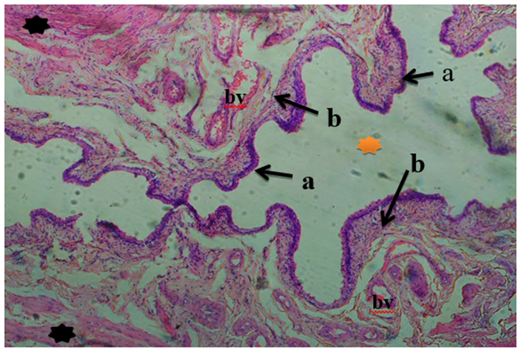
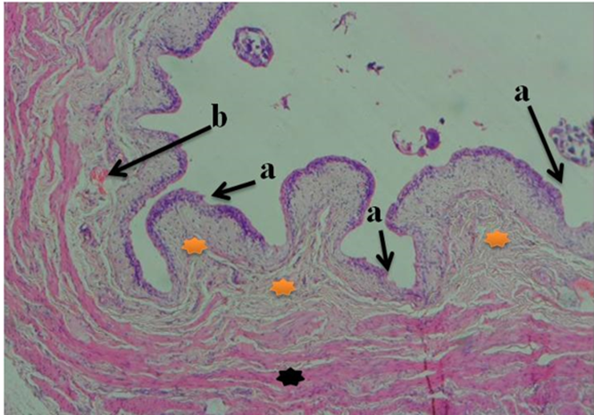
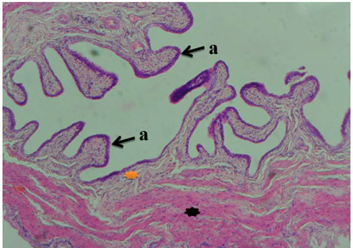
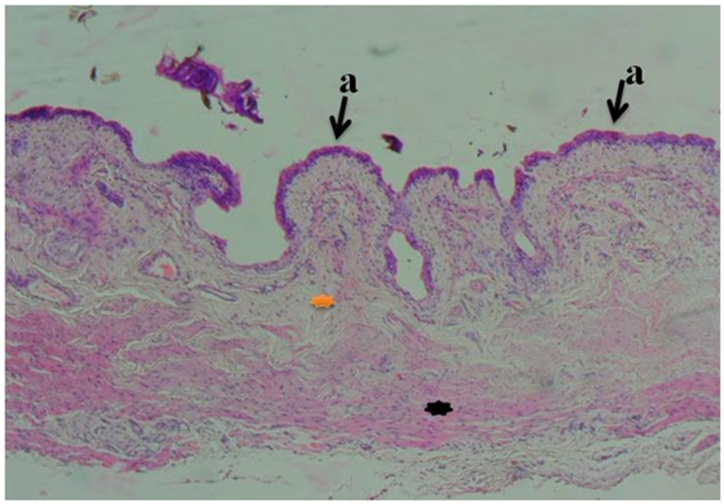
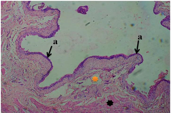
In plate I, the urinary bladder of the control (group 1) animals given feed and water only, showed a well-defined urothelium with transitional epithelium, lamina propria with blood vessels, detrusor muscle layer and normal lumen. The urinary bladder of group 2 animals given 3mg/kg of cadmium only for 4 weeks showed a disorganized epithelium, hemorrhagic cystitis, tissue edema and detrusor muscle hypertrophy (plate II). The urinary bladder of group 2 animals was severely affected due to inflammation. The urinary bladder of group 3 animals given 3mg/kg of cadmium for 4 weeks and 500mg/kg of Uvaria chamae extract for 28 days, showed a well-defined urothelium, reduced tissue edema but with the presence of detrusor hypertrophy in the muscle layer (plate III). This indicated the commencement of the healing process. The urinary bladder of group 4 animals given 3mg/kg of cadmium for 4 weeks and 1000mg/kg of Uvaria chamae extract for 28 days showed a well-defined urothelium, reduced tissue edema and reduced detrusor muscle hypertrophy (plate IV). This indicated the progression of healing process. The urinary bladder of group 5 animals given 3mg/kg of cadmium for 4 weeks and 1500mg/kg of Uvaria chamae extract for 28 days showed a well-defined urothelium, absence of tissue edema in the lamina propria and non-hypertrophied detrusor muscle. Here, the healing process was completed (plate V).
Discussion
This study evaluated the effect of cadmium on the urinary bladder and kidney function parameters of Wistar rats and the role of Uvaria chamae in mitigating these effects. Cadmium is a known contaminant that poses risk to human health. Nearly everyone in the general population is exposed to this contaminant through food supply and it accumulates in the body over a lifetime. Ingestion of cadmium poses a major concern as it is a nonessential trace element that does not play a role in the growth of humans or plants, but is toxic to humans [9-12]. Cadmium toxicity is contingent on exposure route, quantity, and rate. In humans, the kidney bears the brunt of toxic effects, particularly the S1 segment of the proximal tubule. This region becomes a major site for cadmium deposition, manifesting clinically as defects in reabsorption of proteins, amino acids, glucose, bicarbonate, and phosphate. This condition is collectively termed Fanconi syndrome. These defects arise from cadmium-induced oxidative damage to transport proteins and mitochondria, potentially triggering apoptosis of tubular cells [13, 14]. Cadmium could also hinder the metabolism of Vitamin D in the kidney, leading to adverse effects on bone [15]. This, combined with cadmium's direct interference with the absorption of calcium in the gut and disruption of collagen metabolism, may result in osteomalacia and/or osteoporosis [16]. A notable manifestation of this sequence is itai-itai disease in Japan, characterized by intense pain from osteomalacia, osteoporosis, renal tubular dysfunction, anemia, and calcium malabsorption [17]. In contrast, Uvaria chamae is a plant with arrays of medicinal and therapeutic properties. It is a plant that has both medicinal and nutritional values. The root barks, stem barks, and leaves are extensively utilized for their medicinal properties. The root bark is employed in treating respiratory catarrh, and in phytomedicine, the root extract proves beneficial for addressing ailments like piles, menorrhagia, epistaxis, hematuria, and hemolysis [18]. The root extract serves to alleviate abdominal pains, while the juice extracted from roots, stems, or leaves is commonly applied to wounds or sores and this helps in promoting swift and effective healing [19]. Also, in Sierra Leone and Nigeria, the root is renowned for its purgative and febrifugal properties. In traditional medicine, concoctions of roots, barks, and leaves are used to address and treat various ailments such as gastroenteritis, vomiting, diarrhea, dysentery, wounds, sore throats, inflamed gums, etc [20]. Studies on the root extract of Uvaria chamae showed the presence of bioactive components comprising flavonoids, alkaloids, tannins, saponins and phenols [21, 22]. Bioactive compounds, particularly flavonoids, form the core of U. chamae's medicinal properties in Nigerian herbal medicine. Their diverse biological functions, encompassing protection against allergies, platelet aggregation, microbes, ulcers, hepatoxins, viruses, and tumors, underscore the significance of these compounds [23]. By impeding the enzymes involved in estrogen production, flavonoids lower the risk of estrogen-induced cancers. Moreover, specific flavonoids exhibit robust protective properties against inflammatory disorders. They decrease edema formation, suppress the synthesis of prostaglandin E2 and thromboxane B2 [23, 24]. Okwu and Iroabuchi (2009) [21] found that the root extract of U. chamae exhibits a higher concentration of flavonoids in comparison to alkaloids, tannins, saponins, and phenols. Alkaloids are nerve stimulants, convulsants and muscle relaxant [25]. The abundant presence of saponins and tannins in U. chamae roots may contribute to the plant's hemostatic activity, potentially arresting bleeding from damaged or injured vessels through the precipitation of proteins to form vascular plugs. Indicative of its diverse potential, the plant contains phenols that may serve as anti-clotting agents, antioxidants, immune enhancers, and hormone modulators. Extensive research has focused on phenols as bioactive compounds employed in disease prevention [26]. Phenols exhibit the capability to inhibit specific enzymes associated with inflammatory disorders and modify prostaglandin pathways, safeguarding platelets from clumping [26]. The ethanolic extract from Uvaria chamae roots was found to exhibit significant anti-inflammatory properties, possibly attributed to the presence of bioactive compounds like flavonoids, saponins, and phenolic compounds [21]. This current study aimed to evaluate the potency of Uvaria chamae extract at different doses against cadmium induced urinary bladder toxicity and disturbances in the kidney function biomarkers. Evaluating kidney function often relies on blood chemistry analyses, specifically measuring concentrations of serum urea, serum creatinine, and blood urea nitrogen (BUN), which are widely employed for this purpose. Serum urea, serum creatinine, and blood urea nitrogen serve as established clinical indicators reflecting the physiological and functional status of the kidneys, each with its defined normal concentration range. Any deviation from these values is considered indicative of kidney morbidity. Our findings, which is consistent with Bekheet et al., 2011 [27], indicate that exposure to cadmium resulted in significant (p< 0>
Conclusion
In conclusion, the study suggests that Uvaria chamae played ameliorative role in shielding experimental rats from cadmium-induced bladder toxicity. This plant-based product, Uvaria chamae, may offer a certain degree of therapeutic efficacy against acute or bladder impairments in humans. Thus, the ethanolic extract of Uvaria chamae is suggested as a potential remedy for alleviating toxicity induced by cadmium.
Competing Interest
We declare that, no competing interests exist among us.
References
- Lide, D.R. (2005). “Magnetic susceptibility of the elements and inorganic compounds”. CRC Handbook of Chemistry and Physics, 86th edition.
View at Publisher | View at Google Scholar - Page, A.L. and Bingham, F. T. (1973). Cadmium residues in the environment. in residue Reviews, 1-44.
View at Publisher | View at Google Scholar - Goering, P. L., Waalkes, M. P., and Klaassen, C. D. (1995). Toxicology of cadmium. In Toxicology of metals, 189-214.
View at Publisher | View at Google Scholar - Chummy, S. S. (2011). Last’s anatomy, regional and applied anatomy, 12thedition.
View at Publisher | View at Google Scholar - Shermadou, E. S., Rahman, S., and Leslie, S. W. (2022). Anatomy, abdomen and pelvis, bladder. In stat pearls. Retrieved from .
View at Publisher | View at Google Scholar - Oluremi, B., Osungunna, M.and Omafuma, O. (2010). Comparative assessment of antibacterial activity of Uvaria chamae plants. African Journal of Microbiology Research, 4(13): 1391-1394.
View at Publisher | View at Google Scholar - Okokon, J. E., Ita, B. N. and Udokpoh, A. E. (2006). The in-vivo antimalarial activities of Uvariachamae and Hippocratea africana. Annals of Tropical Medicine and Parasitology, 100(7): 585-590.
View at Publisher | View at Google Scholar - Carl, A. B., Edward, R. A. and David E. B. (2008). Tietz fundamentals of clinical chemistry, 6th edition.
View at Publisher | View at Google Scholar - Clemens, S., Aarts, M., Thomine, S. and Verbruggen, M. (2013). Plant science: The key to preventing slow cadmium poisoning. Trends in Plant Science, 18(2): 92-99.
View at Publisher | View at Google Scholar - European Safety Authority (EFSA). (2009). Scientific opinion of the panel on contaminants in the food chain on a request from the European commission on cadmium in food. EFSA Journal, 7(3): 183-232.
View at Publisher | View at Google Scholar - Khan, M., Khan, S., Khan, A. and Alam, M. (2017). Soil contamination with cadmium, consequences and remediation using organic amendments. Science of the Total Environment, 1591-1605.
View at Publisher | View at Google Scholar - Smolders, E. (2001). Cadmium uptake by plants. International Journal of Occupational Medicine and Environmental Health, 14(2): 177–183.
View at Publisher | View at Google Scholar - Thévenod, F., (2003). “Nephrotoxicity and the proximal tubule: insights from Cadmium,” Nephron, 93(4): 87-93.
View at Publisher | View at Google Scholar - Sabath, E. and M. L. Robles-Osorio, (2012). “Renal health and the environment: heavy metal nephrotoxicity,” 32(3): 279-286.
View at Publisher | View at Google Scholar - Kjellstrom, T. (1992). “Mechanism and epidemiology of bone effects of cadmium,” IARC Scientific Publications, 118(14): 301-310.
View at Publisher | View at Google Scholar - Nordberg,G.F., Nogawa,K., Nordberg M. andFriberg,L. (2007). “Cadmium”, in HandbookoftheToxicologyofMetals, 3rd edition, 23(5): 445-486.
View at Publisher | View at Google Scholar - Ogawa,T., Kobayashi, E., Okubo,Y., Suwazono,Y., Kido, T. and Nogawa, K. (2004). “Relationship among prevalence of patients with Itai-itai disease, prevalence of abnormal urinary findings, and cadmium concentrations in rice of individual hamlets in the Jinzu River basin, Toyama prefecture of Japan,” International Journal of Environmental Health Research, 14(4): 243-252.
View at Publisher | View at Google Scholar - Oliver, B. (1986). Medicinal plants in Tropical West Africa, 123-125.
View at Publisher | View at Google Scholar - Irvin,F. R. (1961). Woody plants of Ghana with Special Reference to their Uses. Oxford University Press, London, 19(20): 500-695.
View at Publisher | View at Google Scholar - Okwu, D. E. (2007). Medicinal and Aromatic Plant Science and Biotechnology. International Journal of Biomedical and Pharmaceutical Sciences, 1(1): 90-96.
View at Publisher | View at Google Scholar - Okwu, D. E., Iroabuci, F. (2009). Phytochemical Composition and Biological Activities of Uvaria chamae and Clerodendoron splendens. Electronic Journal of chemistry, 6(2): 553-560.
View at Publisher | View at Google Scholar - Olufunmilayo, E.A, Adelodun, L.K., Oladimeji, P.R. and Lateef, S. K.(2010). In vitro antisickling activities and phytochemical evaluation of Plumbago zeylanica and Uvaria chamae. African Journal of Biotechnology, 9(53):932-936.
View at Publisher | View at Google Scholar - Okwu,D. E. and Omodamiro, O.D.(2005). Phytochemical Composition and Biological Activities of Uvaria chamae and Clerodendoron splendens. Bio-research, 3(2): 40-44.
View at Publisher | View at Google Scholar - Samasundaram, S., and Sadique, J. (2009). Intestinal permeability in the pathogenesis of NSAID-induced enteropathy. Journal of Gastroenterology, 23-29.
View at Publisher | View at Google Scholar - Kenner, D. L. and Yves, R. M. (1996). Botanical Medicine. A European Professional Perspective, 487-490.
View at Publisher | View at Google Scholar - Duke, J. N. (1992). Handbook of biological active phytochemicals and their activities, 99-131.
View at Publisher | View at Google Scholar - Bekheet, S.H., Awadalla, E.A., Salman, M. M.and Hassan, M.K.(2011). Bradykinin potentiating factor isolated from Buthus occitanus venom has a protective effect against cadmium induced rat liver and kidney damage tissue cell, 43(4): 337-343.
View at Publisher | View at Google Scholar - Wang,L.,Chen, D., Cao, J. and Liu, Z.(2009). Protective effect of N-acetylcysteine on experimental chronic cadmium nephrotoxicity in immature female rats, Human Experimental Toxicology, 28(5): 221-229.
View at Publisher | View at Google Scholar - Tormanen, C.D.(2006). Inhibition of rat liver and kidney arginase by cadmium ion, Journal of Enzyme Inhibition and Medicinal Chemistry, 21(1): 119–123.
View at Publisher | View at Google Scholar - Lujambio,I., Sottolano, M., Luzardo,L., Robaina, S., Krul, N., and Thijs, L.(2014). Estimation of glomerular filtration rate based on serum cystatin C versus creatinine in a Uruguayan population. International Journal of Nephrology, 83-106.
View at Publisher | View at Google Scholar - Levey, A.S. (1990). Measurement of renal function in chronic renal disease. Kidney International, 167-184.
View at Publisher | View at Google Scholar - Romaniuk, A., Sikora, Lyndin V., Smiyanov M, Sikora V., V. and Lyndina, Y. (2017). The features of morphological changes in the urinary bladder under combined effect of heavy metal salts. Intervention Medical Applied Science, 9(2): 105-111.
View at Publisher | View at Google Scholar - Feki-Tounsi, M., and Hamza-Chaffai, A. (2014). Cadmium as a possible cause of bladder cancer: a review of accumulated evidence. Environmental Science Pollution Control, 10561-10573.
View at Publisher | View at Google Scholar - Golabek,T., Darewicz,B.,Borawska, M., Markiewicz, R., Socha, K. and Kudelski, J. (2009). Lead concentration in the bladder tissue and blood of patients with bladder cancer, Scandinavian Journal of Urology and Nephrology, 43(6): 467-470.
View at Publisher | View at Google Scholar
