

Review Article | DOI: https://doi.org/10.31579/2835-9232/038
Obtaining Semi-Thin Sections for Electron Microscopic Examination of The Brain
1Candidate of biological science, assistant professor of pathophysiology department named D.A. Maslakov, Grodno State Medical University, 80, Gorky St., 230009, Grodno, Belarus.
*Corresponding Author: Lizaveta I. Bon, Candidate of biological science, assistant professor of pathophysiology department named D.A. Maslakov, Grodno State Medical University.
Citation: E.I. Bon, N.Ye. Maksimovich, S.M. Zimatkin, O.B. Ostrovskaya, A.V. Malykhina, (2023), Obtaining Semi-Thin Sections for Electron Microscopic Examination of The Brain, International Journal of Clinical Epidemiology, 2(5); DOI:10.31579/2835-9232/038
Copyright: © 2023, Lizaveta I. Bon. This is an open-access article distributed under the terms of the Creative Commons Attribution License, which permits unrestricted use, distribution, and reproduction in any medium, provided the original author and source are credited.
Received: 06 October 2023 | Accepted: 16 October 2023 | Published: 25 October 2023
Keywords: semi-thin sections; electron microscopic; brain
Abstract
The study of neuronal ultrastructure is an important and current area of modern science. However, to obtain proper results, it is necessary to strictly follow the methodological rules for making not only ultrathin, but also semi-thin sections.
For the description and morphometry of mitochondria, we selected neurons that had a pyramidal shape with a clearly defined apical dendrite and a maximum transverse width of the perikaryon (usually the width at the base of a pyramidal-shaped perikaryon) of at least 15 am, as well as a nuclear shape close to round or oval. Thus, neurons captured on one grid could not be counted again on adjacent grids.
Summery
The study of neuronal ultrastructure is an important and current area of modern science. However, to obtain proper results, it is necessary to strictly follow the methodological rules for making not only ultrathin, but also semi-thin sections.
For electron microscopic examination fragments of the cortex measuring about 1.0x1.0x2.0 mm, prismatic in shape were taken so that the long axis of the prism was perpendicular to the surface of the cortex and included all layers of gray matter, as well as the thin layer of white matter adjacent to the latter (Figure. 1). When pouring, the samples were oriented in such a way as to obtain sections in a plane perpendicular to the surface of the cortex. Semi-thin (0.35 µm thick) and ultra-thin (40 nm) sections were obtained on a Leica EM UC7 ultramicrotome (Leica, Germany) using glass knives. The first
sections were stained with methylene blue and used to select the area containing cells of the inner pyramidal layer of the cortex. In this case, to obtain ultrathin sections, we selected the area containing the largest (compared to other neurons in the section) pyramidal-shaped neurons along with dendritic profiles of the greatest thickness running perpendicular to the surface of the cortex (Fig. 2). Since the central region of the sample was usually insufficiently fixed, only the lateral areas containing the described neurons and characterized by satisfactory fixation were selected to obtain ultrathin sections (Fig. 1-4). Moreover, in this area no more than 3 pyramidal neurons corresponding to the requirements could be found at the same time (Figure. 4).
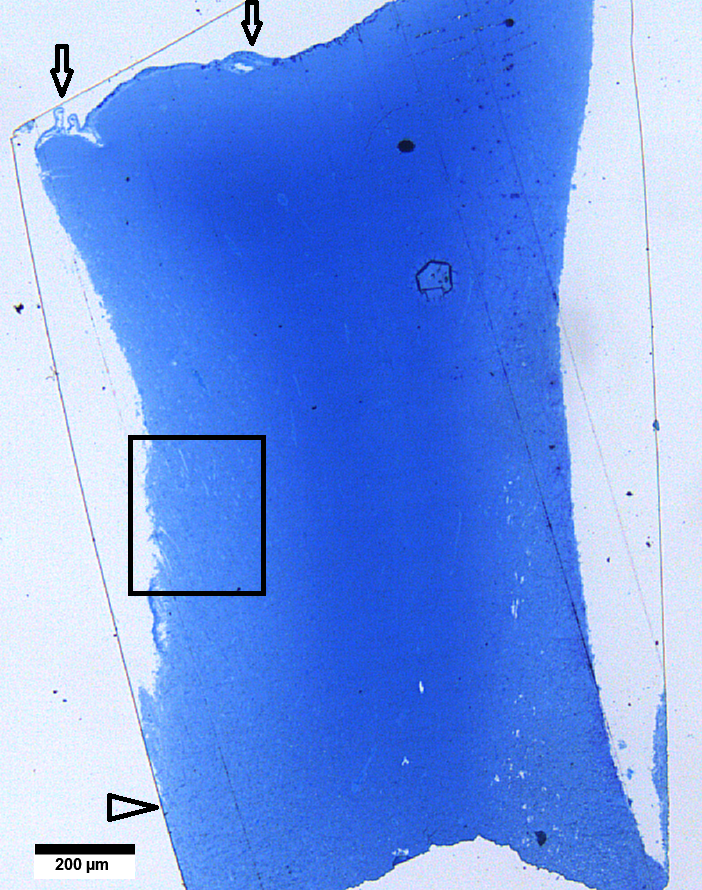
Figure 1. Semi-thin section (350 nm thickness) of a rat cortex sample stained with methylene blue. The surface of the cortex (above) is covered with the pia mater (arrows), below - white matter (arrow head). The central region of the sample has a darker color; individual structures in it are not determined as a result of poor fixation. The rectangle shows the area selected for ultrathin sectioning.
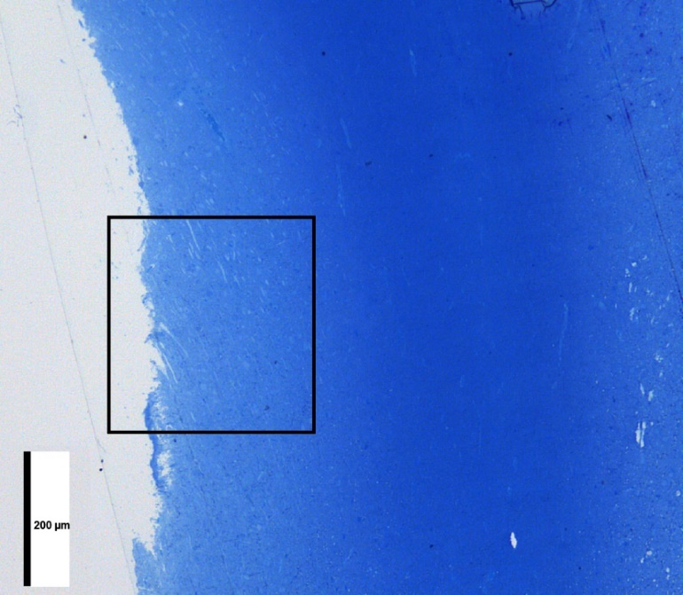
Figure 2. The area indicated in Figure 1 at higher magnification.
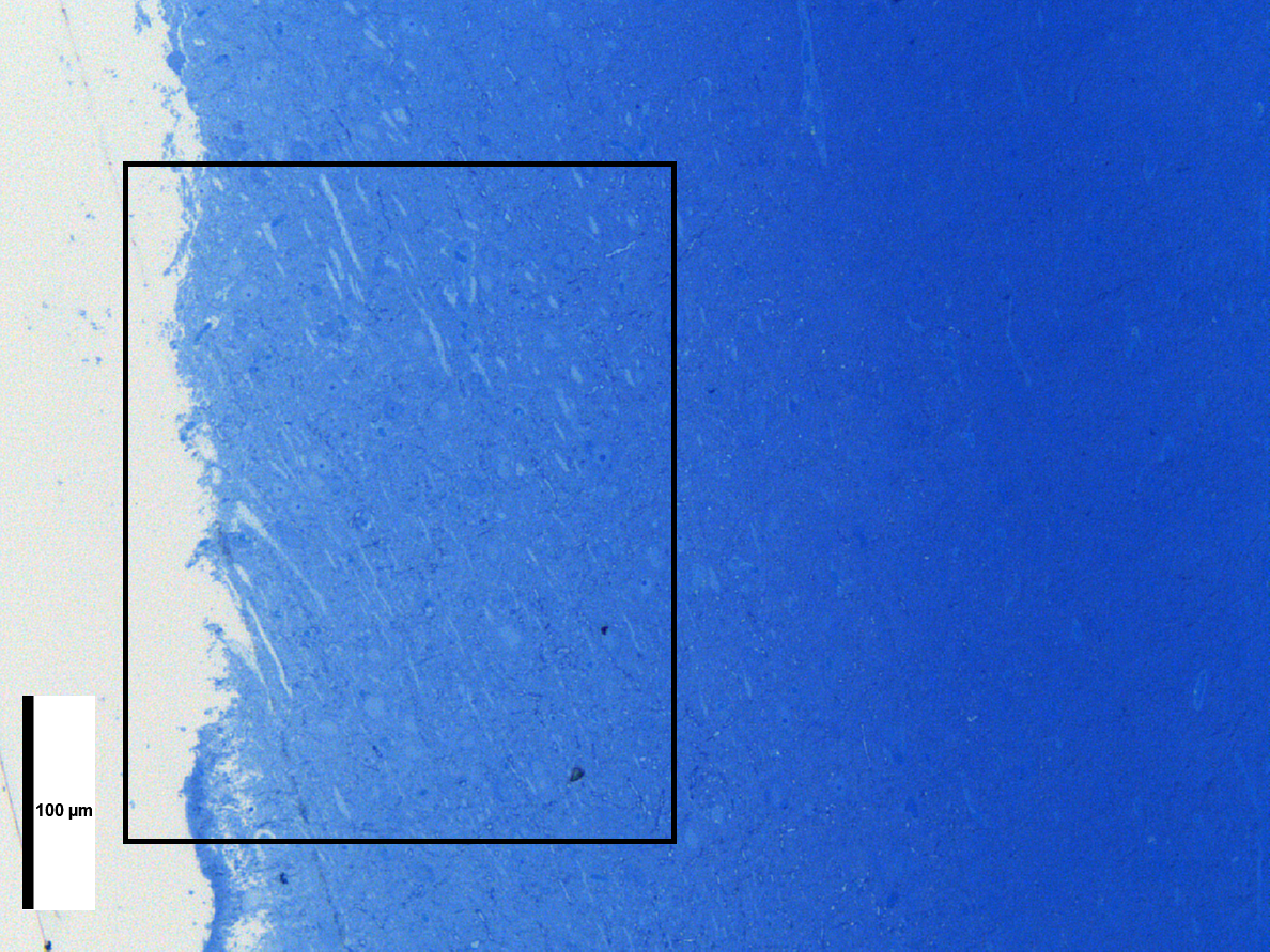
Figure 3. The area indicated in Figure 2 at higher magnification.
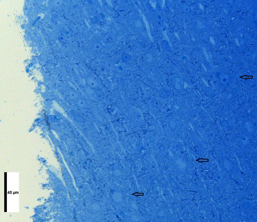
Figure 4. The area indicated in Figure 3 at higher magnification. Arrows indicate neurons that have a pyramidal shape, an apical dendrite, and a maximum perikaryon width of 15-20 µm.
Therefore, to visualize a sufficient number of neurons from the area selected in the described manner, sharpened by a “pyramid,” 5-6 series of ultrathin sections were obtained with an interval of 15 am. Ultrathin sections were prepared with the microtome feed set at 40 nm (occasionally 35 nm) and were always gray or silver in color. Each series (6-8 adjacent sections) was mounted on a copper grid, contrasted with uranyl acetate and lead citrate, and examined in a JEM-1011 electron microscope (JEOL, Japan). To obtain images, a complex consisting of an Olympus Mega View III digital camera (Japan) and the item program (Version 5.0 (Build 1224); Serial Number A3766900-7E852FAB) was used. For the description and morphometry of mitochondria, we selected neurons that had a pyramidal shape with a clearly defined apical dendrite and a maximum transverse width of the perikaryon (usually the width at the base of a pyramidal-shaped perikaryon) of at least 15 am, as well as a nuclear shape close to round or oval. Thus, neurons captured on one grid could not be counted again on adjacent grids.
