

Research Article | DOI: https://doi.org/10.31579/2834-5126/064
Morphological Changes in Pyramidal Neurons of Frontal Cortex of The Rat`S Brain During Experimental Myocardial Ischemia
- Vasilevich M. V
- Khodosovsky M. N
- Bon E. I *
- Maksimovich N. Ye
- Sinkevich E.V
Grodno State Medical University, 80 Gorky St, 230009, Grodno, Belarus.
*Corresponding Author: Elizaveta I Bon, Grodno State Medical University, 80 Gorky St, 230009, Grodno, Belarus.
Citation: Vasilevich M.V., Khodosovsky M.N., Bon E.I., Maksimovich N.Ye., Sinkevich E.V. (2024), Morphological Changes in Pyramidal Neurons of Frontal Cortex of The Rat`S Brain During Experimental Myocardial Ischemia, Clinical Trials and Clinical Research, 3(3); DOI:10.31579/2834-5126/064
Copyright: © 2024, Elizaveta I Bon. This is an open access article distributed under the creative commons’ attribution license, which permits unrestricted use, distribution, and reproduction in any medium, provided the original work is properly cited.
Received: 08 April 2024 | Accepted: 20 May 2024 | Published: 27 May 2024
Keywords: rats; myocardial ischemia; cerebral ischemia; cerebral cortex
Abstract
Objective: To analyze changes in morphological characteristics of rat frontal cortex neurons during myocardial ischemia.
Methodology. Experiments were performed on 12 male non-pedigreed white rats with an initial weight of 240±20 g. Myocardial ischemia in white mongrel rats was modeled by isoprenaline administration. The material was sampled 24 hours after the last injection of the drug.
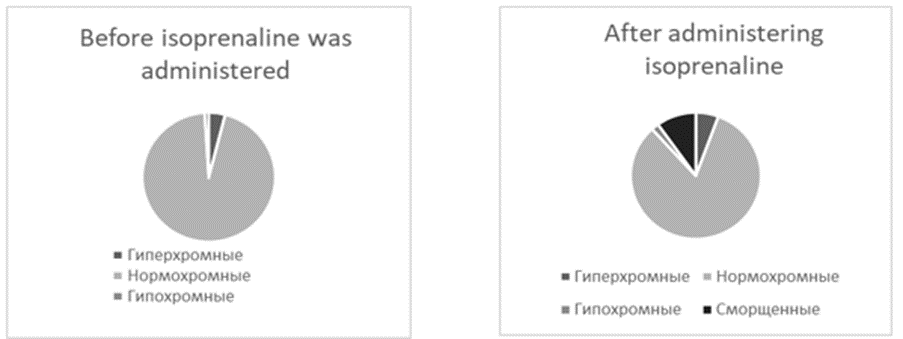
Results. Experimental myocardial ischemia was accompanied by a slight decrease in neuronal size and pericarion deformation. The number of hyperchromic wrinkled neurons increased. Wrinkled neurons constituted the majority of cells in the studied cortex section.
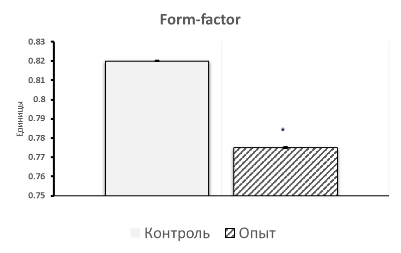
Conclusion: During experimental myocardial ischemia, parietal cortex neurons decreased in size by 3.3% compared to control (p<0.05), the shape of neurons slightly changed - they became more elongated (by 6.4%) (p<0.05), while the form factor decreased by 5.5% (p<0.05) compared to control. The number of normochromic neurons decreased by 8% (p<0.05), while the number of hyperchromic neurons increased 2-fold compared to controls. Hyperchromic shriveled neurons appeared, their number amounted to 4.5% of the number of all neurons.
Background
Acute myocardial infarction is one of the most serious complications of ischemic heart disease, which still remains the main cause of population disability and death [1]. The prognosis of quality of life after acute myocardial infarction is determined not only by its type, localization and prevalence, but also by the development of concomitant cognitive disorders [2]. In patients after myocardial infarction, dementia develops 5 times more often than in the average human population [3]. The possible connection of cognitive disorders in cardiovascular pathology is of interest for further active scientific search of the causes and pathogenetic mechanisms that determine this relationship.
Methods
The experiments were performed on 12 male mongrel white rats with an initial weight of 240±20 g in compliance with the requirements of the Directive of the European Parliament and Council No. 2010/63/EU of 22.09.2010 on the protection of animals used for scientific purposes. Animals were kept in an air-conditioned room (22°C) with mixed lighting on a standard vivarium diet and free access to food and water, in groups of no more than 5 individuals per vivarium cage [4].
Experimental myocardial ischemia was modeled by subcutaneous injection of isoproterenol hydrochloride (Sigma) twice with an interval of 24 hours in doses of 80 mg/kg. Control animals were injected with physiologic sodium chloride solution in equivalent volumes. The animals were divided into 2 groups of 6 each: group I - control; group II "Isoproterenol 80" - subcutaneous injection of isoproterenol at a dose of 80 mg/kg. Material was sampled 24 hours after the last injection of the drug. After decapitation, the brain was quickly extracted, pieces of the anterior cortex were fixed in formalin solution. Serial paraffin sections were stained with 0.1% toluidine blue according to the Nissl method. Histological preparations were studied, microphotographed, and morphometry was performed using an Axioscop 2 plus microscope (Zeiss, Germany), a digital video camera (LeicaDFC 320, Germany), and ImageWarp image analysis program (Bitflow, USA). The localization of the frontal cortex in histological preparations of rat brain was determined using a stereotaxic atlas [5]. At least 30 neurons of the fifth layer of the parietal cortex were evaluated in each animal, which provided a sufficient sample size for further analysis. The number of large pyramidal neurons per unit area of cortical slices was determined on paraffin sections. Among the total number, cells were distinguished by the intensity of cytoplasm coloration (chromatophilia). Several types were distinguished: normochromic - moderately colored; hyperchromic - dark; hyperchromic - very dark, with deformed perikaryons; hypochromic - lightly colored. The number of each cell type was counted. After preliminary check for normality of the distribution of parameters, the obtained data were analyzed by methods of nonparametric statistics using Statistica 10.0 for Windows (StatSoft, Inc., USA). The results are presented as Me(LQ; UQ), where Me is the median, LQ is the value of the lower quartile; UQ is the value of the upper quartile. Differences between the indices of the control and experimental groups were considered reliable at p<0>
Results of the study
During experimental myocardial ischemia, parietal cortex neurons decreased in size, by 3.3% compared to control (p<0>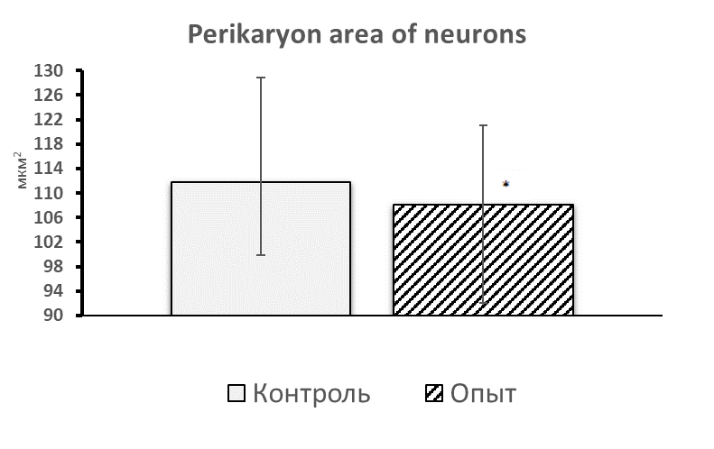
The shape of neurons changed insignificantly - they became more elongated (by 6.4%), (p<0>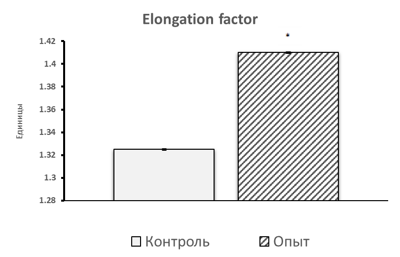
The number of normochromic neurons decreased by 8% (p<0>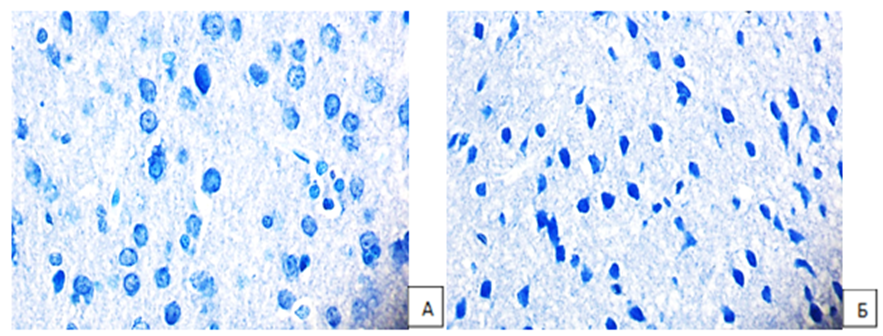
According to the literature, hyperchromic neurons are considered as ischemia-altered cells [7]. The appearance of wrinkled dark cells under
conditions of hypoxia and anoxia is a universal and the most severe form of reactive and pathological changes in neurons, which is accompanied by changes not only in metabolic rate, but also in tinctorial properties of cytoplasm, as well as karyoplasm of cells and various degrees of ultrastructural changes in cytoplasmic organelles of the cell [9].
It is known that intense staining of neuronal cytoplasm characterizes the predominance of protein formation over its utilization [9]. However, a number of authors believe that a hyperchromic neuron, through overexpression of amplified genes, is a cell capable of intensive protein synthesis that goes to their own needs [10].
Shriveled neurons are cells with suppression of functional activity. Their characteristic form is associated with pathological irreversible changes in water-salt metabolism [8].
Depending on the conditions of functioning, neurons with initial signs of hyper- and hypochromia may turn either into shadow cells (hypochromic) or into shriveled hyperchromic neurons with subsequent collisional and coagulation necrosis or apoptosis [11].
Conclusion
The obtained data on histologic changes of frontal cortex neurons in experimental myocardial ischemia provide a basis for further detailed study of brain changes, determination of pathogenetic mechanisms, creating a fundamental basis for studying the properties of neurons, including their transition from one functional state to another.
References
- Gafarov V. V. (2015). World Health Organization program
View at Publisher | View at Google Scholar - Thong, Elizabeth Hui En et al. (2023). Acute Myocardial Infarction and Risk of Cognitive Impairment and Dementia: A Review // Biology. Vol. 12, 8. - P. 1154.
View at Publisher | View at Google Scholar - Derevnina, E. S. (2012). Cognitive disorders in patients with cardiovascular diseases / E. S. Derevnina, D. G. Persashvili, Y. G. Shvarts // Modern problems of science and education. - № 5. - С. 28.
View at Publisher | View at Google Scholar - Karkishchenko N.N., Gracheva S.V. (2010). Manual on laboratory animals and alternative models in biomedical research. - Moscow: Profile-2C. - 241 с.
View at Publisher | View at Google Scholar - Paxinos G., Watson C. The Rat Brain in stereotaxic coordinates. - Academic Press, Australia, 1998. - P. 242.
View at Publisher | View at Google Scholar - Batin N.V. (2008). Computer statistical data analysis: textbook. - Minsk: Institute of scientific personnel training of the National Academy of Sciences of Belarus. - 235 с.
View at Publisher | View at Google Scholar - Popova E.N. (2010). Ultrastructure of the brain, alcohol and offspring: monograph. - Moscow. Izd. Nauchny Mir. - 155 с.
View at Publisher | View at Google Scholar - Bon E.I., Maksimovich N.Ye., Zimatkin S.M., Valko N.A., Kot V.N., et al. (2019). Dynamics of morphological changes in pyramidal neurons of phylogenetically different sections of the rat cerebral cortex at total cerebral ischemia // Bulletin of Smolensk State Medical Academy. №2.
View at Publisher | View at Google Scholar - Gallyas F., Pal J., Bukovics P. (2009). Supravital microwave experiments support that the formation of
View at Publisher | View at Google Scholar - Zimatkin S.M., Bon E.I. (2017). Dark neurons of the brain // Morphology: №6. - С. 81-86.
View at Publisher | View at Google Scholar - Semchenko V.V., Stepanov S.S., Alekseeva G.V. (1999). Postanoxic encephalopathy. - Omsk. - 448 с.
View at Publisher | View at Google Scholar
