

Review Article | DOI: https://doi.org/10.31579/2834-8761/010
Metal Bound Nanoparticles Drug Delivery Methods for Cancer and Tumor Therapeutics: A Review
- Ravi Kant Upadhyay * *
Department of Zoology, Deen Dayal Upadhyaya Gorakhpur University, Gorakhpur, India 273009.
*Corresponding Author: Ravi kant Upadhyay, Department of Zoology, Deen Dayal Upadhyaya Gorakhpur University, Gorakhpur, India 273009.
Citation: Ravi Kant Upadhyay (2023), Metal Bound Nanoparticles Drug Delivery Methods for Cancer and Tumor Therapeutics: A Review, Clinical Endocrinology and Metabolism, 2() DOI:10.31579/2834-8761/010
Copyright: © 2023, Ravi Kant Upadhyay. This is an open access article distributed under the Creative Commons Attribution License, which permits unrestricted use, distribution, and reproduction in any medium, provided the original work is properly cited.
Received: 03 May 2023 | Accepted: 19 May 2023 | Published: 23 May 2023
Keywords: nano-materials/nanostructures; drug delivery; metal based nanoparticles; liposome encapsulation of metals; anticancer compounds
Abstract
Nanotechnology is an interdisciplinary field that includes three highly overlapping areas: nanomaterials, nanoelectronics, and nanobiotechnology. Nanomaterials have revolutionized both therapeutics and diagnostics, as these are emerged as promising candidates which efficiently carry drug molecules. These nanosystems or metallic nanoparticles have increased stability and half-life of drugs, enhanced biodistribution, and carry loaded drug into the required target site. The current review mainly focuses on more environmentally friendly methods to fabricate metal nanocarriers and their surface modifications, such as silver, gold, platinum, ruthenium, palladium, copper, zinc oxide, iron oxide, grapheme oxide. metal sulfides, and nanometal-organic frameworks in drugs. This article signifies role of most efficacious drug delivery systems microemulsions (MEs), cyclodextrin (CD), polymeric nanoparticles (PN), solid lipid nanoparticles (SLNs), nanostructured lipid carriers (NLCs). It focuses on various MNP applications as biological safe drug delivery vehicles. This review describes the applications of various nanocarrier-based delivery systems for more effective treatment of cancer and tumor therapy without side effects.
Introduction
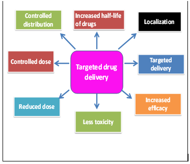
Cancer is currently the second leading cause of morbidity and mortality in the world's population, after communicable diseases. Although cancer and tumor-related deaths have greatly exceeded the past two decades, survival rates for many cancers have improved due to the development of significantly improved diagnosis, cancer screening, treatment, and prevention. Cancer is characterized by uncontrolled cell proliferation and the production of abnormal cell masses or tumors in body tissues or organs. It grows and exhibits neovascularization in the early stages, but in the secondary stages it acquires metastatic potential, spreading to all organ systems of the body and ultimately leading to death. Cancer growth invades and destroys normal body tissue. Major causes of cancer include environmental carcinogens, particulate matter, toxic gases, drugs, DNA damage, or mutations in a cell's genetic material due to environmental or genetic factors. Its treatment does not involve general treatment and treatment. Controlling growth requires potent cytostatics and enzyme inhibitors. A major question is how to deliver drug molecules to the tumor site to localize cancer growth. However, nanoparticles have demonstrated the ability to freely load and transport various anticancer agents [1] (Figure 1). There are numerous classes of nanomaterials that are widely used for the controlled delivery of therapeutic compounds to treat cancer. They are also used in the clinical sciences of diagnostic imaging, drug/gene delivery, regenerative medicine, cancer therapy, and essentially organ transplantation [2]. New processes, molecules and nanomaterials have undoubtedly revolutionized cancer and tumor treatments.
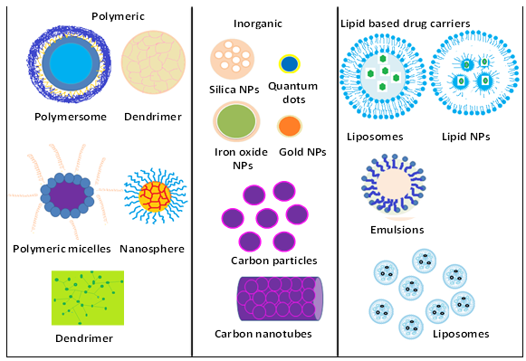
Nanomaterials/nanostructures are multifunctional and are used in biosensing, drug delivery, imaging, medical implants, cancer therapy, and tissue engineering (Figure 1). These support the drug's solubility, its systemic distribution. It also improves the therapeutic efficacy of drugs and the performance of diagnostic procedures. Metal nanoparticles (MNPs) are used in therapeutics such as antibodies, nucleic acids, chemotherapy, and peptides. In general, NPs can be classified into macromolecular NPs, dendrimers, micelles, liposomes, lipid-based NPs and organics such as ferritin. Inorganics, including metals and metal oxide NPs (e.g. silver, gold, iron oxides, zinc oxides, and silicon dioxide). Most commonly used carbon-based NPs are fullerenes, graphene, and carbon nanotubes (Table 1).
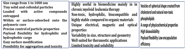
Metallic nanoparticles are a unique class of functional nanomaterials with sizes ranging from 1 to 100 nanometers and large surface areas. They may be used as drug delivery vehicles in pharmacy and biomedicine [2]. These metal complexes possess hydrophilic/lipophilic properties and can readily remove toxic metal ions from extracellular sites or enter intracellular compartments to facilitate removal of toxicity [5]. There are various metal-based nanoparticles made from platinum, silver, gold, palladium, vanadium, ruthenium, titanium, zinc and copper nanoparticles. In addition to nano drug delivery systems, CPT and its derivatives have been developed. Additionally, metal-based nanoparticles have the disadvantage of being toxic to living cells and tissues. However, chelating agents (dimercaptosuccinate (DMSA) and dimercaptopropionate sulfonate (DMPS)) have rarely been used to reduce the cytotoxicity of metal-based nanoparticles. Bodies [5] are carbohydrate-based paramagnetic metal chelators [6] containing manganese or gadolinium that act as more potent relaxants than iron, copper, erbium, or nickel derivatives.
Metal nanoparticles such as gold nanoparticles (AuNPs), silver nanoparticles, and cysteine-capped silver nanoparticles (Cys-AgNPs) utilize the reducing ability of EAB in the presence of organic substrates such as electron donors. synthesized (Lee SH, Jun BH. 2019) [7]. EAB uses metal oxides as electron acceptors for synthesizing various metal nanoparticles. Metal oxides are used as precursors. These metal nanoparticles exhibit potent antimicrobial activity nanoparticles [8]. In addition, carbon-based metallic and semi-metallic nanomaterials have also been prepared for drug delivery. The most useful are carbon allotropes such as graphene oxide (GO), carbon nanotubes (CNT), and nanodiamonds (ND). Some non-CPT organic compounds, indenoisoquinolines and dibenzonaphthyridines, have been used to combat cellular resistance [9] (Figure 1).
Polymeric microspheres and liposomes are also used to deliver anticancer medications. The majority of efficient drug delivery techniques, such as polymeric nanoparticles (PN), solid lipid nanoparticle (SLN), nanostructured lipid carriers (NLCs), and microemulsions (MEs), are well known and used in the treatment of cancer. Additional drug delivery techniques include the use of coordination compounds, magnetic and gold nanoparticles (MNPs / AuNPs), and liquid crystalline systems (LC) [10]. There are numerous industrial applications for metal NPs. A powerful technique for stabilising the nanoparticles and further modifying the properties of the material is surface functionalization with organic molecules. Mercapto derivatives are among the most often used ligands for surface functionalization of nanoparticles because of the strong attraction thiol moieties have for transition metal surfaces, which leads to the formation of (polar) metal-thiolate linkages [11]. Single metal, multimetallic, and multimatrix combinations have all been synthesised, with many more potential uses in the biological and medicinal sciences. On the other hand, it seems that there is a significant demand for drug solubility, a decrease in systemic toxicity through targeted cancer therapy, and rapid clearance of hazardous waste (Figure 2).
In this review, different metal nanoparticle kinds have been described together with how frequently they are used in cancer treatment. Metal toxicity, medication loading, drug delivery, and vascular leakage have all been mentioned as significant risk factors. The goal of this study is to investigate potential applications of additional surface-characterized nanoparticles for secure medication administration in the treatment of cancer. It is important to emphasise the potential of metal NPs, coordinating complexes, and other nanomaterials for the treatment of cancer. With the help of plant extracts, nanoparticles were created in a green manner. In addition to biomedicine, nanoparticles are utilised in energy production, disease treatment, and the reduction of environmental pollutants. These are very advantageous because they don't pose a toxicity risk, are less expensive, cause less pollution, and increase environmental and human health safety [12].
Types of nanoparticles:
Platinum nanoparticles:
Alkene derivatives can self-assemble onto the surface of platinum to create stable platinum nanoparticles, possibly forming interfacial bonds with platinum-vinylidene (Pt-C-CH-) or -acetylide (Pt-C). Due to the dehydrogenation and transformation of the olefin moieties, which is catalyzed by platinum, this bond is created [13]. These have numerous biomedical uses, such as imaging, implants, photothermal therapy, and drug delivery. Indeed, the antimicrobial, antioxidant, and anticancer properties of PtNPs are intrinsic. PtNPs have been widely used in industry, as well as in healthcare and diagnostics (Table 1).
Ruthenium nanoparticles:
Ruthenium nanoparticles are spherical in shape having high surface area. These are available in form for nanodots or nanopowder. Nanoscale Ruthenium Particles typically range in size from 20 to 100 nm, with SSAs between 1 and 3 m2/g. There are also high purity, ultra high purity, coated, and dispersed forms of nano-ruthenium particles. Ruthenium nanoparticles were created by a functional group self-assembling on a bare Ru colloid surface. Ruthenium(II)-arene complexes with biotin-containing ligands affect the endocytosis mediated by tumor-specific vitamin receptors. These are ruthenium (II)-biotin half-sandwich conjugates function as biological carriers for cancer cells [14]. Ruthenium-based nanoparticles (ruthenium complexes, namely NAMI-A, KP1019, KP1339, and TLD1433) demonstrated significant anticancer or anti-metastatic properties with minimal side effects [15]. Ru-containing nanomaterials (Ru-cNMs) have demonstrated applications as anticancer, antimicrobial, and antioxidant agents when combined with metals like platinum and palladium or with non-metals like phosphorus and oxygen. These have numerous biological catalytic applications (Table 1).
Platinum- and non-platinum-based materials have been used to create tumor-inhibiting metal complexes. These medications have excellent anticancer properties. The antimetastatic drug NAMI-A is a ruthenium compound. There are kinetically inert platinum (IV) complexes, pH-sensitive platinum prodrugs, platinum-based drug targeting strategies with low-molecular-weight carriers, and tumor-inhibiting non-platinum anticancer drugs based on ruthenium and gallium [16]. The use of ruthenium complexes on tumors that have developed cisplatin resistance has shown great promise [16]. The most effective anticancer medications currently in use are cisplatin and related compounds. These exhibit extremely low general toxicity(Table 1).
Gold nanoparticles: There are many different sizes, shapes, and structures of gold nanoparticles (Au NPs). The remarkable ability of these nanoparticles to scatter and absorb light, to convert optical energy into heat via non-radiative electron relaxation dynamics, and to take advantage of surface chemistries to act as drug carriers make them very appealing and useful [17]. Gold nanoparticles, or Au NPs, are used to deliver chemotherapy drugs, radiation therapy, and thermal therapy [18]. These nanodevices target and deliver chemotherapeutic drugs precisely to the location of the tumor [7]. Gold nanoparticles' permeability and retention effect, which are caused by their small size and non-toxic, non-immunogenic nature, offer additional advantages by enabling easy penetration and accumulation of drugs at the tumor sites [17]. These are highly stable, biocompatible, and have customizable shapes and sizes. These have a surface that is simple to functionalize, a high drug loading capacity, and little toxicity (Table 1) [18]. These multifunctional gold nanoparticle (AuNP) systems also incorporate monoclonal antibodies against the epidermal growth factor receptor (EGFR) (anti-EGFR D-11) for active targeting cancer cells [19].
Also created are gold nanoparticles coated in folic acid and conjugated with the fluorophore FITC using poly(ethylene glycol) with an amine termination (Au-SMCC-DOX nanoconjugates. These nanoparticles help multi-drug resistant hepG2-R cancer cells accumulate and retain drugs. These are the most promising drug delivery systems, with good biocompatibility and potential for future uses [20]. Wide-ranging medical uses for gold nanoparticles include diagnosis, treatment, prevention, and hygiene. Recently, a variety of nanomaterials have been used, including conventional colloidal gold with quasi-spherical particles (nanospheres), non-spherical particles like nanorods, nanoshells, nanocages, nanostars, and other types of particles (this group of particles were named "plasmon resonance particles of noble metals") (Table 1).
Copper nanoparticles: Cu and Cu-based nanoparticles are chemically and physically stable nanoparticles. For generation of nanoparticles copper metal, is complexed with polymers. They range in size from 1 to 100 nm. These show negligible side effects and are used for delivery of radiopharmaceutical [21, 22]. These are also used as diagnostic tools for central nervous system (CNS) cancers, particularly glioblastoma [23]. However, copper compounds that cause cancer have an impact on epithelial cells. In theranostic applications, copper-based tracers 64Cu [64Cu][Cu(ATSM)] and 67Cu are used [24]. For the diagnosis of tumors and neurodegenerative diseases, polyamines with various substituent groups that are ionizable and have different lengths are used [25]. These are employed to alter and regulate the copper complexes' hydrophilic/lipophilic balance [24].
These are extremely tiny copper nanoparticles with a high surface-to-volume ratio can also act as antifungal and antibacterial agents [26]. Their close interaction with microbial membranes and the metal ions they release into solutions cause the antimicrobial activity. Additionally, native amino acids can be found using nanoparticles [27]. A stable and efficient sensing system for the detection of all 20 amino acids is a carbon electrode screen-printed with copper nanoparticles [28]. Green chemistry can also be used to create copper nanoparticles, minimizing the reaction's negative effects on the environment. Stable copper nanoparticles can be created by reducing copper chloride with only L-ascorbic acid in a heated aqueous solution (Table 1).
Silver nanoparticles:
Silver nanoparticles (AgNPs) are well-known and potentially effective antimicrobial agents.
These metallic nanoparticles are among the most significant and fascinating nanomaterials used in biomedical applications. In particular, AgNPs are crucial to nanomedicine and other fields of nanoscience and nanotechnology. These have multiple biological uses for AgNPs, such as as antibacterial, antifungal, antiviral, anti-inflammatory, anti-angiogenic, and anti-cancer agents [29]. They also have the mechanism underlying AgNPs' anti-cancer activity. These are employed in a variety of ways to treat a wide range of pathogenic agents. AgNPs is a brand-new generation of cancer cell treatment and diagnostic tool [30]. These were found effective methods for cancer diagnosis and drug delivery for its treatment [31]. AgNPs made from C. sativum have a lot of potential for biomedical uses like treating breast cancer, dandruff, and acne [32] (Table 1).
The bio-distribution of the copper complexes in mice is changed when they are modified with different functional groups that contain amines. These are used in the diagnosis of brain imaging of tumors and neurodegenerative diseases [25]. One complex, with a pendent (N, N-dimethylamino)ethane functional group, displayed tumor uptake similar to that of [64Cu]Cu(atsm), but higher brain uptake. In clinical settings, anionic linear globular dendrimers based on nanosilver are used to prevent HIV virus replication [34]. These are widely used in the treatment of cancer and exhibit cytotoxicity to cancer and tumor cells [4].
Iron nanoparticles:
Magnetic iron oxide nanoparticles (IONs) are used for the delivery of anticancer drugs [35]. Superparamagnetic iron oxide nanoparticles (MNPs) with suitable surface chemistry have shown many biomedical applications. Fe3O4 nanoparticles exhibited superparamagnetic behavior and the saturation magnetization of Fe3O4 nanoparticles increased with particle size [36]. Lysosomal MNPs, iron ions, are incorporated into the natural circulation and remain in the bloodstream longer. The mechanism of action of the mononuclear phagocytic system and the half-lives of various nanostructures are presented. Due to their hydrodynamic properties, core size, core morphology and coating molecules, surface charge, they are more useful in clinical medicine [37]. Magnetic iron oxide nanoparticles (MNPs) are widely used in medical theranostics and have promising future potential in medicine [38] (Table 1).
These applications required MNPs such as Fe3O4 magnetic nanoparticles (Fe3O4-MNPs) to have high magnetization values and particle sizes less than 100 nm [39]. The use of magnetic nanoparticles (MNPs) such as iron oxide nanoparticles (IONPs) in biomedicine is viewed as a valuable alternative to conventional materials due to their chemical stability, cost effectiveness, and surface functionalization. MNPs are used in numerous biotechnological procedures such as gene transfection and molecular recognition systems. Characterization and surface functionalization of MNPs, interactions between DNA and IONPs, fabrication of DNA nanoplatforms and biotechnology applications such as DNA magnetic separation, magnetic fection, DNA vaccine fabrication, and molecular recognition tools [39] (Table 1).
Calcium-siRNA nanocomplexes:
Calcium ions play a key role in the formation of siRNA nanocomplexes, facilitating cellular uptake or internalization and endosomal escape. Ca2+-siRNA nanocomplexes are used to deliver siRNA to the cytoplasm of cells [40]. These are based on reversible calcium-siRNA interactions, strong carriers, strong complex formation, siRNA, cellular uptake and cytoplasmic export of their cargo. These have been successfully targeted for gene silencing by siRNA [40].
Reducing the toxicity, immunogenicity, and cost of small interfering RNA (siRNA) support are key goals for moving RNA interference (RNAi) technology from the lab to the bedside. Calcium ions (Ca2+) have recently attracted attention as a new alternative for delivering siRNAs to cells. However, their in vivo application is limited due to cell type-dependent tolerance to Ca2+ concentrations. Bovine serum albumin (BSA) allows him to bind Ca2+ by chelation. Moreover, BSA is an inexpensive coating material for nanoparticles due to its excellent biocompatibility. We therefore hypothesized that coating Ca2+ siRNA with BSA would help buffer Ca2+ toxicity in vivo. BSA-Ca2+ may be used for siRNA delivery that is not only highly efficient and inexpensive, but also biocompatible with host tissues due to the BSA coating [41]. The use of BSA coating enables convenient and efficient siRNA delivery for periodontal therapy. By using BSA coating convenient and efficient SiRNA delivery is possible for periodontoitis therapy (Table 1).
The siRNA-loaded BSA nanoparticles resulted in nanoparticles with diameters less than 200 nm and negative zeta potential, demonstrating optimal properties for in vivo applications. KRAS is a gene that directs the production of a protein called K-Ras, which is part of the RAS/MAPK signaling pathway. KRAS is the most frequently mutated gene in human cancers. It is possible to directly inhibit KRAS gene activity using pharmacological inhibitors. RNAi-induced knockdown using siRNA against mutant KRAS alleles is an important tool for selective therapeutic silencing in KRAS-mutant lung cancer.
Biomimetic calcium phosphate nanaoparticles: Biomimetic nanoparticles made from calcium phosphate nanoparticles (CaP) are more extensively used in biomedicine due to their biodegradability and biocompatibility. These non-viral vectors are used in gene therapy as agents such as plasmid DNA, micro RNAs (miRNAs), and silencing RNAs (siRNAs). These can be easily machined into the surface. Both K- and N-doped nanoparticles are available in amorphous form (calcium phosphate) or in the form of nanocrystalline apatite. They are also used in the slow release of nanofertilizers to enhance their effectiveness [43]. They are biologically much harder than bone, dentin, and deer antler [44]. These biominerals can be used to form various nanomaterials with subtle functional properties [45]. Its synthetic nanocrystalline form apatite (Ap) and amorphous calcium phosphate form biocompatible, nontoxic and biodegradable nanoparticles [46]. These are physiologically "biomimetic". In addition, they exhibit excellent adsorption capacities for organic molecules (drugs, proteins, or other active species) [47] and various cations (Ca, K, Mg, Zn, etc.) and anion substitutions [48]. These carbonates predominantly found in bone [49]. Calcium phosphate nanoparticles are commonly used for loading and delivery of therapeutic agents at the site of bone injury [50]. It is used in combination with scaffolds and hydrogels for tissue healing [50] (Table 1).
Arsenic trioxide release and delivery:
ATO-based multifunctional drug delivery systems have also been developed to efficiently deliver ATO to treat tumors [51]. Arsenic trioxide anticancer drug is delivered to the cancer site for efficient cancer treatment. Arsenic trioxide (ATO) drug delivery system has been proven to be an effective treatment for hepatocellular carcinoma (HCC) [52]. Compared to As2O3, As2O3 nanoparticles (As2O3 NPs) showed better inhibition, promoted more LDH release and induced cell morphology indicative of pyroptosis in vitro. As2O3-NP suppressed tumor growth more than As2O3 or controls. This is likely due to downregulation of PCNA and DNMT-associated proteins and upregulation of GSDME-N [53] (Table 1).
Engineered polypeptide-based drug delivery system
Engineered polypeptide-based drug delivery systems (so-called EPP-DDS) deliver cargo to target sites via specific recognition effects and subsequent release across barriers such as the blood-brain barrier (BBB). It can respond to microenvironmental cues to improve therapeutic efficacy and reduce side effects. They have been used as promising candidates in the biomedical field and have been extensively studied, and their high biocompatibility, good biodegradability, diverse structures, and diverse functions make them useful in the treatment of diseases, especially cancer. It is widely used as a drug delivery vehicle for [54] (Table 1).
Coordination complexes:
In addition to the above nanoparticle categories, coordination compounds are also used for drug loading. These are substances in which a central metal atom is attached to groups of atoms called nonmetallic atoms or ligands. Examples include vitamin B12, hemoglobin, chlorophyll, dyes and pigments, and catalysts used in organic synthesis. In these unique particles, metal complexes are combined with specific mechanisms of action of naturally-extracted cooperating bioligands [55]. The most common examples of these complexes are Zn(ii) and Cu(ii) complexes containing bioactive O,O-chelating ligands, mainly homoleptic and heteroleptic metal-based biomolecules. [55]. Au-SMCC-DOX nanoconjugates showed improved drug accumulation and retention in multidrug-resistant hepG2-R cancer cells [56] (Table 1).
Doxorubicin-metal complex
For safe drug release, DSPC/Chol liposomes or doxorubicin conjugates are manufactured using the transition metal manganese (Mn(2+)). Drugs are primarily available as Mn(2+) complexes (MnSO(4) or MnCl(2)) or as 1,2-dimyristoyl-sn-glycero-3-phosphocholine (DMPC)/cholesterol (Chol) or 1 ,2-Distearoyl-sn-glycero-3-phosphocholine (DSPC)/Chol liposomes, where citrate is the encapsulating salt. a less stable bridged alginate complex of terephthalic acid forming metal ions [57].
Liposomes (1,2-distearoyl-sn-glycero-3-phosphocholine (DSPC) and cholesterol (CH) (55:45, molar ratio) were loaded with manganese (Mn), copper (Cu), zinc (Zn), or Cobalt (Co) ion gradient (metal inside). Topotecan was then added to the outside of the liposomes (final drug-to-lipid ratio (mol/mol) 0.2) [58] paramagnetic lanthanides ( III) Liposomes encapsulating complexes are also these nanoparticles pH-responsive, i.e. stable at pH 7.4 and soluble under acidic conditions due to the nature of their coordination complexes [59] as well as mono- or bimetallic, core/shell Alternatively, branched nanostructures or dendrimers can also be prepared [60] (Table 2) (Figure 3).
Metal-Phenolic
A new class of pH-responsive capsules is manufactured based on metal-phenol networks (MPN). They are used for the loading of anticancer drugs, their delivery and normal release. In the manufacture of drug-filled MPN capsules, natural polyphenols and metal ions are used on drug-coated templates. These have robust and versatile drug carriers [61]. In addition, pH-responsive multifunctional nanoparticles based on the encapsulation of the antioxidant tannic acid (TA) use flash nanoprecipitation, a polymer-directed self-assembly method. The formation of an insoluble coordination complex of tannic acid and iron during mixing facilitates nanoparticle assembly (Table 2).
Porous materials
A compound of yttrium-90 and ethylenediaminetetramethylenephosphonate (EDTMP) is used as a potential candidate for the treatment of bone metastases. This (90)y-(carrier-loaded)-edtmp is also an effective agent for relieving bone pain because it is rapidly cleared from the blood, readily absorbed by bone, and poorly absorbed by soft tissue [ 62]. Similarly, gelatin-encapsulated Fe3O4 nanoparticles have potential applications in multifunctional drug delivery systems for disease therapy, MR imaging, and fluorescence sensors [63]. Pure sterically stabilized micelles (SSM) of DSPE-PEG2000 and sterically stabilized mixed micelles (SSMM ) is used as the developed gold delivery system. Based on the cytostatic Au(III) dithiocarbamate conjugate AuL12 [64] (Table 2).
Organic nanoparticles for the treatment of bone metastases are small particles of aggregated molecules or polymers. These materials are of great interest due to their ease of fabrication and the ability to achieve a wide range of aggregate structures. Aggregated molecular/polymer morphologies are not readily accessible in annealed thin films and provide a useful platform for fundamental photophysical studies. It is also of interest in photovoltaic applications where its small size reduces the distance that must be traveled to extract the change.
Small, agglomerated molecules or polymers make up organic nanoparticles. Due to their simplicity of fabrication and wide range of achievable aggregated structures, these materials are of widespread interest. Thin films that have been annealed make it difficult to see the morphology of the aggregated molecules or polymers, making them a useful platform for basic photophysical research. They are also useful for photovoltaic applications because of how quickly changes can be extracted thanks to their small size.
Manufacturing of organic nanomaterials have wider biomedical applications. These are popularly used for drug delivery. Studies that use nanomaterials with organic structures to regenerate bone, cartilage, skin, or dental tissues are becoming more and more prevalent. Utilizing organic nanostructures, whether they are natural or artificial, has been shown to have several benefits in a number of dental specialties, including implantology, endodontics, periodontics, regenerative dentistry, and wound healing. Though new nanocomposites are constantly being created, the majority of research is focused on nanoforms of chitosan, silk fibroin, synthetic polymers, or their combinations. The current work provides a thorough review of recent studies on organic nanoparticles and their potential dental applications [65].
Theragnostic vesicles
The theranostic vesicle was quickly created using bovine serum albumin-gadolinium (BSA-Gd) complexes and poly(ethylene glycol)-block-poly(l-lactic-co-glycolic acid) (PEG-b-PLGA). PEG-b-PLGA is an amphiphilic, biocompatible, and biodegradable diblock copolymer. These biodegradable and biocompatible vesicles have a bright future in theranostic uses [66].
Collagen matrix and other complexes: In a rat excisional full-thickness skin wound model, composites of a collagen matrix and dinitrosyl iron complexes with glutathione (DNIC-GS) (in a dose of 4.0 moles per item) were made and then topically applied. It has been established that metal-organic contact is crucial for controlling the optical and electronic characteristics of nanoparticles. The synthesis of ruthenium nanoparticles with various metal-ligand interfacial linkages and examination of their electronic and optical properties have received a great deal of attention in this thesis [67] (Table 2).
Polysaccharides and oligosaccharides inclusion complexes for carotenoid delivery: By increasing the solubility and stability of lipophilic drugs, polysaccharide and oligosaccharide based inclusion complexes play a significant role in pharmacology. Additionally, they are utilized as drug delivery systems to improve the drugs' bioavailability and rate of absorption. Increased bioavailability and stability against radiation and reactive oxygen species are the primary goals of using carotenoids. Carotenoids join forces with cyclodextrin, arabinogalactan, and glycyrrhizin form inclusion complexes. The physical and chemical characteristics of carotenoids are significantly altered as a result of this incorporation into the "host" macromolecule. Carotenoids in water solutions exhibit improved photostability, particularly in polysaccharide complexes (Figure 3). When carotenoids are used in food processing, these inclusion complexes with polysaccharides and oligosaccharides like arabinogalactan, cyclodextrins, and glycyrrhizin minimize the drawbacks of these compounds (colors and antioxidant capacity) as well as for production of therapeutic formulations [68,69] (Table 2).
Poly-β-cyclodextrin-Chitosan: Chitosan (CHT), a naturally occurring, positively charged polyelectrolyte, was used as a barrier to control the rate of drug delivery, and a synthetic, negatively charged -cyclodextrin-based polymer (PCD) that is well known for forming stable and reversible complexes with hydrophobic therapeutic agents was used as a multidrug reservoir [70]. Obtainable from the alkaline deacetylation of chitin, chitosan is a polyaminosaccharide with useful properties such as biodegradability, biocompatibility, low toxicity, and good miscibility with other polymers(Figure 3). It is widely utilized in a wide range of applications in the biological, medical, agricultural, environmental, food, and pharmaceutical industries [70] (Table 2).
Carbon nanotubes: Carbon-based nanomaterials or carbon nanotubes (CNTs) are effective for both diagnosing and drug delivery for treating cancer. Due to their distinctive structures and characteristics, such as high aspect ratios, sizable surface areas, a wealth of surface chemical functionalities, and size stability on the nanoscale, carbon nanotubes (CNTs) have attracted increasing attention in the biomedical fields [71]. Carbon nanotubes (CNTs) are cylindrical molecules made up of sheets of graphene, a single layer of carbon atoms, that have been rolled up. The diameter of the nanotube is 1-3 nanometers. They can be single-walled (SWCNT) and have a diameter of less than one nanometer (nm), or they can be multi-walled (MWCNT) and have diameters of more than one hundred nm. Sp2 bonds, a very powerful type of molecular interaction, are used to chemically bond carbon nanotubes (CNTs) (Figure 3).
These exhibit a high level of biocompatibility and functionalization for industrial and commercial applications [73]. Inflammation, fibrosis, and cancer are among the negative effects that CNTs have on lab animals. These effects are consistently seen for some CNTs. A significant portion of human disease is caused by the complex pathological processes of inflammation, fibrosis, and cancer. The adverse effects caused by CNT have some similarities to human disease conditions like pneumoconiosis, idiopathic pulmonary fibrosis (IPF), and mesothelioma. Nanomaterials inhaled through the air can cause inflammation, fibrosis, and cancer. CNT adverse effects and pathogenesis offer new targets for monitoring exposure and developing drugs to combat these effects [74] (Figure 3).
Particularly with chemotherapy drugs, nanoparticles like CNTs could be used as carriers to direct drug delivery. The majority of these carriers were multi-wall carbon nanotubes (MWCNT) that were intended to target cancerous cells. It will be necessary to conduct a thorough assessment that considers the CNT dose, duration, and induction method [75]. The ability of CNTs to enter different types of cells through a variety of mechanisms using energy-dependent and/or passive routes of cell uptake, the potential advantages of using these cylindrical nano-vectors as cancer vaccine delivery systems, and the challenges their clinical application is currently facing are all discussed in detail in [76].
Nanomaterials with special properties, like carbon nanotubes (CNTs), quantum dots, and dendrimers, can be used for cancer drug delivery, thermal ablation, and diagnostic purposes. Additionally, CNTs may be used to deliver medications directly to tissues and cells that need them. [77]. Proteins and immunotherapy components can also be delivered by CNTs. They have also been used as mediators for photothermal therapy and photodynamic therapy to destroy cancer cells directly without seriously harming healthy tissue [71]. CNTs are expected to develop into one of the most powerful tools for treating cancer as well as other biomedical conditions [71]. For anticancer medications like doxorubicin, camptothecin, carboplatin, cisplatin, paclitaxel, Pt(II), and Pt(IV), as well as genes like plasmid, CNTs have been used as nanocarriers(Figure 3).
Carbon nanotubes are one of the most researched nanomaterials because of their exceptional mechanical, electrical, and thermal properties. Carbon nanotubes are also known as buckytubes. Nanotubes are made by folding or rolling two-dimensional graphite into a cylindrical shape. Systemic effects of metastases are frequently fatal [77]. However, carbon nanotubes have emerged as promising candidates for physical ablation and cancer imaging in addition to drug delivery. These exhibit potential cytotoxicity and biocompatibility and are utilized in both gene therapy and direct physical metastasis ablation [78]. Carbon nanotubes (CNTs) are a special type of carbon with a length of micrometers and a length to diameter ratio of more than 1000. CNT is made of cylindrical graphitic sheets (also known as graphene) that are rolled up and wrapped into a seamless cylinder with a diameter of a few nanometers. CNT-based radiosensitive nanodrug delivery systems were purposefully created to combat hepatocellular carcinoma's multidrug resistance. Additionally, the nanosystem successfully extends in vivo blood circulation and reduces the toxic side effects of loaded drugs [79]. Carbon nanotubes (CNTs) have a high mechanical strength and are both electrically and thermally conductive(Figure 3).
Acknowledgments
We gratefully thank to HOD Zoology, for the facilities.
Conflict of Interest
The authors declare that there is no conflict of interest that could be perceived as prejudicing the impartiality of the re-search reported
Informed Consent
Not applicable
Author Contributions
Ravi Kant Upadhyay writing and edition, suggesting the idea and responsible for conceptualization.
References
- Pérez-Herrero E, Fernández-Medarde A. (2015). Advanced targeted therapies in cancer: Drug nanocarriers, the future of chemotherapy. Eur J Pharm Biopharm.93:52-79.
View at Publisher | View at Google Scholar - Diez-Pascual AM, Rahdar A. (2022). Functional Nanomaterials in Biomedicine: Current Uses and Potential Applications. ChemMedChem.17(16): e202200142
View at Publisher | View at Google Scholar - Xiaobin Li, Junyu Liu, Haihong Chen, Yaxin Chen, Yi Wang, et.all., (2022). Multi-functional engineered polypeptide-based drug delivery systems for improved cancer therapy, Green Chemical Engineering, 2022.1-16.
View at Publisher | View at Google Scholar - Miranda RR, Sampaio I, Zucolotto V. (2022). Exploring silver nanoparticles for cancer therapy and diagnosis. Colloids Surf B Biointerfaces.210:112254.
View at Publisher | View at Google Scholar - Yadav A, Flora SJ. (2016). Nano drug delivery systems: a new paradigm for treating metal toxicity, Expert Opinion on Drug Delivery 13(6):1-
View at Publisher | View at Google Scholar - Rongved P, Klaveness J. (1991). Water-soluble polysaccharides as carriers of paramagnetic contrast agents for magnetic resonance imaging: synthesis and relaxation properties. Carbohydr Res.214(2):315-323.
View at Publisher | View at Google Scholar - Lee SH, Jun BH. (2019).Silver Nanoparticles: Synthesis and Application for Nanomedicine. Int J Mol Sci.20(4):865.
View at Publisher | View at Google Scholar - Zhang XF, Liu ZG, Shen W, Gurunathan S. (2016). Silver nanoparticles: Synthesis, characterization, properties, applications, and therapeutic approaches. Int. J. Mol. Sci. 17:1534.
View at Publisher | View at Google Scholar - Zhang X.-F., Liu Z.-G., Shen W., Gurunathan S. (2016). Silver nanoparticles: Synthesis, characterization, properties, applications, and therapeutic approaches. Int. J. Mol. Sci. 17:1534.
View at Publisher | View at Google Scholar - Gokduman K, Bestepe F, Li L, Yarmush ML, Usta OB. (2018). Dose-, treatment- and time-dependent toxicity of superparamagnetic iron oxide nanoparticles on primary rat hepatocytes. Nanomedicine (Lond). 13(11):1267-1284.
View at Publisher | View at Google Scholar - Sato MR, da Silva PB, de Souza RA, dos Santos KC, Chorilli M. (2015). Recent advances in nanoparticle carriers for coordination complexes. Curr Top Med Chem. 15(4):287-297.
View at Publisher | View at Google Scholar - Hu P, Chen L, Kang X, Chen S. (2016). Surface Functionalization of Metal Nanoparticles by Conjugated Metal-Ligand Interfacial Bonds: Impacts on Intraparticle Charge Transfer. Acc Chem Res. 49(10):2251-2260.
View at Publisher | View at Google Scholar - Shuaixuan Ying, Zhenru Guan, PolycarpC. Ofoegbu, Preston Clubb, Cyren Rico, Feng He, Jie Hong, Green synthesis of nanoparticles: Current developments and limitations. Environmental Technology & Innovation, 26, 102336.
View at Publisher | View at Google Scholar - Hu P, Duchesne PN, Song Y, Zhang P, Chen S. (2015). Self-assembly and chemical reactivity of alkenes on platinum nanoparticles. Langmuir.;31(1):522-528.
View at Publisher | View at Google Scholar - Maria V Babak et al, (2015) Babak MV, Plażuk D, Meier SM, Arabshahi HJ, Reynisson J, Rychlik B, Błauż A, Szulc K, Hanif M, Strobl S, Roller A, Keppler BK, Hartinger CG. Half-sandwich ruthenium (II) biotin conjugates as biological vectors to cancer cells. Chemistry. 21(13):5110-5117.
View at Publisher | View at Google Scholar - Chanchal Sonkar et al, (2022). Chanchal Sonkar, Sayantan Sarkar and Suman Mukhopadhyay, 2022, Ruthenium (II)–arene complexes as anti-metastatic agents, and related techniques, RSC Med. Chem., 13, 22-38
View at Publisher | View at Google Scholar - Galanski M, Arion VB, Jakupec MA, Keppler BK. Galanski M, et all. (2003). Recent developments in the field of tumor-inhibiting metal complexes. Curr Pharm Des. ;9(25):2078-2089.
View at Publisher | View at Google Scholar - Singh P, Pandit S, Mokkapati VRSS, Garg A, Ravikumar V, et. all. (2018). Gold Nanoparticles in Diagnostics and Therapeutics for Human Cancer. Int J Mol Sci.19(7):1979.
View at Publisher | View at Google Scholar - Connor et al, (2018). Connor DM, Broome AM. Gold Nanoparticles for the Delivery of Cancer Therapeutics. Adv Cancer Res. 2018; 139:163-184
View at Publisher | View at Google Scholar - Fernandes AR, Jesus J, Martins P, Figueiredo S, Rosa D, (2017). Multifunctional gold-nanoparticles: A nanovectorization tool for the targeted delivery of novel chemotherapeutic agents. J Control Release. 245:52-61.
View at Publisher | View at Google Scholar - Cheng J, Gu YJ, Cheng SH, Wong WT. (2013). Surface functionalized gold nanoparticles for drug delivery. J Biomed Nanotechnol. 9(8):1362-1369.
View at Publisher | View at Google Scholar - da Silva DA, De Luca A, Squitti R, Rongioletti M, Rossi L, (2022). Copper in tumors and the use of copper-based compounds in cancer treatment. J Inorg Biochem. 226:111634
View at Publisher | View at Google Scholar - Paterson BM, Donnelly PS, Paterson BM, Donnelly PS. (2011). Copper complexes of bis(thiosemicarbazones): from chemotherapeutics to diagnostic and therapeutic radiopharmaceuticals. Chem Soc Rev. 40(5):3005-3018.
View at Publisher | View at Google Scholar - Pasquali M, Martini P, Shahi A, Jalilian AR, Osso JA Jr, (2020 ). Copper-64 based radiopharmaceuticals for brain tumors and hypoxia imaging. Q J Nucl Med Mol Imaging. Dec;64(4):371-381.
View at Publisher | View at Google Scholar - Dhas NA, Raj CP, Gedanken A. (1998). Synthesis, Characterization, and Properties of Metallic Copper Nanoparticles. Chem. Mater. 1998, 10 (5): 1446–1452.
View at Publisher | View at Google Scholar - Paterson BM, Cullinane C, Crouch PJ, White AR, Barnham KJ, (2019). Modification of Biodistribution and Brain Uptake of Copper Bis(thiosemicarbazonato) Complexes by the Incorporation of Amine and Polyamine Functional Groups. Inorg Chem. 58(7):4540-4552.
View at Publisher | View at Google Scholar - Wei Y, Chen S, Kowalczyk B, Huda S, Gray TP, Synthesis of Stable, Low-Dispersity Copper Nanoparticles and Nanorods and Their Antifungal and Catalytic Properties. J. Phys. Chem. C. 114 (37): 15612–15616.
View at Publisher | View at Google Scholar - Zen JM, Hsu, CT, Kumar AS, Lyuu HJ, (2004) . Amino acid analysis using disposable copper nanoparticle plated electrodes. Analyst. 129 (9): 841–845.
View at Publisher | View at Google Scholar - Zhang XF, Liu ZG, Shen W, Gurunathan S. (2016). Silver Nanoparticles: Synthesis, Characterization, Properties, Applications, and Therapeutic Approaches. Int J Mol Sci. 17(9):1534.
View at Publisher | View at Google Scholar - Huy TQ, et al, (2020). Huy TQ, Huyen PTM, Le AT, Tonezzer M. Recent Advances of Silver Nanoparticles in Cancer Diagnosis and Treatment. Anticancer Agents Med Chem. 20(11):1276-1287.
View at Publisher | View at Google Scholar - Huy TQ et al, (2020).
View at Publisher | View at Google Scholar - Sathishkumar P, Preethi J, Vijayan R, Mohd Yusoff AR, Ameen F, (2016). Anti-acne, anti-dandruff and anti-breast cancer efficacy of green synthesised silver nanoparticles using Coriandrum sativum leaf extract. J Photochem Photobiol B. 163:69-76.
View at Publisher | View at Google Scholar - Ardestani MS, Fordoei AS, Abdoli A, et al. (2015). Nanosilver based anionic linear globular dendrimer with a special significant antiretroviral activity. Journal of Materials science. Materials in Medicine. 26(5):179.
View at Publisher | View at Google Scholar - Miranda RR et al, (2022). Miranda RR, Sampaio I, Zucolotto V. Exploring silver nanoparticles for cancer therapy and diagnosis. Colloids Surf B Biointerfaces.210:112254.
View at Publisher | View at Google Scholar - Vangijzegem T et al, (2019). Vangijzegem T, Stanicki D, Laurent S. Magnetic iron oxide nanoparticles for drug delivery: applications and characteristics. Expert Opin Drug Deliv. 16(1):69-78.
View at Publisher | View at Google Scholar - Mahdavi M et al, (2013). Mahdavi M, Ahmad MB, Haron MJ, Namvar F, Nadi B, Rahman MZ, Amin J. Synthesis, surface modification and characterisation of biocompatible magnetic iron oxide nanoparticles for biomedical applications. Molecules. 18(7):7533-7548.
View at Publisher | View at Google Scholar - Nowak-Jary J, Machnicka B. Nowak-Jary J, Machnicka B. (2022). Pharmacokinetics of magnetic iron oxide nanoparticles for medical applications. J Nanobiotechnology. 20(1):305.
View at Publisher | View at Google Scholar - El-Boubbou K. El-Boubbou K. (2018). Magnetic iron oxide nanoparticles as drug carriers: clinical relevance. Nanomedicine (Lond). 2018:13(8):953-971.
View at Publisher | View at Google Scholar - Sosa-Acosta JR ,Sosa-Acosta JR, Iriarte-Mesa C, Ortega GA, Díaz-García AM. et al, (2020). DNA-Iron Oxide Nanoparticles Conjugates: Functional Magnetic Nanoplatforms in Biomedical Applications. Top Curr Chem (Cham). 378(1):13.
View at Publisher | View at Google Scholar - Ruvinov E et al, (2015). Ruvinov E, Kryukov O, Forti E, et al. Calcium-siRNA nanocomplexes: what reversibility is all about. Journal of Controlled Release : Official Journal of the Controlled Release Society.203:150-160.
View at Publisher | View at Google Scholar - Wang Y, Song W, Cui Y, Zhang Y, Mei S, (2020). Calcium-siRNA Nanocomplexes Optimized by Bovine Serum Albumin Coating Can Achieve Convenient and Efficient siRNA Delivery for Periodontitis Therapy. Int J Nanomedicine. 15:9241-9253.
View at Publisher | View at Google Scholar - Mehta A, Dalle Vedove E, Isert L, Merkel OM. (2019). Targeting KRAS Mutant Lung Cancer Cells with siRNA-Loaded Bovine Serum Albumin Nanoparticles. Pharm Res. 36(9):133.
View at Publisher | View at Google Scholar - Gloria B, Ramírez-Rodríguez, Gregorio Dal Sasso, Francisco J. Carmona, Cristina Miguel-Rojas, et al, (2020). Delgado-López Engineering Biomimetic Calcium Phosphate Nanoparticles: A Green Synthesis of Slow-Release Multinutrient (NPK) Nanofertilizers, ACS Applied Bio Materials 3 (3):1344-1353.
View at Publisher | View at Google Scholar - Wegst, U. G. K, Bai, H. Saiz, E. Tomsia, (2015). O. Bioinspired Structural Materials. Nat. Mater. 14, 23,
View at Publisher | View at Google Scholar - Nudelman, F, Sommerdijk, N. A. J. M Nudelman, F Sommerdijk, N. A. J. M. ( 2012) Biomineralization as an Inspiration for Materials Chemistry. Angew. Chem., Int. Ed. 51: 6582– 6596,
View at Publisher | View at Google Scholar - Epple, M (2018). Epple, M. Review of Potential Health Risks Associated with Nanoscopic Calcium Phosphate. Acta Biomater.77: 1– 14.
View at Publisher | View at Google Scholar - Gómez-Morales J, Iafisco M, Delgado-López JM, Sarda S, Drouet C. (2013). Progress on the Preparation of Nanocrystalline Apatites and Surface Characterization: Overview of Fundamental and Applied Aspects. Prog. Cryst. Growth Charact. Mater. 59, 1– 46,
View at Publisher | View at Google Scholar - Uskoković V; Tang S; Wu, V. M. (2018). On Grounds of the Memory Effect in Amorphous and Crystalline Apatite: Kinetics of Crystallization and Biological Response. ACS Appl. Mater. Interfaces 10, 14491– 14508.
View at Publisher | View at Google Scholar - Mann, (2001). S. Biomineralization: Principles and Concepts in Bioinorganic Materials Chemistry; Oxford University Press: Oxford.
View at Publisher | View at Google Scholar - Levingstone TJ, Herbaj S, Dunne NJ. (2019). Calcium Phosphate Nanoparticles for Therapeutic Applications in Bone Regeneration. Nanomaterials (Basel). 9(11):1570.
View at Publisher | View at Google Scholar - Zhao Z, Wang X, Zhang Z, Zhang H, Liu H, (2015). Real-time monitoring of arsenic trioxide release and delivery by activatable T (1) imaging. ACS Nano. 9(3):2749-2759.
View at Publisher | View at Google Scholar - Jiang Sun, Mengying Cheng,Tingxian Ye, Bin Li, Yinghui Wei, (2023). Nanocarrier-based delivery of arsenic trioxide for hepatocellular carcinoma therapy. Nanomedicine: 17:26.
View at Publisher | View at Google Scholar - Hu, J., Dong, Y., Ding, L. et al. (2019). Local delivery of arsenic trioxide nanoparticles for hepatocellular carcinoma treatment. Sig Transduct Target Ther, 4, 28.
View at Publisher | View at Google Scholar - Xiaobin Li, Junyu Liu, Haihong Chen, Yaxin Chen, Yi Wang, (2022). Multi-functional engineered polypeptide-based drug delivery systems for improved cancer therapy, Green Chemical Engineering, 1-16.
View at Publisher | View at Google Scholar - Mendiguchia BS, Aiello I, (2015). Crispini A Zn(
ii ) and Cu(ii ) complexes containing bioactive O, O-chelated ligands: homoleptic and heteroleptic metal-based biomolecules Dalton transactions, 44, 9321-9334.
View at Publisher | View at Google Scholar - Cheng Z, Dai Y, Kang X, Li C, Huang S, (2014). Gelatin-encapsulated iron oxide nanoparticles for platinum (IV) prodrug delivery, enzyme-stimulated release and MRI. Biomaterials. 35(24):6359-6368.
View at Publisher | View at Google Scholar - Al-Otoum R, Abulateefeh SR, Taha MO. (2014). Preparation of novel ionotropically crosslinked beads based on alginate-terephthalic acid composites as potential controlled release matrices. Pharmazie. 69(1):10-18.
View at Publisher | View at Google Scholar - Taggar AS, Alnajim J, Anantha M, Thomas A, Webb M, et. all. (2006). Copper-topotecan complexation mediates drug accumulation into liposomes. J Control Release. 114(1):78-88.
View at Publisher | View at Google Scholar - Tang C, Livingston MJ, Safirstein R, Dong Z. (2023). Cisplatin nephrotoxicity: new insights and therapeutic implications. Nat Rev Nephrol. 19(1):53-72.
View at Publisher | View at Google Scholar - An K, Musselwhite N, Kennedy G, Pushkarev VV, Robert Baker L, et, all. (2013). Preparation of mesoporous oxides and their support effects on Pt nanoparticle catalysts in catalytic hydrogenation of furfural. J Colloid Interface Sci. 392:122-128.
View at Publisher | View at Google Scholar - Ping Y, Guo J, Ejima H, Chen X, Richardson JJ, et. all, (2015). pH-Responsive Capsules Engineered from Metal-Phenolic Networks for Anticancer Drug Delivery. Small. 11(17):2032-2036.
View at Publisher | View at Google Scholar - Khalid MF, Iqbal Khan R, Jawaid MZ, Shafqat W, Hussain S, et. all, (2022). Alina Marc R. Nanoparticles: The Plant Saviour under Abiotic Stresses. Nanomaterials (Basel). 12(21):3915.
View at Publisher | View at Google Scholar - Cheng Z, Al Zaki A, Hui JZ, Muzykantov VR, Tsourkas A. (2012). Multifunctional nanoparticles: cost versus benefit of adding targeting and imaging capabilities. Science. 338(6109):903-910.
View at Publisher | View at Google Scholar - Ringhieri P, Mannucci S, Conti G, Nicolato E, Fracasso G, (2017). Liposomes derivatized with multimeric copies of KCCYSL peptide as targeting agents for HER-2-overexpressing tumor cells. Int J Nanomedicine. 12:501-514.
View at Publisher | View at Google Scholar - Virlan MJ, Miricescu D, Radulescu R, Sabliov CM, Totan A, et. all. (2016). Organic Nanomaterials and Their Applications in the Treatment of Oral Diseases. Molecules. 21(2):207.
View at Publisher | View at Google Scholar - Liu Q, Zhu H, Qin J, Dong H, Du J. (2014). Theranostic vesicles based on bovine serum albumin and poly (ethylene glycol)-block-poly (L-lactic-co-glycolic acid) for magnetic resonance imaging and anticancer drug delivery. Biomacromolecules. 15(5):1586-1592.
View at Publisher | View at Google Scholar - Cheng Z, Dai Y, Kang X, Li C, Huang S, (2014). Gelatin-encapsulated iron oxide nanoparticles for platinum (IV) prodrug delivery, enzyme-stimulated release and MRI. Biomaterials. 35(24):6359-6368.
View at Publisher | View at Google Scholar - Polyakov, Nikolay E; Lowell D. Kispert; (2015). Water soluble biocompatible vesicles based on polysaccharides and oligosaccharides inclusion complexes for carotenoid delivery, AGRIS, 128:207-219.
View at Publisher | View at Google Scholar - Focsan, A.L, Polyakov, N.E.; Kispert, L.D. (2019). Supramolecular Carotenoid Complexes of Enhanced Solubility and Stability—The Way of Bioavailability Improvement. Molecules 24, 3947.
View at Publisher | View at Google Scholar - Campos EVR, Oliveira JL, Fraceto LF. (2017). Poly (ethylene glycol) and Cyclodextrin-Grafted Chitosan: From Methodologies to Preparation and Potential Biotechnological Applications. Front Chem. 5:93.
View at Publisher | View at Google Scholar - Son KH, Hong JH, Lee JW. (2016). Carbon nanotubes as cancer therapeutic carriers and mediators. Int J Nanomedicine. 11:5163-5185.
View at Publisher | View at Google Scholar - Zhang Y, Petibone D, Xu Y, Mahmood M, Karmakar A, (2014). Toxicity and efficacy of carbon nanotubes and graphene: the utility of carbon-based nanoparticles in nanomedicine. Drug Metab Rev. 46(2):232-246
View at Publisher | View at Google Scholar - Ahmadian E, Janas D, Eftekhari A, Zare N. (2022). Application of carbon nanotubes in sensing/monitoring of pancreas and liver cancer. Chemosphere.302:134826.
View at Publisher | View at Google Scholar - Dong J, Ma Q. (2019). Integration of inflammation, fibrosis, and cancer induced by carbon nanotubes. Nanotoxicology. 13(9):1244-1274.
View at Publisher | View at Google Scholar - Sheikhpour M, Naghinejad M, Kasaeian A, Lohrasbi A, Shahraeini SS, (2020). Zomorodbakhsh S. The Applications of Carbon Nanotubes in the Diagnosis and Treatment of Lung Cancer: A Critical Review. Int J Nanomedicine. 15:7063-7078.
View at Publisher | View at Google Scholar - Hassan HAFM, Diebold SS, Smyth LA, Walters AA, Lombardi G, et, all, (2019). Application of carbon nanotubes in cancer vaccines: Achievements, challenges and chances. J Control Release. 297:79-90.
View at Publisher | View at Google Scholar - Madani SY, Naderi N, Dissanayake O, Tan A, Seifalian AM. (2011). A new era of cancer treatment: carbon nanotubes as drug delivery tools. Int J Nanomedicine. 6:2963-2979.
View at Publisher | View at Google Scholar - Sanginario A, Miccoli B, Demarchi D. (2017). Carbon Nanotubes as an Effective Opportunity for Cancer Diagnosis and Treatment. Biosensors (Basel). 7(1):9.
View at Publisher | View at Google Scholar - Wang Y, Song W, Cui Y, Zhang Y, Mei S, et. all, (2020). Calcium-siRNA Nanocomplexes Optimized by Bovine Serum Albumin Coating Can Achieve Convenient and Efficient siRNA Delivery for Periodontitis Therapy. Int J Nanomedicine.;15:9241-9253.
View at Publisher | View at Google Scholar
