

Research Article | DOI: https://doi.org/10.31579/2834-8508/031
Insulin Like Growth Factor II mRNA-Binding Protein 3 (IMP3) Immunohistochemical Expression in Salivary Glands Tumors- Local Study
1Ibn Sina University of Medical and Pharmaceutical Sciences, Iraq, Baghdad
2Ibn Sina University of Medical and Pharmaceutical Sciences, Iraq, Baghdad
3Ghazy Al-Hariri Hospital for Surgical Specialties/Medical City, Baghdad, Iraq
*Corresponding Author: Rand M. Anwer, Ibn Sina University of Medical and Pharmaceutical Sciences, Iraq, Baghdad.
Citation: Rand M. Anwer, Faaiz Alhamdani, Saba A. Abdulkareem (2024), Insulin Like Growth Factor II mRNA-Binding Protein 3 (IMP3) Immunohistochemical Expression in Salivary Glands Tumors- Local Study. Archives of Clinical and Experimental Pathology. 3(4); Doi:10.31579/2834-8508/031
Copyright: © 2024 Rand M. Anwer, This is an open access article distributed under the Creative Commons Attribution License, which permits unrestricted use, distribution, and reproduction in any medium, provided the original work is properly cited.
Received: 13 June 2024 | Accepted: 01 July 2024 | Published: 15 July 2024
Keywords: insulin like growth factor ii, imp3, benign, malignant, pleomorphic adenoma.
Abstract
Objective: To identify the relationship between the expression of IMP3 in benign and malignant tumors of salivary glands and other clinic-pathological variables.
Materials and Methods: Twenty-six cases of salivary gland tumors were collected from the histopathology department, Gazi-Hariri hospital/ Baghdad Medical City from January 2021 to June 2021. Patients’ reports were included for diagnosis and clinicopathological data. Three histological sections were taken from the paraffin blocks for each case. The first section was stained with H&E stain, and the other two sections were stained with IMP3 (Dako-code M3626) monoclonal antibody by the immunohistochemical method.
Results: Twenty-six cases of salivary gland tumor, with equal male/female distribution; the mean age for the patients was 45 years (19-60 years). There was no significant relationship between gender (P=.0.115), tumor size (P=0.359), tumor location (P=0.335), and tumor type (P=0.673) with positive expression of IMP3. Regarding malignant cases, no significant relation between IMP3 expression and tumor stage (P=1.000), and lymphovascular invasion (P=1.000). On the other hand, there was a highly significant relationship (P=0.000) between IMP3 expression and cell type (epithelial/mesenchymal).
Conclusion: Imp3 might not be conclusive in differentiation between benign and malignant salivary gland tumors. Further studies with follow-up are needed to assess the prognostic value of this marker in salivary gland pathologies.
Introduction
Salivary gland tumors are uncommon tumors. They account for 3% of the total body tumors and 2% to 6.5% of all neoplasms of the head and neck. Salivary glands tumors are rare in general [1], in Iraq, it represents 0.37% of all cancers in 2020 [2, 3]
They show a wide variety of presentations and behaviours with 24 types and subtypes of both benign and malignant salivary glands [1, 4-8]. Tumor variation appears in the cell of origin (epithelial, acinar, ductal, myoepithelial cells, and stromal elements [1, 4-8].
The behaviour of salivary gland tumors, malignant tumors in particular is a matter of debate. It seems that the microscopic grade of salivary gland carcinoma is an independent predictor oftumour behavior [2]. The current knowledge of the genetic changes in salivary gland carcinomas opens the doors widely for the study of different biomarkers for their diagnostic and prognostic values [9, 10].
One of these markers is Insulin-like growth factor-II mRNA-binding protein-3 (IMP3). It has been considered as a prognostic factor in some cancers and a tool that differentiates between benign and malignant pancreatic lesions.”2, 3”: However, its value in salivary gland tumors is yet to be confirmed.
This case series study aimed to identify the relationship between the expression of IMP3 in benign and malignant tumors of salivary glands and other clinic-pathological variables.
Materials and Method
Twenty-six cases of salivary gland tumors were collected from the histopathology department, Gazi-Hariri hospital/ Baghdad Medical City from January 2021 to June 2021.Patients’ reports were included for diagnosis and clinicopathological data.
Three histological sections were taken from the paraffin blocks for each case. The first section was stained with H&E stain, and the other two sections were stained with IMP3(Dako-code M3626) monoclonal antibody by the immuno- histochemical method.
Slides Dewaxing, antigen iniucei-heat retrieval with ph.9(Dako DM828) solution for 20 minutes in awater bath at 95 C⁰. Immune histochemical staining protocol using HRPT/ chromogranin detection kit (Dako K8023) was followed as in the reference method [11].
Both H&E and immuno-histochemical stained slides were examined independently by two pathologists positive and negative control was included.
The expression of IMP3 sasrecorded as positive shen brown signal is found within the cytoplasm /nucleus of tumor cells, theintensity of the signal was scored as 1+ when faint focal cytoplasmic stain, (2+) when intense diffuse cytoplasmic/ nuclear stain, and 3+ for very deep brown diffuse stain, and the percentage of cells also included [7].
Both descriptive and inferential statistics were performed using SPSS Ver.25. Chi-Square Test was used to determine the association between the variables. P<0>
Results
Twenty-sixcases of salivary gland tumor, with equal male/female distribution; the mean age for the patients was 45 years (19-60 years). Table 1 shows the distribution of tumors among cases.
| No. of cases | Type of tumor | Affected gland |
| 12 | Pleomorphic adenoma | Parotid gland |
| 6 | Warthin’s tumor | Parotid gland |
| 3 | Muco-epidermoid carcinoma | Parotid gland,tong |
| 3 | Low-grade Adenocarcinoma | The floor of the mouth,hard palate, softpalate |
| 1 | Cyto-adenocarcinoma | Parotid gland |
| 1 | Malignant pleomorphic adenoma | Submandibular gland |
Table 1: The distribution of the tumors in the cases
Chi-Square Tests | ||||||
| Value | Df | Asymptotic Significance (2-sided) | Exact Sig. (2-sided) | Exact Sig. (1-sided) | Point Probability | |
| Pearson Chi-Square | .722a | 1 | .395 | .673 | .336 | |
| Continuity Correctionb | .181 | 1 | .671 | |||
| Likelihood Ratio | .728 | 1 | .394 | .673 | .336 | |
| Fisher's ExactTest | .673 | .336 | ||||
Linear-by-Linear Association | .694c | 1 | .405 | .673 | .336 | .236 |
| N of Valid Cases | 26 | |||||
| No significant relationship between tumortype and IMP3 expression | ||||||
Table 2: Relationship between tumor type and IMP3 expression
Chi-Square Tests | ||||||
| Value | df | Asymptotic Significance (2-sided) | Exact Sig. (2- sided) | Exact Sig. (1- sided) | Point Probability | |
| Pearson Chi-Square | 26.000a | 2 | .000 | .000 | ||
| Likelihood Ratio | 36.044 | 2 | .000 | .000 | ||
| Fisher's ExactTest | 28.521 | .000 | ||||
Linear-by-Linear Association | 20.014b | 1 | .000 | .000 | .000 | .000 |
| N of Valid Cases | 26 | |||||
| a. 4 cells(66.7%) have expected count less than 5. Theminimum expected countis 2.00. Highly significant relationship the tissue involved and IMP3 expression | ||||||
Table 3: Relationship the tissue involved and IMP3 expression
Both epithelial and mesenchymal cells were evaluated normal salivary tissue shows anegative stain. The presence of brown stain (cytoplasmic and/or nuclear) within the tumor cells isregarded as positive (Figure 1-1). Thestaining intensity on a scale of (1+,2+, and 3+).
Regarding the benign cases, four cases of Warthin tumor show weak 1+ focal signal in few cells. All the cases of pleomorphic adenoma showed apositive 2+ signal within the chondromyxoid areas (Figure 1-2) while the epithelial part show only aweak few scattered positive cells in two cases.
During the scoring process, some cases showed just afew scattered positive cells, which was confusing, only positive groups of cells (more than 5) were considered positive. All three cases of mucoepidermoid carcinoma showed positive 1+ focal (5-10%) stains of the tumor cells (Figure 3&4), while the other types of malignant tumors were negative.
There was no significant relationship between gender (P=.0.115), tumor size (P=0.359), tumor location (P=0.335), and tumor type (P=0.673) with positive expression of IMP3. Regarding malignant cases, no significant relation between IMP3 expression and tumor stage (P=1.000), and lymphovascular invasion (P=1.000).
There was ahighly significant relationship (P=0.000) between IMP3 expression and cell type (epithelial/mesenchymal).
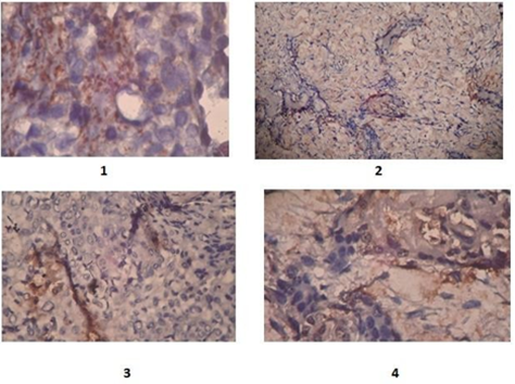
Figure 1: 1)- Medium power view showing positive cytoplasmic 2+ stain of IMP3 in Mucoepidermoied carcinoma; 2)- Medium power view of weak postive1+ Imp3, in pleomorphic adenoma; 3&4)- Medium power view, focal positivity 1-2+ of IMP3 in mucoepidermoied carcinoma
Discussion
IMP3 is a binding-RNA protein required for ribosomal RNA processing, which has been suggested to be a prognostic marker in a large variety of cancers in humans. However, available data on the prevalence of IMP3 expression is largely debatable. IMP3 was frequently expressed in many different tumor types and was typically associated with aggressive tumor features [4, 7].
The pathogenesis and molecular changes in salivary gland tumors arestill under study. In this case series study, the immunohistochemical expression of the fetal oncogene IMP3 has been investigated for its relation to tumor classical clinicopathological criteria.
The neoplastic cells exhibit differentiation of epithelial cells (luminal), myoepithelial cells و as well as a very characteristic stromal tissue comprising chondroid, myxoid, osseous and myxo-chondroidelements [8], which were evaluated also as part of Imp3 assessment.
In this study, most the cases were benign pleomorphic adenomas which is the most common tumor type [1, 3, 8,12] it is histologically characterized by the complex intermingling of mesenchymal-epithelial areas. There was an
interesting finding, which was the positive stain within the chondroid areas in pleomorphic adenoma. This finding can be explained by the presence ofthe cells. Despite they are normal cells, they show positive stain for IMP3”8”.
The absence of significant association between IMP3 expression with 'patients age, gender, and anatomical location, in benign variants, agrees with Mohamed R et al., However, this study's findings do not agree with Mohamed R et al., [4] and Burdelski C et al., studies [7] in the insignificant association between mucoepidermoid carcinoma and IMP3 expression may due to the low case number, especially the malignant ones.
One important limitation of this study was the low number of cases was a major restriction in the evaluation of the IMP3. The low number of collected cases might be related to data collection, which was affected by the COVID19 epidemic where theoral pathology department was closed.
The significant association between IMP3 expression and epithelial/mesenchymal cells similar to Christoph Burdelski et al., [7] may bedue to the variation in expression between normal tissue and tumor since most ofthe cases were benign pleomorphic adenoma like Faris k.”12” and Omar ShebliMuseedi et al., [2] and this tumor contains mixed tissue, while other tumor types especially carcinomas are predominantly epithelial.
Conclusion
There is variation in IMP3 expression between normal, benign, and malignant tissue. IMP3 might not be conclusive in differentiation between benign and malignant salivary gland tumors. Further studies with follow-up are needed to assess the prognostic value of this marker in salivary gland pathologies.
References
- Amezcua, C. A. eds. (2009). Modern Surgical Pathology. pdf. 2009.
View at Publisher | View at Google Scholar - Museedi, O. S., & Younis, W. H. (2014). Oral cancer trends in Iraq from 2000 to 2008. The Saudi Journal for Dental Research, 5(1), 41-47.
View at Publisher | View at Google Scholar - Organization, W. H. (2019). International agency for research on cancer.
View at Publisher | View at Google Scholar - Elshafey, M. R., Ahmed, R. A., Mourad, M. I., & Gaballah, E. T. (2016). The oncofetal protein IMP3 is an indicator of early recurrence and poor outcome in mucoepidermoid carcinoma of salivary glands. Cancer Biology & Medicine, 13(2), 286.
View at Publisher | View at Google Scholar - Ismerim, A. B., Ferreira, S. V., Lessa, A. M., Pereira Junior, A. S., Gurgel, C. A., Coutinho-Camillo, C. M., ... & Santos, J. N. (2016). Insulin-like growth factor II messenger RNA-binding protein 3 in Salivary Gland tumors. Applied Immunohistochemistry & Molecular Morphology, 24(6), 422-426.
View at Publisher | View at Google Scholar - Ibrahim, T. R., Ahmed, M. M., & Hegazy, A. A. (2020). Diagnostic Utility of Immunohistochemical Expressions of IMP3 Versus DOG1 and p63 in Salivary Gland Tumors. Turkish Journal of Pathology, 36(3), 227-236.
View at Publisher | View at Google Scholar - Burdelski, C., Jakani-Karimi, N., Jacobsen, F., Möller-Koop, C., Minner, S., Simon, R., ... & Wilczak, W. (2018). IMP3 overexpression occurs in various important cancer types and is linked to aggressive tumor features: A tissue microarray study on 8,877 human cancers and normal tissues. Oncology reports, 39(1), 3-12.
View at Publisher | View at Google Scholar - Purkait, S. K. (2011). Essentials of oral pathology. JP Medical Ltd.
View at Publisher | View at Google Scholar - Santana, T., Pavel, A., Martinek, P., Steiner, P., Grossmann, P., Baněčková, M., & Skálová, A. (2019). Biomarker immunoprofile and molecular characteristics in salivary duct carcinoma: clinicopathological and prognostic implications. Human Pathology, 93, 37-47.
View at Publisher | View at Google Scholar - Takase, S., Kano, S., Tada, Y., Kawakita, D., Shimura, T., Hirai, H., ... & Nagao, T. (2017). Biomarker immunoprofile in salivary duct carcinomas: clinicopathological and prognostic implications with evaluation of the revised classification. Oncotarget, 8(35), 59023.
View at Publisher | View at Google Scholar - Boenisch, T. J. G. (2001). Denmark: DAKO, Handbook on immunohistochemical staining methods.
View at Publisher | View at Google Scholar - Al-Khiro, F. I. (2014). Salivary gland tumors: A review of 171 cases, with particular reference to histological types, site, age and gender distribution. Journal of Baghdad College of Dentistry, 26(1), 88-91.
View at Publisher | View at Google Scholar
