

Opinion | DOI: https://doi.org/10.31579/2834-8761/016
Hypovolemia with Peripheral Edema: What is Wrong?
- Ahmed N. M. Ghanem * *
NHS The UK and Mansoura University, Faculty of Medicine Egypt Consultant Urologist Surgeon- Retired Independent Investigator & Scientist, Free Lance Author, Dreamer & White Revolutionary in Science.
*Corresponding Author: Ahmed N. M. Ghanem, NHS The UK and Mansoura University, Faculty of Medicine Egypt Consultant Urologist Surgeon- Retired Independent Investigator & Scientist, Free Lance Author, Dreamer & White Revolutionary in Science.
Citation: Ahmed N. M. Ghanem (2023), Hypovolemia with Peripheral Edema: What is Wrong?, Clinical Endocrinology and Metabolism, 2(2) DOI:10.31579/2834-8761/016
Copyright: © 2023, Ahmed N. M. Ghanem. This is an open access article distributed under the Creative Commons Attribution License, which permits unrestricted use, distribution, and reproduction in any medium, provided the original work is properly cited.
Received: 06 March 2023 | Accepted: 15 March 2023 | Published: 24 March 2023
Keywords:
Abstract
Introduction
I read with great interest this recently published article by Professors Dull, R.O. and Hahn, R.G. [1]. The authors are commended on this review based on evidence from published studies that represent the current understanding of the condition and its scientific basis. The authors have faithfully and factually summarized the evidence based on published reports, including some of the commonly received errors and misconceptions on the scientific foundation that identifying and correcting may help to answer the vitally important question in the title of the report.
The authors acknowledge that Starling’s law represents the scientific foundation of the volume-pressure relationship of the vascular, capillary, and
interstitial fluid compartments. It thus underlies the rules that govern fluid therapy in shock management. This is the subject on which both authors are among the top world authority. My research has demonstrated clearly and completely the substantial evidence that Starling’s law is wrong, and the correct replacement is the hydrodynamics of the porous orifice (G) tube [2]- that has been gathered in a book [3]. This will revolutionize our understanding of the condition and related issues, particularly on the path-etiology and management of acute respiratory distress syndrome (ARDS). The hydrodynamics of the G tube in a surrounding chamber mimics the capillary-ISF transfer (Figure 1).
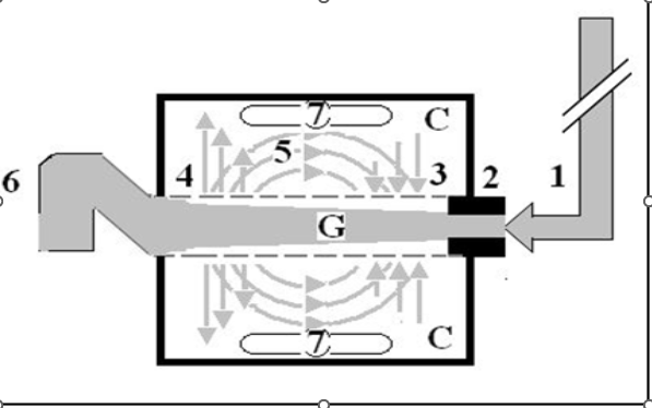
Figure 1: Shows a diagrammatic representation of the hydrodynamic of G tube based on G tubes and chamber C. This 38-years old diagrammatic representation of the hydrodynamic of G tube in chamber C is based on few photographs. The G tube is the plastic tube with narrow inlet and pores in its wall built on a scale to capillary ultra-structure of precapillary sphincter and wide inter cellular slit pores. The chamber C around it is another bigger plastic tube to form the G-C apparatus. The chamber C represents the ISF space. The diagram represents a capillary-ISF unit that should replace Starling’s law in every future physiology, medical and surgical textbooks, and added to chapters on hydrodynamics in physics textbooks. The numbers should read as follows:
1. The inflow pressure pushes fluid through the orifice
2. Creating fluid jet in the lumen of the G tube**.
3. The fluid jet creates negative side pressure gradient on the G tube’s wall causing suction maximal over the proximal part of the G tube near the inlet that sucks fluid into lumen.
4. The side pressure gradient turns positive pushing fluid out of lumen over the distal part maximally near the outlet.
5. Thus, the fluid around G tube inside C moves in magnetic field-like circulation (5) taking an opposite direction to lumen flow of G tube.
6. The inflow pressure 1 and orifice 2 induce the negative side pressure creating the dynamic G-C circulation phenomenon that is rapid, autonomous, and efficient in moving fluid and particles out from the G tube lumen at 4, irrigating C at 5, then sucking it back again at 3,
7. Maintaining net negative energy pressure inside chamber C that is always lower than the distal pressure at 6.
**Note: The shape of the fluid jet inside the G tube (Cone shaped), having a diameter of the inlet on right hand side and the diameter of the exit at left hand side (G tube diameter). I lost the photo on which the fluid jet was drawn, using tea leaves of fine and coarse sizes that runs in the center of G tube leaving the outer zone near the wall of G tube clear. This may explain the finding in real capillary of the protein-free (and erythrocyte-free) sub-endothelial zone in the Glycocalyx paradigm.
I also noted that fine tea leaves exit the distal pores in small amount maintaining a higher concentration in the circulatory system- akin to plasma proteins.
Hypovolaemia and peripheral edema refer to the condition that affects acutely ill patients presenting with any shock then suffer clinically with ARDS induced by excessive fluid therapy in whom there is massive volumetric overload with hypotension shock (? Hypovolaemia) and massive fluid creep on the interstitial fluid space causing generalized edema. It complicates fluid therapy for shock resuscitation of burns, sepsis, haemorrhage, trauma, and acute pancreatitis [4,5]. It initially presents and seamlessly occurs as volume kinetic or volumetric overload shock (VOS) [6], among new scientific discoveries in physics, physiology, and medicine [7]. It has high morbidity and mortality and affects thousands of patients every year all over the world. Although there is hypotension shock here it is probably incorrect to assume hypovolaemia exists. In other words, and contrary to what is generally believed, hypotension is not synonymous with hypovolaemia. It is a simple physics: if the cardiovascular system is overfilled to above its maximum capacity, the surplus will simply spill into the ISF space!
Starling’s law has proved wrong on both of its hydrostatic and oncotic pressure forces [2] However, it continues to dictate the current faulty rules on fluid therapy in the management of shock. It thus misleads physicians into giving too much fluid during shock resuscitation [8]. More than 21 reasons were reported to show that Starling’s law is wrong [9]. The correct replacement is the hydrodynamic of the porous orifice (G) tube) that was built on the capillary ultrastructure anatomy of the precapillary sphincter [10] and a porous wall [11] that allow the passage of plasma proteins to nullify the oncotic pressure in Vivo. It follows that the extended Starling Principle is wrong, and a misnomer, and all the equations are also wrong. Commonly received but erroneous concepts and laws represent fraud in modern science.
The clinical significance is that Starling’s law dictates the faulty rules on fluid therapy causing many errors and misconceptions that mislead physicians into giving too much fluid infusions of albumin and crystalloids for the resuscitation of shock [8] which both cause edema of ISF space and vital organs as well as “hypervolemia” with hypotension [5,12]. This shock is mistaken for septic shock or any known shock and is wrongly treated with further huge volume expansion, occurring with both liberal and conservative approaches of fluid therapy. This has been newly recognized as volume kinetic or volumetric overload shocks (VOS).
Volumetric overload inducing VOS is of 2 types [6,12]: VOS 1 is induced by sodium-free fluid that causes the transurethral resection of the prostate (TUR) syndrome. VOS 2 is induced by sodium-based fluids of crystalloids and plasma proteins and causes ARDS and acute kidney injury (AKI) as part of the multiple organ dysfunction syndrome (MODS) with high morbidity and mortality [11]. Volumetric overload shock induced by persistence to elevate CVP to a high level of 20-22 mmHg during shock resuscitation is also based on the faulty Starling’s law. ARDS was originally reported by Ashbaugh et al in 1967 in which the dead patients had 12-14 Litres of fluid creep retained in their bodies [13]. In recent huge prospective multicentre clinical trial studies, fluid retention is 7-10 L in surviving ARDS patients [14].
I trust the respected authors, and invite the world authorities, to kindly fulfil their authority and responsibility by writing an update on the subject that summarises the results of my recently reported research for the awareness of the doctors’ readers and the benefit of their patients.
Conflict of Interest: None declared.
Funds Received: None declared.
Declarations
Ethical Approval
Is not applicable. I consent to participate in and consent to publish this article.
Availability of data and materials
The datasets used can be accessed from the given references of published articles.
References
- undefined
View at Publisher | View at Google Scholar - undefined
View at Publisher | View at Google Scholar - undefined
View at Publisher | View at Google Scholar - undefined
View at Publisher | View at Google Scholar - undefined
View at Publisher | View at Google Scholar - undefined
View at Publisher | View at Google Scholar - undefined
View at Publisher | View at Google Scholar - undefined
View at Publisher | View at Google Scholar - undefined
View at Publisher | View at Google Scholar
