

Case Report | DOI: https://doi.org/DOI:10.31579/2834-5134/044
Huge Mucinous Cystadenoma in a 14 years old female: A Case Report
General Surgeon, Assistant professor, Department of Surgery, Medical Faculty, Zahedan University of Medical Sciences and Health Services, Zahedan, Iran.
*Corresponding Author: Ahmad Reza Shahraki, General Surgeon, Assistant professor, Department of Surgery, Medical Faculty, Zahedan University of Medical Sciences and Health Services, Zahedan, Iran.
Citation: Ahmad R. Shahraki, (2024), Huge Mucinous Cystadenoma in a 14 years old Female: A Case Report, Journal of Clinical Anatomy, 3(2); DOI:10.31579/2834-5134/044
Copyright: © 2024, Ahmad Reza Shahraki. This is an open access article distributed under the Creative Commons Attribution License, which permits unrestricted use, distribution, and reproduction in any medium, provided the original work is properly cited.
Received: 08 March 2024 | Accepted: 13 March 2024 | Published: 05 April 2024
Keywords: mandibular ameloblastoma; odontogenic tumors; pathogenesis; diagnosis; surgical management; recurrence; molecular therapy
Abstract
Giant ovarian cysts are rarely described in the literature, owing to the availability of advanced imaging technologies in developed countries leading to early treatment. In resource-limited settings, various factors lead to late presentation. Huge ovarian masses are mostly benign, but malignancy should be ruled out by investigations and clinical assessment. Giant cysts require resection because of compressive symptoms or risk of malignancy and their management invariably requires laparotomy to prevent perforation and spillage of the cyst fluid into peritoneal cavity.
This case is about a 14 years old female with abdominal pain and abdominal distention that in surgery shows Huge mucinous cystadenoma that resected.
The surgical management of these huge tumors is associated with many life-threatening complications. Transvaginal ultrasound should be used in resource-limited settings to delineate ovarian masses. Community health workers must be involved in scouting and follow up of community members with unusual abdominal swellings in developing countries to avoid delays in care.
The main aim of this report is to draw attention to huge ovarian epithelial cysts with unsuspected presentation contributing to a decrease in any under diagnosis, misdiagnosis, and mismanagement that might occur.
Introduction
Tumors of the ovary presenting with diameters greater than 10 cm are referred to as giant ovarian cysts (2). These are rarely seen in high-income countries and consequently are rarely described in the literature, owing to availability of resources and advanced imaging technologies leading to early diagnosis of small or medium-sized tumors (3). These tumors are generally asymptomatic at early stages, causing symptoms only after reaching enormous dimensions, and consequently are diagnosed late in low- and middle-income countries (LMICs) (2). Compressive symptoms or a visible abdominal mass are the most frequent presenting complaints (3). The surgical management of these masses is associated with many life-threatening complications, which arise predominantly after surgery owing to rapid changes in body circulation, and with pulmonary edema. The former includes severe hypotension, increased venous return, cardiac failure, respiratory failure, and intestinal distention (3). Most of the patients who have large tumors present mainly with the pressure symptoms over the genitourinary system leading to urinary complaints and also pressure over respiratory system leading to respiratory embarrassment. The role of imaging modalities such as computed tomography (CT) scan and magnetic resonance imaging gives better idea about the extension of the tumor in the various quadrants of the abdomen and consistency of the tumor. Management of ovarian cysts depends on the patient's age, the size of the cyst, and its histopathological nature. Conservative surgery as ovarian cystectomy and salpingo-oophorectomy is adequate for benign lesions. Four frozen section is very important to know the malignant variation of this tumor and that helps in the management of the patient. Surgical expertise is required to prevent complications as in huge tumors the anatomical planes get distorted (5).
Here are four major categories of ovarian tumors:
1-Epithelial tumors (65-75%) — serous or mucinous cystadenoma/carcinoma, clear cell carcinoma, and Brenner tumor;
2-Germ cell tumors (15%) — dysgerminoma, embryonal cell cancer, choriocarcinoma, and teratoma;
3-Sex-cord-stromal tumors (5-10%) — granulosa cell tumor, thecoma, and fibroma;
4-Metastatic tumors (10%) — uterine, stomach, colon, breast, and lymphoma. (6)
Extra-large benign and malignant cysts of the ovary (5) are uncommon and involve diagnostic and management challenges, and determination of cancer antigen (CA)-125 can help to identify epithelial tumors of the ovary (4). Ovarian mucinous cystadenoma is a benign tumour that arises from the surface epithelium of the ovary. It is a multilocular cyst with smooth outer and inner surfaces. It tends to be huge in size. Of all ovarian tumours, mucinous tumours comprise 15% (7,8). About 80% of mucinous tumours are benign, 10% are border-line and 10% are malignant. Although benign ovarian mucinous tumours are rare at the extremities of age, before puberty and after menopause (9). they are common between the third and the fifth decades (10). The most frequent complications of benign ovarian cysts, in general, are torsion, haemorrhage and rupture. As it contains mucinous fluid, its rupture leads to mucinous deposits on the peritoneum (pseudo-myxoma peritonei) (11).
We describe this case that any abdominal pain or size changing is important.
Case presentation:
Our case was a 14 years old female with abdominal pain and abdominal distention (figure1) from 3 months before. We admit her and takes lab data that all of them was in normal range, because of our examination we did Ultrasonography that reports a huge mass in abdominal cavity. We did Abdominopelvic CT for her that reports a huge mass in right adnexa. We schedule a surgery by midline laparotomy and at first, we face with a huge mass (figure 2).
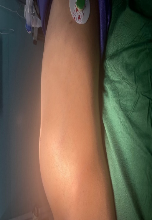
Figure 1: Abdominal distention

Figure 2: Huge mass
We start to separate this mass from intra-abdominal organs and prevent of rupture of mass. (figure 3).
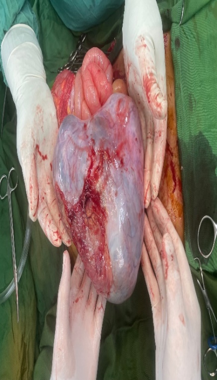
Figure 3: Clear and unruptured mass
After exclusion of mass, we start evaluation of mass origin that shows it is from right ovary (figure 4).
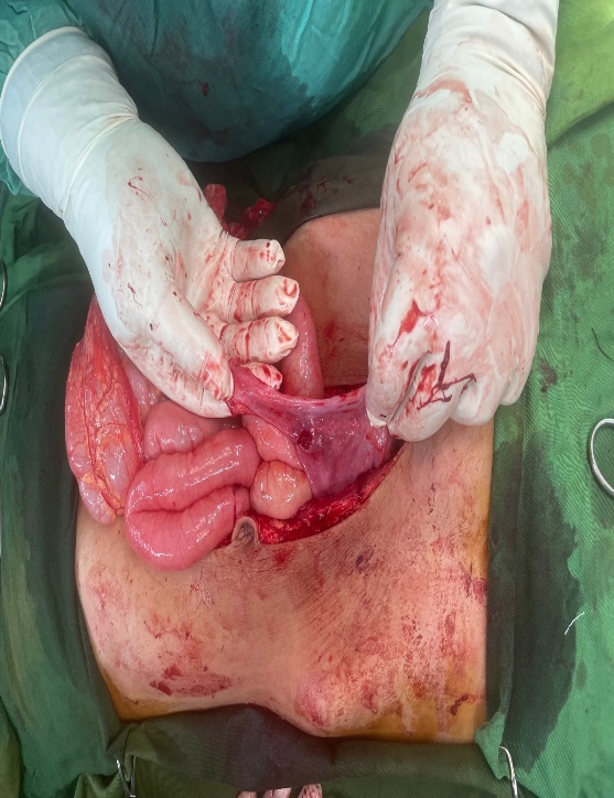
Figure 4: origin of mass
We exams liver, spleen and omentum, abdominal wall for spreadin of mass that was clear and we end surgery successfully. After surgery we exams the mass that weights was7.100 gr and diameter was 15*12*10 (figure 5,6). One days after surgery we send tumor markers and start eating of patient and 3 days after admission, we discharge her healthy.
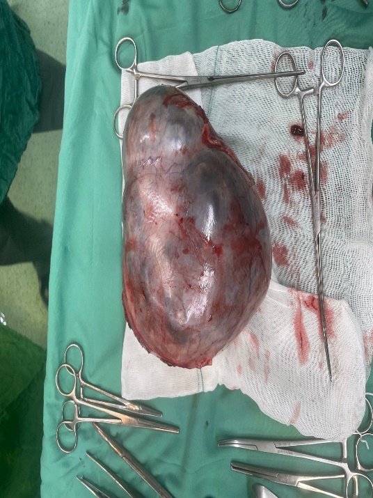
Figure 5: mass size
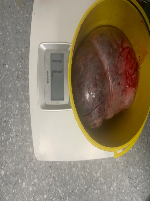
Figure 6: Mass weight
Discussion:
In young people the majority of ovarian cysts decreases in size or even disappears and therefore should be dealt with a careful expectant follow-up by ultrasonography. Benign cysts of <8>
Women with abdominal-pelvic masses constitute a challenging condition in general practice because the clinical features and findings from physical examination are usually nonspecific. Moreover, concomitance with overweight and obesity can be additional diagnostic pitfalls. Imaging studies of the abdomen can contribute in ruling out the main alternative hypotheses. Although tumor markers can be a useful tool for differential diagnosis of malignant cysts, some authors have described elevated levels of these markers in patients with benign tumors (4). Major diagnostic difficulties are often posed if inner nodules are disclosed in these cystic cavities because this finding must be considered as indicative of a malignant tumor. In our present case study, the giant ovarian tumor was multilocular with diverse inner solid masses, and the histopathology evaluation characterized the diagnosis of cystadenocarcinoma. In our case, the woman described a progressive infraumbilical swelling and abdominal pain. Malignant tumors (anaplastic carcinoma, carcinosarcoma, fibrosarcoma, rhabdomyosarcoma, undifferentiated sarcoma), mixed nodules, and leiomyoma among others, were ruled out (4).Based on cell origin, ovarian tumors are classified as Germ cell tumors (undifferentiated and extraembryonic); stromal tumors (granulosa-theca, Sertoli, and Leydig cells); and epithelial tumors (cystadenoma, borderline cystadenoma, and cystadenocarcinoma) (4).The interval between the symptoms' onset and clinical presentation of ovarian cancer is a major concern and diagnostic challenge involving this malignancy. Ovarian tumors are included among malignancies with shorter symptom-to-visit interval. Nevertheless, symptoms frequently develop insidiously and with intervals usually longer in the localized disease. Noteworthy is that delay in ovarian cancer detection has a direct relationship with poor outcome, but some examples of longstanding localized evolution have been reported (4). However, CA 125 should not be used in isolation, because it is nonspecific for ovarian cancer. There is not enough evidence to suggest that panels including multiple tumor markers will offer any further advantage in the initial assessment of ovarian cysts in postmenopausal women, because all the markers show low sensitivity and wide variation in specificity when used in isolation or in combination with CA 125; hence, their routine use is not recommended (13).
Transvaginal ultrasonography is the recommended and most commonly used imaging modality to evaluate an adnexal mass (14).
Malignant characteristics on ultrasound include a size greater than 10 cm, irregularity, papillary or solid structures, ascites, and high color Doppler flow on ultrasound. Benign characteristics include simple, thin-walled structures with no solid components or internal blood flow on Doppler ultrasound (15,16). Masses believed to be benign, which are asymptomatic, may be observed with repeated transvaginal ultrasound rather than surgery (14,17). Giant ovarian tumours have become rare in current medical practice, as most cases are discovered early during routine check-ups. Detection of ovarian cysts causes considerable worry for women because of fear of malignancy, but fortunately the majority of ovarian cysts are benign, Mucinous cystadenoma is a benign ovarian tumour. It is reported to occur in middle-aged women. It is rare among adolescents (19) or in association with pregnancy (20).
Conclusion:
The surgical management of these huge tumors is associated with many life-threatening complications. Transvaginal ultrasound should be used in resource-limited settings to delineate ovarian masses. Community health workers must be involved in scouting and follow up of community members with unusual abdominal swellings in developing countries to avoid delays in care (1). The main aim of this report is to draw attention to huge ovarian epithelial cysts with unsuspected presentation contributing to a decrease in any under diagnosis, misdiagnosis, and mismanagement that might occur (4). Adnexal masses can be difficult to evaluate and manage because of the large differential diagnosis and variable urgency associated with differing etiologies (18). A CT scan was done in our ED after ultrasonography to better assess the source and size of the mass. CT scans can be helpful in detecting lymph node enlargement, metastases, ascites, and possible alternative primary sites of tumors (19). Adnexal masses have a large variety of etiologies that can be difficult to diagnose on initial presentation. Mucinous cystadenomas are a type of benign adnexal neoplasm that can grow much larger than other masses. This caused the abdominal distension and pain seen in the patient discussed. The management of benign masses depends on the concern for malignancy, size, and patient’s symptoms. Point of care or bedside ultrasound can guide in medical decision-making or diagnostics, many times enhancing our process by helping us decide which is the best next test or step in care (18).
Declarations:
Ethical Approval and Consent to participate:
The content of this manuscript is in accordance with the declaration of Helsinki for Ethics. No committee approval was required. Oral and written consent to participate was granted by her family.
Consent for publication:
“Written informed consent was obtained from the patient's legal guardian for publication of this case report and any accompanying images. A copy of the written consent is available for review by the Editor-in-Chief of this journal.”
- Availability of supporting data
It is available.
- Competing interests:
The author declares that they have no competing financial interests and nothing to disclose.
- Funding:
There is no funding.
- Authors' contributions:
Ahmad Reza Shahraki is the surgeon of patient and writes this paper.
The author declares that they have no competing financial interests and nothing to disclose.
- Acknowledgements:
Giant ovarian cysts are rarely described in the literature, owing to the availability of advanced imaging technologies in developed countries leading to early treatment. In resource-limited settings, various factors lead to late presentation. The main aim of this report is to draw attention to huge ovarian epithelial cysts with unsuspected presentation contributing to a decrease in any underdiagnosis, misdiagnosis, and mismanagement that might occur.
References
- Gwanzura C, Muyotcha AF, Magwali T, Chirenje ZM, Madziyire MG. (2019). Giant mucinous cystadenoma: a case report. J Med Case Rep. Jun 14;13(1):181.
View at Publisher | View at Google Scholar - Yeika EV, Efie DT, Tolefac PN, Fomengia JN. (2017). Giant ovarian cyst masquerading as a massive ascites: a case report. BMC Res Notes.;10(1):749.
View at Publisher | View at Google Scholar - Pilone V, Tramontano S, Picarelli P, Monda A, Romano M, Renzulli M, et al. (2018). Giant mucinous ovarian borderline tumor. A good lesson from an asymptomatic case. Int J Surg Case Rep.; 50:25–27.
View at Publisher | View at Google Scholar - Katke RD. (2016). Giant mucinous cystadenocarcinoma of ovary: A case report and review of literature. J Midlife Health. Jan-Mar;7(1):41-4.
View at Publisher | View at Google Scholar - Katke RD, Kiran U, Saraogi M, Sarode S, Thawal R. Giant borderline mucinous cystadenoma with previous 3 caesareans. J Postgraduate Gynaecol Obstet. 2014; 1:1–4.
View at Publisher | View at Google Scholar - Katke RD, Usha K, Mohit S, Smita S, Ravindra T. “Giant borderline mucinous cystadenoma with previous 3 caesareans” J Postgraduate Gynaecol Obstet. 2014;1:1–4.
View at Publisher | View at Google Scholar - Vizza E, Galati GM, Corrado G, Atlante M, Infante C, Sbiroli C: (2005). Voluminous mucinous cystadenoma of the ovary in a 13-year-old girl. J Ped Adoles Gynecol., 18 (6): 419-422.
View at Publisher | View at Google Scholar - Mittal S, Gupta N, Sharma A, Dadhwal V: (2008). Laparoscopic management of a large recurrent benign mucinous cystadenoma of the ovary. Arch Gynecol Obstet., 277 (4): 379-380.
View at Publisher | View at Google Scholar - Crum CP, Lester SC, Cotran RS: (2007). Pathology of female genital system and breast. Robbins' Basic pathology. Edited by: Kumar V, Abbas A, Fausto N, Mitchell R., Elsevier Company, USA, Ch 19: 8.
View at Publisher | View at Google Scholar - Ioffe OB, Simsir A, Silverberg SG: Pathology. (2000). Practical Gynaecologic Oncology. Edited by: Berek JS, Hacker NF., Lippincott Williams & Wilkins Company, 213-214.
View at Publisher | View at Google Scholar - Kamel, R.M. A massive ovarian mucinous cystadenoma: a case report. Reprod Biol Endocrinol 8, 24 (2010).
View at Publisher | View at Google Scholar - Gorgone S, Minniti C, Ilaqua A, Barbuscia M. (2008). Giant mucinous cystadenoma in a young patient. A case reports. G Chir.;29:42–4.
View at Publisher | View at Google Scholar - Nolen B, Velikokhatnaya L, Marrangoni A, De Geest K, Lomakin A, BastBDR92102 RC Jr, et al. Serum biomarker panels for the discrimination of benign from malignant cases in patients with an adnexal mass. Gynecol Oncol. 2010;117(3):440–445.
View at Publisher | View at Google Scholar - Eskander R, Berman M, Keder L, (2016). Practice bulletin no. 174: evaluation and management of adnexal masses. Obstet Gynecol., 128:1193-1195.
View at Publisher | View at Google Scholar - Sokalska A, Timmerman D, Testa AC, et al.: (2009). Diagnostic accuracy of transvaginal ultrasound examination for assigning a specific diagnosis to adnexal masses. Ultrasound Obstet Gynecol., 34:462-470.
View at Publisher | View at Google Scholar - Timmerman D, Testa AC, Bourne T, et al.: (2008). Simple ultrasound-based rules for the diagnosis of ovarian cancer. Ultrasound Obstet Gynecol., 31:681-690.
View at Publisher | View at Google Scholar - Froyman W, Landolfo C, Cock BD, et al.: (2017). OC01.01: Risk of complications in conservatively managed adnexal masses initially thought to be benign at subjective impression by the ultrasound examiner. Ultrasound Obstet Gynecol., 50:1.
View at Publisher | View at Google Scholar - Craen A M, Lebowitz D, Amico K, et al. (November 30, 2018) Mucinous Cystadenoma Causing Abdominal Distension: A Case Report. Cureus 10(11): e3657.
View at Publisher | View at Google Scholar - Micco M, Sala E, Lakhman Y, Hricak H, Vargas HA: (2014). Role of imaging in the pretreatment evaluation of common gynecological cancers. Women’s Health (Lond)., 10:299-321.
View at Publisher | View at Google Scholar - Mucinous cystadenoma is a benign ovarian tumour. It is reported to occur in middle-aged women. It is rare among adolescents [5] or in association with pregnancy [6].
View at Publisher | View at Google Scholar
