

Review Article | DOI: https://doi.org/10.31579/2835-9232/009
Histological Changes in The Neurons of The Parietal Cortex and Hippocampus of Rats with Subtotal Cerebral Ischemia Against the Background of The Introduction of Omega-3 Polyunsaturated Fatty Acids
Grodno State Medical University, 80, Gorkogo St., 230009, Grodno, Republic of Belarus.
*Corresponding Author: Lizaveta I. Bon, Candidate of biological science, assistant professor of pathophysiology department named D.A. Maslakov, Grodno State Medical University, Grodno State Medical University, 80, Gorky St., 230009, Grodno, Belarus.
Citation: Bon E.I, Maksimovich N.Ye, Zimatkin S.M and Lelis A.R, (2023), Histological Changes in The Neurons of The Parietal Cortex and Hippocampus of Rats with Subtotal Cerebral Ischemia Against the Background of The Introduction of Omega-3 Polyunsaturated Fatty Acids, International Journal of Clinical Epidemiology, 2(1); DOI:10.31579/2835-9232/009
Copyright: © 2023 Bon E.I. This is an open-access article distributed under the terms of the Creative Commons Attribution License, which permits unrestricted use, distribution, and reproduction in any medium, provided the original author and source are credited.
Received: 30 January 2023 | Accepted: 03 February 2023 | Published: 17 February 2023
Keywords: neurons; parietal cortex; hippocampus; rats, cerebral ischemia; Omega-3 polyunsaturated fatty acids
Abstract
The administration of omega-3 polyunsaturated fatty acids has a corrective effect on the hippocampus under conditions of subtotal ischemia, reducing the number of shadow cells and hyperchromic shriveled neurons, without having a significant effect on the size and shape of neurons in the parietal cortex of the brain.
Introduction
Known for the role of Omega-3 polyunsaturated fatty acids (Omega-3 PUFAs) in exercising control over the work of the immune and reproductive systems as precursors for the biosynthesis of prostaglandins, leukotrienes and thromboxanes [1-5].
Omega-3 PUFAs ensure the functioning of cell membranes, transmembrane ion channels,participate in the regulation of physiological processes and the implementation of the main functions of neurons - the transmission of impulses and the operation of receptors. Brain neurons, being electrically active cells rich in ion channels, are most sensitive to deficiency PUFAs [1-6,9].
The experiments were carried out on 30 male outbred white rats weighing 260±20 g in compliance with the requirements of the Directive of the European Parliament and of the Council № 2010/63/EU dated 22.09.2010 on the protection of animals used for scientific purposes.
The choice of experimental animals is due to the similarity of the angioarchitectonics of the brain of rats and humans. Modeling of cerebral ischemia (CI) was performed under conditions of intravenous thiopental anesthesia (40-50 mg/kg).
Morphological features of neurons in the parietal cortex and hippocampus of rats with subtotal cerebral ischemia (SCI) were studied against the background of intragastric administration of Omega-3 PUFAs «Omegamed» at a dose of 5 g/kg of body weight during a week was carried out. A morphological study of the size and shape of neuronal perikaryons was carried out, as well as the number of neurons with different degrees of cytoplasmic chromatophilia was determined [2-8].
Statistical data processing
To prevent a systematic measurement error, brain samples from the compared control and experimental animals were treated under the same conditions.
As a result, of morphometric and cytophotometric studies, quantitative continuous data were obtained, which were processed using the licensed computer program Statistica 10.0 for Windows (StatSoft, Inc., USA).
Since the experiment used small samples that had an abnormal distribution, the analysis was carried out using nonparametric statistics methods. The data are presented as Me (LQ;UQ), where Me is the median, LQ is the value of the lower quartile; UQ is the value of the upper quartile. Differences between groups were considered significant at p<0>
Results
With the introduction of Omega-3 PUFAs (SCI + Omega-3 group) there were no differences in the morphometry of neuronal perikarya compared with those in the group without PUFAs administration in both studied sections of the cerebral cortex of rats with SCI (p>0.05), Table 1.

Note: * - p<0>
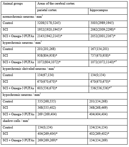
Table 1. Sizes and shape of neuronal perikaryas in the parietal cortex and hippocampus of rats with 1-hour subtotal cerebral ischemia (SCI) and administration of Omega-3 PUFAs, Me (LQ; UQ)
In the parietal cortex, there were no differences in the distribution of neurons according to the degree of cytoplasmic chromatophilia in rats of the SCI group with the introduction of Omega-3 PUFAs and in rats with SCI that did not receive this drug (Table 2., Figure 1).
In the hippocampus of animals of the SCI + Omega-3 group, compared with the SCI group, there was a decrease in the number of hyperchromic wrinkled neurons by 20% (p<0>0.05).
Note: * - p<0>
Table 2: The number of different forms of neurons according to the degree of cytoplasmic chromatophilia in the parietal cortex and hippocampus of rats with 1-hour subtotal cerebral ischemia (SCI) and the introduction of Omega-3 PUFAs, Me (LQ; UQ)
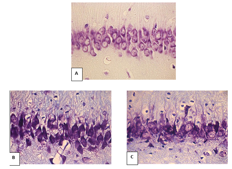
Figure 1. Neurons of the CA1 pyramidal layer of the rat hippocampus. A – control, B – SCI, C – SCI+Omega-3. Digital micrograph. Nissl stain
The beneficial effect of polyunsaturated fatty acids on the state of hippocampal neurons under conditions of subtotal cerebral ischemia may be due to an improvement in the rheological properties of blood due to a decrease in the production of thromboxane A by platelets and an increase in the level of tissue plasminogen activator, as well as an improvement in the fluidity of the neuron membrane, a decrease in blood viscosity. Omega-3 PUFAs also have an anti-inflammatory effect due to their incorporation into the phospholipid layer of cell membranes of monocytes, leukocytes, and endothelial cells, which is accompanied by a decrease in the production of inflammatory mediators and a decrease in leukocyte adhesion to the endothelial wall. In addition, polyunsaturated fatty acids affect the synthesis of prostaglandins that regulate vascular tone and prevent vascular vasoconstriction under the influence of catecholamines, which causes a moderate hypotensive effect [1-9]. The neurons of the hippocampus, as a phylogenetically older part of the cortex, are less sensitive to hypoxia, which could be the reason for the therapeutic effect of Omega-3 PUFAs (a decrease in the number of pathological forms of neurons - hyperchromic wrinkled and shadow-cells).
Thus, the administration of omega-3 polyunsaturated fatty acids has a corrective effect on the hippocampus under conditions of subtotal ischemia, reducing the number of shadow cells and hyperchromic shriveled neurons, without having a significant effect on the size and shape of neurons in the parietal cortex of the brain.
References
- Bon, L.I. Evaluation of neurological deficiency in rats with cerebral ischemia following the administration of omega polyunsaturated fatty acids / L. I. Bon, N. Ye. Maksimovich // Journal of Medical Science. – 2021. – V. 90(3) – P. 137-143.
View at Publisher | View at Google Scholar - Belyaeva, L.E. Early programming of human diseases and the use of nutraceuticals for preventive purposes: focus on fish oil / L.E. Belyaeva, A.N. Pavlyukevich // Bulletin of VSMU. - 2019. - No. 4. - P. 7-16.
View at Publisher | View at Google Scholar - Belyaeva, L.E. Early programming of human diseases and the use of nutraceuticals for preventive purposes: focus on fish oil / L.E. Belyaeva, A.N. Pavlyukevich // Bulletin of VSMU. - 2019. - No. 5. - S. 12-25.
View at Publisher | View at Google Scholar - Bon, L.I. Evaluation of neurological deficiency in rats with cerebral ischemia following the administration of omega polyunsaturated fatty acids / L. I. Bon, N. Ye. Maksimovich // Journal of Medical Science. – 2021. – V. 90(3) – P. 137-143.
View at Publisher | View at Google Scholar - Bon, L.I. Histological disorders of neurons of phylogenetically different parts of the cerebral cortex in partial, subtotal, stepwise subtotal, and total
View at Publisher | View at Google Scholar - cerebral ischemia. / L. I. Bon, N. Ye. Maksimovich // Journal of Medical Science. – 2021. – V. 90(1) – P. 108-115.
View at Publisher | View at Google Scholar - Bon, L.I. Morphological features of neurons in the parietal cortex and hippocampus of rats after subtotal cerebral ischemia against the background of the introduction of omega-3 polyunsaturated fatty acids / L.I. Bon, N.E. Maksimovich, S.M. Zimatkin // Siberian medical journal. - 2020. - No. 3. - P. 34-40.
View at Publisher | View at Google Scholar - Bon, L.I. Neurological Deficit and Corrective Effect of Omega-3 Polyunsaturated Fatty Acids in Cerebral Ischemia in Rats: A case-control study / L. I. Bon, N. Ye. Maksimovich // Biotechnology and Bioprocessing. – 2021. – V. 2 – P. 3-6.
View at Publisher | View at Google Scholar - Kaliannan, K. Multi-omic analysis in transgenic mice implicates omega-6/omega-3 fatty acid imbalance as a risk factor for chronic disease / K. Kaliannan, X.Y. Li, B. Wang, Q. Pan // Commun Biology. - 2019. - Vol. 2(1). - P. 276-280.
View at Publisher | View at Google Scholar - Khunt, D. Role of Omega-3 Fatty Acids and Butter Oil in Targeting Delivery of Donepezil Hydrochloride Microemulsion to Brain via the Intranasal Route: a Comparative Study / D Khunt, M Shrivas, S Polaka, P Gondaliya, M Misra // Pharmacology Sciencific Technology. – 2020. – Vol.21(2). – P. 45-50.
View at Publisher | View at Google Scholar - Maksimovich, N. Ye. Rat brain and its response to ischemia: monograph / N. Ye. Maksimovich, E. I. Bon, S. M. Zimatkin. - Grodno: GrGMU, 2020. - 240 p.
View at Publisher | View at Google Scholar
