

Research Article | DOI: https://doi.org/10.31579/2834-5177/009
Helicobacter Pylori Assay and Urine Bacteriology of Patients with Gastritis
1 Department of Public Health, National Open University of Nigeria, Uromi Community Study Centre, Uromi, Edo State, Nigeria.
2 Lassa Fever Enable Study CEPI/ISTH Irrua, Edo State, Nigeria.
3 Health Initiatives for Safety and Stability in Africa (HIFASS), 68 Nigeria Army Reference Hospital, Yaba, Lagos State, Nigeria.
4 Quality Assurance Department, R-Jolad Hospital, Lagos State, Nigeria.
*Corresponding Author: Iyevhobu, K.O, Department of Public Health, National Open University of Nigeria, Uromi Community Study Centre, Uromi, Edo State, Nigeria. Lassa Fever Enable Study CEPI/ISTH Irrua, Edo State, Nigeria.
Citation: Iyevhobu, K.O, Airefetalor, Irobonosen, I.O., Abinokhauno, S.O, (2023), Helicobacter Pylori Assay and Urine Bacteriology of Patients with Gastritis. International Journal of Clinical Infectious Diseases, 2(1); DOI:10.31579/2834-5177/009
Copyright: © 2023, Iyevhobu, K.O. This is an open access article distributed under the Creative Commons Attribution License, which permits unrestricted use, distribution, and reproduction in any medium, provided the original work is properly cited.
Received: 23 December 2022 | Accepted: 05 January 2023 | Published: 11 January 2023
Keywords: helicobacter pylori; urine, bacteriology;patients; gastritis
Abstract
Helicobacter pylori are a non-spore-forming Gram-negative bacterium. The cellular morphology may be curved, spiral, or fusiform, typically 0.5 to 1.0 μm in width and 2.5 to 5.0 μm long. The aim of this study is to determine and compare the prevalence of H. pylori infection and urinary tract infections among gastritis patients. The subjects used in this project work comprised of patients with gastritis. A total number of twenty-five (25) patients with gastritis were recruited for this study. The predominant isolates were Escherichia coli (4), Klebsiella spp (2), Enterobacter spp (2), Staphylococcus aureus (3), Streptococcus spp (3) and Proteus vulgaris (2) with Escherichia coli having the highest prevalence of 25%. The antibiotic susceptibility patterns of the various isolates were read using their zones of inhibitions on the sensitivity culture plates, which shows that Ciprofloxacin, Gentamycin, Streptomycin and Refampicin were the most sensitive antibiotics against the gram-positive bacteria isolates (Streptococcus spp and Staphylococcus aureus) while other drugs were found to be intermediate and resistant. The gram-negative organisms (Enterobacter spp, Escherichia coli, Klebsiella spp and Proteus vulgaris) were more sensitive to Augmentin and Gentamycin, while Ofloxacin, Peflacine, Ciprofloxacin, Septrin and Ampicillin were intermediate while the other drugs were resistant. The noninvasive test-and-treat strategy for H. pylori infection is reasonable for younger patients who have upper gastrointestinal symptoms but not alarm symptoms, like the patient in the vignette. Noninvasive testing can be performed with the use of the urea breath test, fecal antigen test, or serologic test; the serologic test is the least accurate.
Introduction
Helicobacter pylori are a non-spore-forming Gram-negative bacterium. The cellular morphology may be curved, spiral, or fusiform, typically 0.5 to 1.0 μm in width and 2.5 to 5.0 μm long. The spiral wavelength may vary with the age, growth conditions, and species identity of the cells. In old cultures or those exposed to air, cells may become coccoid (Shirai et al., 2000).
The genus Helicobacter belongs to the ε subdivision of the Proteobacteria, order Campylobacterales, family Helicobacteraceae. This family also includes the genera Wolinella, Flexispira, Sulfurimonas, Thiomicrospira, and Thiovulum. To date, the genus Helicobacter consists of over 20 recognized species, with many species awaiting formal recognition (Fox, 2002). Members of the genus Helicobacter are all microaerophilic organisms and in most cases are catalase and oxidase positive, and many but not all species are also urease positive. Helicobacter species can be subdivided into two major lineages, the gastric Helicobacter species and the enterohepatic (nongastric) Helicobacter species. Both groups demonstrate a high level of organ specificity, such that gastric helicobacters in general are unable to colonize the intestine or liver, and vice versa (Fox, 2002).
Helicobacter pylori (H. pylori) infection is accepted as the primary cause of chronic gastritis (Suerbaum and Michetti, 2002). Moreover, severe atrophic gastritis, accompanying intestinal metaplasia caused by persistent H. pylori infection, is closely related to the development of gastric cancer (Correa,1992). Although H. pylori was discovered more than 30 years ago by Marshall and Warren (1984), which method should be considered as a gold standard for detection of H. pylori infection, especially for epidemiological studies, remains unclear.

Plate 1: A 10,000x computer-aided design image of H. pylori showing curved shape and flagellae that enable the bacteria to propel themselves into the mucus lining of the stomach. (Shirai et al., 2000) Currently, several direct diagnostic tests, including histopathology and/or immunohistochemistry (IHC), rapid urease test (RUT), and culture are frequently used as they provide genotype and antibiotic resistance information. However, due to the small amount of bacteria that colonizes the stomach, the direct test sensitivity decreases. Thus, several indirect tests, including antibody-based tests such as serology and urine test, urea breath test (UBT), and stool antigen test (SAT) have been developed to diagnose H. pylori infection (Burucoa et al., 2013). Among the indirect tests, UBT is one of the most accurate to determine H. pylori infection with a sensitivity and specificity of 99% and 98%, respectively (Gisbert and Pajares, 2004). Together with SAT, UBT became the best method to identify active infection, which cannot be detected by serology (Malfertheiner et al., 2012).
The public health importance of the discovery of H. pylori and its role in stomach diseases was recognized in 2005 by the attribution of the Nobel Prize in Physiology or Medicine to B. Marshall and R. Warren. In the history of Nobel prizes, this is only the third time that the discovery of a bacterium has been acknowledged (Megraud, 2005). For the correct management of peptic ulcer disease and gastric MALT lymphoma, as well as obtaining information on a wide range of diseases associated with H. pylori infection, effective diagnostic methods including susceptibility testing are mandatory. Most of the many different techniques involved in diagnosis of H. pylori infection are performed in microbiology laboratories.
Urinary tract infection (UTI) is a bacterial infection that affects any part of the urinary tract: kidneys, ureters, bladder and urethra (Stamm and Hooton, 1993; Iyevhobu et al., 2020). Stamm and Hooton, (1993) referred to UTI as a clinical (symptomatic) or subclinical (asymptomatic) disease that may involve just the lower tract or both the lower and upper tracts. Although urine contains a variety of salts and waste products, it usually does not have bacteria in it. But when bacterial gets into the bladder or kidney and multiply in the urine, they cause UTI (Nicolle et al., 1992). Infection may involve only a single site, such as urethra (urethritis), prostrate (prostatitis), bladder (cystitis), kidney (pyelonephritis) but the whole system is always at risk of invasion by bacteria once any part is infected. Urinary tract infections (UTIs) are caused by the presence and growth of micro-organisms anywhere in the urinary tract and are perhaps the commonest bacterial infections of mankind (Morgan and Markenzie, 2001; Adeyeba et al., 2002). Urinary tract infection occurs when bacteria is introduced into the urinary system usually through the urethra. When bacteria get into the urinary system they multiply and travel up the urinary tract causing inflammation and irritation along the way (Ayoade et al., 2013; Iyevhobu et al., 2020). It is one of the most common causes of hospitalization and referral to outpatient, having an estimated figure of 150 million per annum worldwide (Hvidberg et al., 2000; Stamm and Norrby, 2001; Fakhrossadat et al., 2009). Urinary tract infection (UTI) is one of the current infections among teenagers and adults who are sexually active. Screening of asymptomatic subjects for bacteriuria is therefore necessary as bacteriuria has adverse outcomes that can be prevented by antimicrobial therapy (USPSTF, 1996). Apart from that, even the progression of the asymptomatic bacteriuria to the symptomatic UTI in later life can be prevented, which emphasizes the fact that, “prevention is better than cure” (Iyevhobu et al., 2020). Furthermore, untreated asymptomatic bacteriuria can lead to the development of cystitis in approximately 30% of cases, and can lead to the development of pyelonephritis in about 50% of cases. Microbiologically, urinary tract infection exists when significant growth of microorganisms is detected in the urinary tract (Iyevhobu et al., 2020). The infection is generally considered significant and requires treatment when more than 105 colony forming units per milliliter (105cfu/ml) of urine are present in a properly collected specimen (Brooks et al., 2004).
Infections of the urinary tract are one of the most common infections for which antibiotics are prescribed and are among the most frequently occurring infections arising in the hospital setting (Iyevhobu et al., 2020). Each year UTIs account for more than five to seven million hospital visits, 20 percent of all prescriptions, and require or complicate more than one million hospital admissions in the United States (Schleupner, 1997). UTI affects all age groups, but women are more exposed than men due to the short urethra, absence of prostatic secretion, pregnancy and easy contamination of the urinary tract with faecal flora (Awaness et al., 2000). Infection particularly in pregnancy and in elderly can be asymptomatic (Al-Dujiaily, 2000) and is associated with an increased risk of intrauterine growth retardation and low birth weight (Iyevhobu et al., 2020).
Materials and Methods
This study was carried out in Ekpoma, The Headquarter of Esan West Local Government area of Edo State. It is located at latitude 6o 45IN and longitude 6o 08IE. It is moderately populated with the peoples’ occupation being farming and trading (World Gazetter, 2007).
The subjects used in this project work comprised of patients with gastritis. A total number of twenty-five (25) patients with gastritis were recruited for this study.
The research was designed to evaluate the urine bacteriology and Helicobacter pylori assay of patients with gastritis. This study was carried out within two (2) months. A record of patients’ age and gender was obtained for each subject. Patients with gastritis were recruited for the study. Apparently healthy individuals and patients with no diagnosis of gastritis were excluded for the study.
Sample Collection: Twenty-five (25) Blood and Urine samples was aseptically collected from each patient using sterile, wide-mouthed, leak-proof universal bottles for urine bacteriology and 3 - 5ml of Blood was collected into Ethylinediaminetetraacetic acid (EDTA) bottle for H. pylori assay. The age as well as gender of each patient was obtained from the subjects before the collection of samples.
Samples collected from the patients were taken to the Microbiology Laboratory of St Kenny Research Consult, Ekpoma, Edo State for analysis.
H. pylori Assay: Blood samples (3-5 ml) were taken. The blood was kept at room temperature for 1 h; the clot was removed by centrifuging at 1,000-2,000rpm for 2 min in the centrifuge. The resulting supernatant was designated serum. Following centrifugation, the samples were immediately transferred into a clean, sterile universal bottle using a Pasteur pipette. The samples were maintained at 2-8°C while handling. Then the serum was stored at-20°C until use for H. pylori assay. Samples that are hemolyzed, icteric or lipemic were excluded from the study. The serum specimens were tested for H. pylori using the “HelicotecUT®Plus” test kit.
Microscopic Examination of Urine for Urine bacteriology: A drop of uniformly mixed uncentrifuged urine samples was aseptically placed on a clean grease-free slide and covered with a cover slip. It was then examined microscopically to detect the presence of pus cells, epithelial cells, red blood cells, yeast cells, crystal cells and cast cells using 10x and 40x objectives with condenser iris closed sufficiently to give good contrast (Cheesbrough, 2000).
Antibiotics Susceptibility Test: In vitro susceptibility tests of the bacterial isolates to antibiotics were done using disc diffusion technique. Zero-point one (0.1) ml of each bacterial isolates prepared directly from an overnight broth culture and adjusted to 0.5 McFarland Standard (NCCLS, 2004) was inoculated using sterile pipette onto each of the nutrient agar media. The commercially available discs containing the following antibiotics: - Penicillin (Pen, 10ug), Ceftazidime, (Caz, 30ug), Streptomycin (Stp, 30ug) Ciprofloxacin (Cpf, 5ug), Gentamycin (Gen, 10ug), Ofloxacin (Ofl, 5ug) Ceftriaxone (Cef, 30ug) and Cotrimoxazole (Cot, 30ug) of oxoid products were aseptically placed on the surfaces of the sensitivity agar plates using a sterile forceps and gently pressed to ensure even contact. The plates were incubated at 37ºC for 24 hours and the zones of inhibition after incubation were observed and the diameters of inhibitory zones were measured in millimeters (mm) using a ruler. The interpretation of the measurement as sensitive and resistant were made according to the manufacturer’s standard zone size interpretative manual. The intermediate readings were considered as sensitive for the assessment of the data.
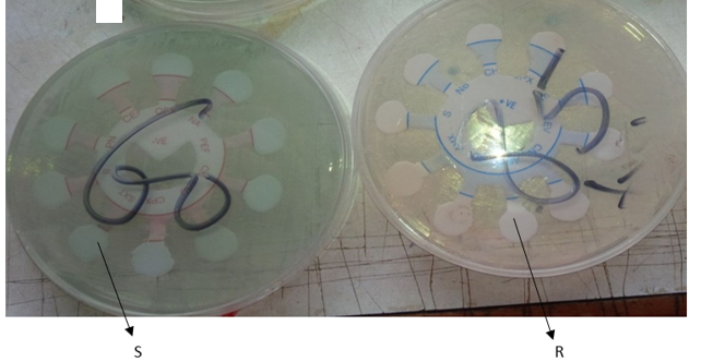
Antibiotic susceptibility test plate showing zones of inhibition[S] and no zone of inhibition[R] around the discs
References
- Adeyeba, O., Adekoya, J., Lowed, A. and Adesiji, Y. (2002). Urinary tract infections among patients attending sexually transmitted disease clinic in Ibadan, Nigeria. J. Sci. 4:4552-4560.
View at Publisher | View at Google Scholar - Ahmed, K. & Imran, I. (2008). Prevalence and antibiogram of uncomplicated lower urinary tract infections in human population of Gilgit, northern areas of Pakistan. Pakistan J Zool. 40(4):295-301.
View at Publisher | View at Google Scholar - Al-Dujiaily, A.A. (2000). Urinary tract infection during pregnancy in Tiknit; Med. J. Tiknit, 6: 220-224.
View at Publisher | View at Google Scholar - Ayoade, F., Osho, A., Fayemi, S.O., Oyejide, N.E. and Ibikunle A.A. (2013). Unusually high prevalence of asymptomatic bacteriuria among male University students on Redemption Camp, Ogun State, Nigeria.Afr. J. Clin. Expert. Microbiol., 14(1): 19-22.
View at Publisher | View at Google Scholar - Bashir, M.F., Qazi, J.I., Ahmad, N. and Riaz, S. (2008). Diversity of urinary tract pathogens and drug resistant isolates of Escherichia coli in different age and gender groups of Pakistanis. Tropical Journal of Pharmaceutical Research. 7 (3): 1025-1031
View at Publisher | View at Google Scholar - Brooks, G.F, Butel, J.S. and Morse, S.A. (2004). Urinary tract infection; case and clinical correlations. In; Jaewetz, Melnick and Adelberg’s medical microbiology, 23rdedition.Brooks, G.F., Butel, J.S. and Morse, S.A.(Editors). McGraw-Hill, New York. (Publisher). Pp. 734-770.
View at Publisher | View at Google Scholar - Broutet, N., Sarasqueta, A.M., Sakarovitch, C., Cantet, F. and Lethuaire, D. (2001). Helicobacter pylori infection in patients consulting gastroenterologists in france: Prevalence is linked to gender and region of residence. Eur. J. Gastroenterol. Hepatol., 13: 677-684.
View at Publisher | View at Google Scholar - Brown, L.M. (2000). Helicobacter pylori epidemiology and routes of transmission. Epidemiol. Rev., 22:283-297.
View at Publisher | View at Google Scholar - Burucoa, C., Delchier, J.C. and Courillon-Mallet, A. (2013). “Comparative evaluation of 29 commercial Helicobacter pylori serological kits,” Helicobacter. 18(3): 169–179.
View at Publisher | View at Google Scholar - Cheesbrough, M. (2000). antimicrobial sensitivity testing in: District laboratory practice in Tropical countries. Cheesbrough, M. (ed). Part 2, Cambridge university press. p. 319-335.
View at Publisher | View at Google Scholar - Chin, B.S., Kim, M.S. and Han, S.H. (2011). Risk factors of all-cause in-hospital mortality among Korean elderly bacteremic urinary tract infection patients. Archives of Gerontology and Geriatrics. 52:50–55.
View at Publisher | View at Google Scholar - Correa, P. (1992). “Human gastric carcinogenesis: a multi-step and multifactorial process—first American cancer society award lecture on cancer epidemiology and prevention,” Can. Res. 52(24): 6735–6740.
View at Publisher | View at Google Scholar - Dielubanza, E.J. and Schaeffer, A.J. (2011). “Urinary tract infections in women.” Med. Clin. North Amer. 95 (1): 27–41.
View at Publisher | View at Google Scholar - Fakhrossadat, M. and Narges, S. (2009). Changing patterns in sensitivity of bacterial uropathogens to antibiotics in children. Pakis. J. Med. Sci. 25(5):801-805.
View at Publisher | View at Google Scholar - Fox, J.G. (2002). The non-H. pylori helicobacters: their expanding role in gastrointestinal and systemic diseases. Gut. 50:273–283.
View at Publisher | View at Google Scholar - Getenet, B. and Wondewosen, T. (2011). Bacterial Uropathogens in Urinary tract infections and Antibiotic susceptibility pattern in JIMMA University specialized hospital, Southwest Ethiopia. Ethiop J Health Sci. 21(2):141-146.
View at Publisher | View at Google Scholar - Gisbert, J.P. and Pajares, J.M. (2004). Stool antigen test for the diagnosis of Helicobacter pylori infection: a systematic review. Helicobacter 9: 347-368.
View at Publisher | View at Google Scholar - Goodman, K.J. and Cockburn, M. (2001). The role of epidemiology in understanding the health effects of helicobacter pylori. Epidemiol., 12: 266-271.
View at Publisher | View at Google Scholar - Gupta, K.D., Scholes, W.E. and Stamm, M. (1999). Increasing prevalence of antimicrobial resistance among uropathogens causing acute uncomplicated cystitis in women. Journal of the American Medical Association. 281: 736-738.
View at Publisher | View at Google Scholar - Hazel, M. & Francis, M. (2002). Epidemiology and diagnosis of H. pylori infection. Helicobacter, 7: 8-16.
View at Publisher | View at Google Scholar - Hvidberg, H., Struve, C., Krogfelt, K.A., Christensen, N., Rasmussen, S.N. and Frimodt- Møller, N. (2000). Development of a long-term ascending urinary tract infection mouse model for antibiotic treatment studies. Antimicrob Agents Chemother., 44:156-163.
View at Publisher | View at Google Scholar - Iyevhobu K.O., Obodo B.N., Obhiozele B., Omolumen L.E., Irobonosen O.I. (2020). Urinary Tract Infection among Male and Female Students in Ambrose Alli University, Ekpoma, Edo State, Nigeria. Global Scientific Journal, Volume 8, Issue 4, 712 – 717.
View at Publisher | View at Google Scholar - Khameneh, Z.R. and Afshar, A.T. (2009). Antimicrobial susceptibility pattern of urinary tract pathogens. Saudi J Kidney Dis Transpl. 20:251-253.
View at Publisher | View at Google Scholar - Malfertheiner, P., Megraud, F. and O’Morain, C.A. (2012). “Management of Helicobacter pylori infection—the maastricht IV/ florence consensus report,” Gut, 61(5):646–664.
View at Publisher | View at Google Scholar - Marshall, B.J. and Warren, J.R. (1984) Unidentified curved bacilli in the stomach of patients with gastritis and peptic ulceration. Lancet 1: 1311-1315.
View at Publisher | View at Google Scholar - Megraud, F. (2005). A humble bacterium sweeps this year’s Nobel Prize. Cell. 123:975–976.
View at Publisher | View at Google Scholar - Moges, A.F., Genetu, A. and Mengistu, G. (2002). Antibiotic sensitivities of common bacterial pathogens in urinary tract infections at Gondar Hospital, Ethiopia. East Afr. Med. J. 79: 140-142.
View at Publisher | View at Google Scholar - Morgan, M. & Markenzie, H. (2001): Controversies in the laboratory diagnosis of community acquired urinary tract infection. European Journal of Clinical Microbiology and Infectious Diseases 12:491-504.
View at Publisher | View at Google Scholar - Nicolle, L.E., Godfrey, K.M., Harding, J.P. and Ronald, A.R. (1992): The association of urinary tract infection with sexual intercourse; J. Infect. Dis,146(5): 579-581.
View at Publisher | View at Google Scholar - Pilotto, A., Franceschi, M., Di Mario, F., Leandro, G. and Bozzola, L. (1998). The long-term clinical outcome of elderly patients with helicobacter pyloriassociated peptic ulcer disease. Gerontology, 44: 153-158.
View at Publisher | View at Google Scholar - Poon, S.K., Chang, C.S., Su, J., Lai, C.H. and Yang, C.C. (2002). Primary resistance to antibiotics and its clinical impact on the efficacy of helicobacter pylori lansoprazole-based triple therapies. Aliment. Pharmacol. Ther., 16: 291-296.
View at Publisher | View at Google Scholar - Salomaa, R.A., Kosunen, T.U., Mattila, J., Sarna, S. and Rautlin, H. (2004). Age-dependent accuracy of helicobacter pylori antibody assays for adults, with special emphasis on atrophic gastritis. Clin. Diagn. Lab. Immunol., 11: 1185-1188.
View at Publisher | View at Google Scholar - Schleupner, C. (1997): Urinary tract infections; Postgrad. Med, 101(6): 231-237.
View at Publisher | View at Google Scholar - Shirai, M., Kakada, J., Shibata, K., Morshed, M.G., Matsushita, T. and Nakazawa, T. (2000). Accumulation of polyphosphate granules in Helicobacter pylori cells under anaerobic conditions. J Med Microbiol. 49: 513-519.
View at Publisher | View at Google Scholar - Sibi, G., Devi, A.P., Fouzia, K. and Patil, B.R. (2011). Prevalence, microbiologic profile of urinary tract infection and its treatment with trimethoprim in diabetic patients. Research Journal of Microbiology. 6: 543-551.
View at Publisher | View at Google Scholar - Stamm, W.E. & Hooton, T.M. (1993): Management of urinary tract infection in adults; N. Engl. J. Med, 329:1328.
View at Publisher | View at Google Scholar - Stamm, W.E. and Norrby, S.R. (2001): Urinary tract infections: disease panorama and challenges. Journal of Infectious Diseases., 183(1):1-4.
View at Publisher | View at Google Scholar - Suerbaum, S. and Michetti, P. (2002). Helicobacter pylori infection. N Engl J Med. 347:1175–1186.
View at Publisher | View at Google Scholar - Susan, A.M.K. (2005). Diagnosis and management of uncomplicated urinary tract infections American Family Physician. 1;72(3):451-456.
View at Publisher | View at Google Scholar - Wong, W.M., Wong, B.C., Lu, H., Gu, Q. and Yin, Y. (2002). One-week omeprazole, furazolidone and amoxicillin rescue therapy after failure of Helicobacter pylori eradication with standard triple therapies. Aliment. Pharmacol. Ther., 16: 793-798.
View at Publisher | View at Google Scholar
