

Research Article | DOI: https://doi.org/10.31579/2835-9232/045
Evidence-Based Comparism of Conventional Sandwich Enzyme-Linked Immunosorbent Assay (Elisa) And Lateral Flow (Lat) Method in The Detection of Human Rotavirus Pathogen for Prompt Monitoring and Robust Surveillance Outcome
1 Department of Medical Laboratory Sciences, Medical Bacteriology/Virology/Parasitology Unit, Rivers State University, Nkpolu–Oroworukwo, Port Harcourt, Rivers State, Nigeria.
2 Department of Medical Laboratory Science, Chemical Pathology Unit, Rivers State University, Nkpolu–Oroworukwo, Port Harcourt, Rivers State, Nigeria.
3 Xteem Laboratory and Diagnostics Centre, No 24 Evo Road, GRA, Port Harcourt, Rivers State, Nigeria
*Corresponding Author: Azuonwu, Department of Medical Laboratory Sciences, Medical Bacteriology/Virology/Parasitology Unit, Rivers State University, Nkpolu–Oroworukwo, Port Harcourt, Rivers State, Nigeria.
Citation: Azuonwu, Elekima I, Sigalo B (2023) Evidence-Based Comparism of Conventional Sandwich Enzyme-Linked Immunosorbent Assay (ELISA) and Lateral Flow (LAT) Method in the Detection of Human Rotavirus Pathogen for Prompt Monitoring and Robust Surveillance Outcome, International journal of clinical epidemiology, 2(6); Doi: 10.31579/2835-9232/045
Copyright: © 2023, Azuonwu O. This is an open-access article distributed under the terms of the Creative Commons Attribution License, which permits unrestricted use, distribution, and reproduction in any medium, provided the original author and source are credited.
Received: 01 December 2023 | Accepted: 14 December 2023 | Published: 21 December 2023
Keywords: human rotavirus; elisa; lateral flow; gastroenteritis; virus detection; sensitivity; monitoring; surveillance
Abstract
Rotavirus has been known to be the causative agent of some cases of gastroenteritis across the world. A disease common in infants and young children ≤5 years of age, mostly in developing countries. This study was aimed at comparing the effectiveness of the sandwich ELISA and lateral flow method for the detection of rotavirus infection. The human rotavirus sample (Code Number: BO218) at a concentration of 1x10⁸/ml) was obtained from Dako A/S, Denmark and subsequently, a 1/4 serial dilution was performed to obtain varying concentrations. The already confirmed positive rotavirus samples obtained from the Nimi Briggs Hospital of the Rivers State University, Port Harcourt were used to evaluate and validate the sensitivity of both methods. Data obtained from ELISA methods were analysed statistically using Microsoft excel and the spearman test to analyse the linear relationship between the absorbance and concentration of rotavirus expressed in number of rotavirus particles per ml. The evidence-based results outcome showed that the ELISA approach was more sensitive to detecting the presence of rotavirus in the samples at the concentration of 381.5 rotavirus particle per ml, while that of LAT detected the presence of rotavirus at the concentration of 1,562,500 rotavirus particle per ml. The plot of absorbance and rotavirus particles following the serial dilution was plotted using Microsoft excel and the relationship between concentration and rotavirus was established. The result of the plot of the mean absorbance values against the rotavirus concentration (number of rotavirus per ml) showed an exponential increase in the absorbance value until a plateau was established as the concentration approached 1.0 × 10⁸/ml of rotavirus in the solution. Also, the correlation of the absorbance and rotavirus concentration and a straight-line graph was plotted showing a correlation value of R2 =0.4505 with an intercept at 1.4236. However, the ELISA is more robust, though very expensive for monitoring and surveillance of diarrhoea cases unlike the LAT method which is very cheap, with less expertise required, thus would be recommended for health care facilities in our remote communities.
Introduction
Gastroenteritis is one of the diseases that commonly affect humans, especially infants and young Children ≤5 years of age [1]. Diarrhoea, nausea, vomiting, weakness and weight loss are common signs and symptoms that characterise the disease [2]. Rotavirus has been implicated as a leading etiologic agent of gastroenteritis [2]. It is a double stranded RNA virus, belonging to the Reoviridae viral family with a characteristic Icosahedral protein structure that is not enveloped [3]. Rotaviruses are made up of nine species (Rotavirus A to Rotavirus I) based on their different immunogenic properties expressed through their antigenicity and genetic makeup [4-5] Species A (RVA) infects mammals and birds, RVI, RVH, RVC, RVE and RVB infect mainly domestic mammals while RVG, RVF and RVD have been identified in birds. However, RVA species have been known as the most important members of the genus [6].
The disease burden induced by rotavirus called gastroenteritis is highly and hugely expressed in developing nations [7]. Annually, rotavirus causes over 120 million gastroenteritis cases resulting in over 1.2 million deaths among infants and young children ≤ 5years of age [8]. Nevertheless, developing Countries contribute over 82% of the total rotavirus induced death cases [9]. There is thus, the need for a rapid, accurate and precise diagnostics tool. As a highly contagious viral pathogen, rotavirus unlike most other viral particles possesses the capacity to survive on hands, exudates, vomits, surfaces for a longer time. In addition, this virus tends to resist several antiseptic solutions and infection can result from the inoculation by few rotavirus particles [10]. Bruijning-Verhagen et al. [10] also stated that rotavirus infection can be transmitted nosocomially in paediatric ward, making this medium a huge contributor to the increase in morbidity and mortality recorded. Furthermore, it has been found to also cause diarrhoea in animals such as pigs, goats, horses, lambs and calves respectively [11].
The analysis of stool samples through microscopic and culture technique for bacteria and parasites are relevant in the differential diagnosis of gastroenteritis [12]. However, in the detection of rotavirus and in order to rule out other possible causes of gastroenteritis, methods such as the Enzyme-linked Immunosorbent Assay (ELISA), Polyacrylamide Gel Electrophoresis (PAGE), lateral flow immunoassay, and Immunoradio assay (RIA) are used and should not be undermined for precise diagnosis and improvement of the patient’s outcome [12-13]. ELISA is commonly used and is often done within 2 to 8 hrs [14]. The indirect ELISA is often used in diagnosis, although other types include the competitive, double antibody, sandwich and blocking ELISA methods could also be useful [15-14]. The lateral flow immunoassay on the hand is simple, less expensive, portable and fast and widely used in environmental, food and biomedical sciences [16]. It is paper-based and can be done within 3 to 13 minutes and requires a small amount of sample [17-18]. Samples such as tears, urine, plasma, serum and whole blood can be used for the assay of rotavirus pathogens
Nonetheless, this study is thus aimed at comparing the effectiveness and sensitivity of conventional sandwich ELISA and Lateral flow (LAT) techniques (commonly used in Nigeria) in the diagnosis of rotavirus in a given samples, and to critically evaluate the two immunological approaches for rotavirus detection stating the merits and demerits of these techniques in practical terms, during active surveillance and contact tracing in our remote communities. The sandwich ELISA and LAT methods are based on the principle of antigen-antibody reactions. However, the request of rotavirus screening by clinicians in the case of gastroenteritis in Nigeria is very low, and there is paucity of awareness on the need for the application of differential diagnosis to enhance the diagnosis and prognosis of the cases of gastroenteritis, hence the key focus of diagnosis has been on the stool microscopy for the detection of Entamoeba histolytica and Giardia labia in the cases of parasitic evidence, while shigella, E. coli and Salmonella remain the focus of interest for bacteriological pathogens that causes gastroenteritis in this part of the globe. Thus, not much attention has been paid to the roles played by viral particle such as rotavirus in the promotion of gastroenteritis in children especially in developing communities of the world. Hence, it is firmly believed that the outcome of this study would promote the need with strong impetus for differential diagnosis of rotavirus in our health facilities and also provide reliable and sustainable method of assay for robust monitoring and surveillance of the pathogen in our region
Methodology
Experimental Set-Up, Sample Acquisition and other Materials Used
The human rotavirus sample (Code Number: BO218) at a concentration of 1x10⁸/ml) was obtained from Dako A/S, Denmark and subsequently, 1/4 serial dilution was performed to obtain varying concentrations (rotavirus particle per ml). Also, the already confirmed positive rotavirus samples were obtained from the sample stored bank of Nimi Briggs Hospital of the Rivers State University, Port Harcourt which were also used to evaluate and validate the sensitivity of both methods. Other materials used include Rotavirus Lateral flow test strips (Bioconcept Rotastrip) from Pall Gelman, Portsmouth, UK, microfuge tubes, Rotavirus ELISA kits, Varo Skam Microplate reader (Thermoscientific, USA), micropipettes, and absorbent towel respectively.
Conventional sandwich Enzyme-Linked Immunosorbent Assay
The methodology for ELISA involves the use of 90 µl of PBS buffer at a pH of 7.4 (containing 10mM phosphate buffer made up of 2.7mM Potassium Chloride (KCl) and 137mM of Sodium Chloride (NaCl)). More so, 1 in 4 serial dilutions of the stock solution containing 120µl of the neat sample was performed using the PBS buffered solution to produce varying concentrations of rotavirus antigens; 100,000,000; 25,000,000; 6,250,000; 1,562,500; 390, 625; 97, 656.25; 24, 414.06; 6103.5; 1,525.88; 381.5; 95.37; 23.85; 5.96; 1.49; 0.37; 0.09; 0.002; and 0.00 concentration per ml. Following the manufacturer’s instructions, the dilutions were dispensed into microplate coated with a polyclonal anti-rotavirus antibody from a rabbit incubated over a period of 24 hours at 4oC, content of microwell washed 4 times using PBS buffer and absorbent towel to remove unbounded antigen, peroxidise-conjugate anti-rotavirus antibody from rabbit diluted 1 in 250 in blocking solution (Boehringer) was added to each well and incubated for 50 minutes at 180C, followed by another washing using PBS buffer and blot-drying using absorbent towel, followed by the addition of 100µl of peroxidise substrate to each well, and then incubated for another 10 minutes at room temperature. The absorbance of the solution was determined using the Varo Skam microplate reader (Thermoscientific, USA) at 450nm within 30minutes of adding 100µl of 0.16M of Sulphuric acid (H2SO4) as stop reagent, obtaining a yellow colouration of the solution (see figure2).
Lateral Flow (LAT) Immunological Approach
In this approach a total of 19 samples were also prepared by ¼ serial dilution as seen in the case of the ELISA method. The 1 in 4 serial dilutions of the stock solution containing 120µl of the neat sample was also performed using the PBS buffered solution to produce varying concentrations of rotavirus antigens; 100,000,000; 25,000,000; 6,250,000; 1,562,500; 390, 625; 97, 656.25; 24, 414.06; 6103.5; 1,525.88; 381.5; 95.37; 23.85; 5.96; 1.49; 0.37; 0.09; 0.002; and 0.00 rotavirus particles per ml.
After the preparation of the ¼ serial dilutions, 100µl of each dilution from their respective microfuge tube were transferred into separate caps of microfuge tubes, and rapid rotavirus test strips was dipped into each respective sample according to manufacturer’s instruction. The setup was allowed at 180C for duration of 15 minutes to obtain results. In interpreting the result of the test, a single red line at the control region indicated negative, double red lines at the test and control region indicated positive and if there was no red line at the control region, the test was reported as invalid. In addition to this, two control samples (a known positive and negative samples, labelled A and B respectively) were used to confirm the potency of the lateral flow assay test strips to detect the presence or absence of rotavirus in two samples labelled A and B.
Data Collection and Analysis
Absorbance and corresponding concentrations of rotavirus from the ELISA were collected read at 450nm while for the lateral flow approach, data were collected based on visualisation and documentation of the presence or absence of single or double colour band(s) on the test or control region, and was used to classify result obtained as negative or positive.
Data obtained from ELISA methods were analysed statistically using Microsoft excel and the spearman test was used to analyse the linear relationship between the absorbance and concentration of rotavirus expressed in number of rotavirus particles per ml. The concentration of rotavirus was calculated by dividing the initial concentration of rotavirus (1.0 × 10⁸/ml) by 4 to obtain the next concentration and so on until the last concentration was established.
Results
Conventional Sandwich Enzyme-Linked Immunosorbent Assay
The results of the ELISA technique indicated the presence and concentration of rotavirus to be 100,000,000; 25,000,000; 6,250,000; 1,562,500; 390, 625; 97, 656.25; 24, 414.06; 6103.5; 1,525.88; and 381.5. However, in the well with rotavirus concentration of 95.37; 23.85; 5.96; 1.49; 0.37; 0.09; 0.002; and 0.00 did not indicate the presence of these antigens as indicated by the reading of the microplate.
The plot of absorbance and rotavirus particles following the serial dilution was plotted using Microsoft excel and the relationship between concentration and rotavirus was established (figure 1 and 2). The result of the plot of the mean absorbance values against the rotavirus concentration (number of rotavirus per ml) showed an exponential increase in the absorbance value until a plateau was established as the concentration approached 1.0 × 10⁸/ml of rotavirus in the solution (Figure 1). In addition, the correlation of the absorbance and rotavirus concentration, a straight-line graph was plotted showing a correlation value of R2 =0.4505 with an intercept at 1.4236. The relationship between the absorbance and rotavirus is given as Y=3.8753x + 1.4236, where Y is the absorbance and X, the rotavirus concentration (Figure 2).

Figure 1: Graph showing absorbance against rotavirus concentration
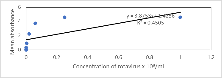
Figure 2: Graph showing the relationship between the absorbance and rotavirus concentration
Lateral Flow (LAT) Immunological Approach
The results from the lateral flow method using Bioconcept Rotastrip indicated that only 4 samples were positive for rotavirus while the remaining 14 test stripes were observed to be negative (Table 1). It was further observed that as the ¼ serial dilution progressed towards the 4th tube (1,562,500 rotavirus particle per ml), the intensity of the colour (indicating that the test was positive) became very faint and finally negative result was observed when the ¼ serial dilution reached 1:256 which indicates 390, 625 rotavirus particle per ml as seen in Table 1.
| Tubes | Rotavirus Conc./mL | Observation | Inference (Result Qualification) | Interpretation |
| 1 | 100,000,000 | Double Lines | Strongly Positive | Presence of Rotavirus |
| 2 | 25,000,000 | Double Lines | Moderately Positive | Presence of Rotavirus |
| 3 | 6,250,000 | Double Lines | Mildly Positive | Presence of Rotavirus |
| 4 | 1,562,500 | Double Lines | Very Weak Positive | Presence of Rotavirus |
| 5 | 390,625 | Single Line | Negative | Absence of Rotavirus |
| 6 | 97,656.25 | Single Line | Negative | Absence of Rotavirus |
| 7 | 24,414.06 | Single Line | Negative | Absence of Rotavirus |
| 8 | 6103.5 | Single Line | Negative | Absence of Rotavirus |
| 9 | 1,525.88 | Single Line | Negative | Absence of Rotavirus |
| 10 | 381.5 | Single Line | Negative | Absence of Rotavirus |
| 11 | 95.37 | Single Line | Negative | Absence of Rotavirus |
| 12 | 23.85 | Single Line | Negative | Absence of Rotavirus |
| 13 | 5.96 | Single Line | Negative | Absence of Rotavirus |
| 14 | 1.49 | Single Line | Negative | Absence of Rotavirus |
| 15 | 0.37 | Single Line | Negative | Absence of Rotavirus |
| 16 | 0.09 | Single Line | Negative | Absence of Rotavirus |
| 17 | 0.002 | Single Line | Negative | Absence of Rotavirus |
| 18 | 0.00 | Single Line | Negative | Absence of Rotavirus |
| Sample A (Positive Control) | - | Double Lines | Positive | Presence of Rotavirus |
| Sample B (Negative Control) | - | Single Line | Negative | Absence of Rotavirus |
Table 1: Result (Visual) Qualification of the Lateral Flow Test Strips for Varying Dilutions for Rotavirus Particles per ml
Note: The dilutions were done in ¼ serial dilution resulting in 1:4; 1:16; 1:64; 1:256; 1:1024, and so on dilutions Resulting in varying concentration front a virus particle per ml.
Discussion
The study focused on the comparison between the use of ELISA and the lateral flow immunoassay methods in the detection of rotavirus in positive samples, both of which are serological methods used in the detection of antibodies in blood samples [19]. The LAT method is cheaper, whereas ELISA is expensive and takes longer time to produce results compared to LAT. In terms of the test procedure and simplicity, the lateral flow approach has shown to be simpler to perform and fast in terms of obtaining results which is basically qualitative. Thus, testing the presence of rotavirus in a sample using this method may not necessarily require much training and subsequently a very useful tool in the quick detection of rotavirus and in outbreaks of gastroenteritis caused by rotavirus in the population in the remote communities. Although, the conventional sandwich ELISA technique is more technical to perform, takes more time, and good laboratory skills are required, even the use of electricity would be needed, which may pose a very huge strong challenge in the remote areas of the world with no access to electricity and paucity of fund.
From the result obtained in the ELISA technique it was shown that there is a linear relationship (R2 = 0.4505) between the absorbance and concentration of rotavirus in the solution. The relationship is positive indicating that the absorbance increases with a corresponding increase in the rotavirus particle in the solution and vice versa. However, there was a plateau at the peak of the plot indicating saturation of the antibody binding sites by the rotavirus antigen. This also indicates a steady state of antibody–antigen reactions irrespective of the number of rotavirus antigens present in the solution.
Rotavirus particles from the study were detected and quantified at dilutions lower than 1: 65,536 with viral particles of 381.5 rotavirus particle per ml using the ELISA method, while in the lateral flow approach, the antigen was detected at a maximum dilution of 1:64 with a viral load of 1,562,500 particles/ml. This indicates that the ELISA approach is far more sensitive and specific to detecting and quantifying rotavirus in a given sample than the lateral flow approach. Also, it could be said that the LAT approach is limited with low rotavirus concentration in the sample, which means that there is a tendency of obtaining false negative results even when rotavirus is present in the sample (Table 1). This also implies that individuals with gastroenteritis caused by rotavirus could be misdiagnosed with negative results especially at the early stage of the disease when the viral load in the individual is quite low. Consequently, this may not be a good tool for managing children with signs and symptoms of gastroenteritis with suspicious of rotavirus infection. However, it could be a good tool for population screening for asymptomatic carriers in the population following it turnaround time and reproducibility, especially in the remote communities with lack of electricity and poor purchasing power to procure ELISA kits and other expensive methods like PCR.
In order to provide statistical interpretation and extrapolations, the ELISA approach will be more useful since it involves quantitative analysis, unlike the lateral flow method which is qualitative. The findings in this study is in consonance with reports by Mohit et al. [20] who also demonstrated that the ELISA method was more effective in the detection of other viral infections like SARS-CoV-2 compared to Rapid test kits. The study by Ha et al. [21] also affirmed the suitability of the ELISA for quantification and not just detection of the virus, alone.
Nonetheless, one critical and fundamental challenge reportedly and obviously facing the use of ELISA kits to strengthen the control and prevention of infectious disease in the developing communities of the world has been its high cost and availability as the kits are imported from abroad with no visible plans of hosting the manufacturing plants in the Nigeria. This strongly distort the gains of using these kits for differential diagnosis and improving accuracy and precision during patient’s care and management. Even, as the use of electricity to power the machine which is very epileptic, has also remained a huge challenge to drive the process of differential diagnosis in our region
Conclusion
The use of Lateral flow test kits can result in misdiagnosis of rotavirus due to their relatively poor sensitivity compared to the conventional sandwich ELISA technique. The ELISA technique is sensitive enough to pick 381.5 rotavirus particle per ml in a sample unlike LAT which detected the presence of rotavirus at the concentration of 1,562,500 particles per ml. Furthermore, the study has also shown that the absorbance is exponentially proportional to the rotavirus antigen present in the sample until a steady state is attained due to antibody-antigen saturation. The equation Y=3.8753x + 1.4236 can be employed to find the unknown concentration of rotavirus antigens when the absorbance is known.
Conflict of Interest
No conflict of interest was reported among researcher’s and no fund was received from any organisation or individual in form of grant to conduct the research
Acknowledgement
The authors are eternally grateful to the laboratory staff of Nimi Briggs Hospital of the Rivers State University, for all their technical support during the practical laboratory analysis of the samples. We are also grateful to the ICT Department of the Rivers State University for providing the enabling ground and technical support for an adequate and effective literature review of the subject matter in question.
References
- Matson, O. D., Staat, A. M., Azimi, P., Itzler, R., Bernstein, I. D., Ward, L. R., Dahiya, R., DiNubile, J. M., Barnes-Eley, M., and berke, T. (2012), Burden of rotavirus hhospitalisations in young children in three paediatric hospitals in the United States determined by active surveillance compared to standard indirect methods. Journal of paediatrics and child Health, 48: 698 - 704.
View at Publisher | View at Google Scholar - Kasi, G. S. (2012), Rotavirus vaccine revisited 2012. Pediatric infectious Disease, 4(4):172 - 177.
View at Publisher | View at Google Scholar - Hu, L., Crawford, E. S., Hyser, M. J., Estes, K. M. and Prasad, V.B. V. (2012), Rotavirus non- structural proteins: Structure and function. Current opinion in virology, 2:380 – 388.
View at Publisher | View at Google Scholar - Matthijnssens J, Otto PH, Ciarlet M, Desselberger, U., Ranst, M.V., Johne, R. (2012). VP6-sequence-based cutoff values as acriterion for rotavirus species demarcation.Arch Virol. 157(6), 1177-1182
View at Publisher | View at Google Scholar - Mihalov-Kova´cs E, Gellert A ´, Marton, S., Farkas, S.L., Feher E., Oldal, M., Jakab, F., Martella, V., Banyai, K. (2015). Candidate new rotavirus species insheltered dogs, Hungary. Emerg Infect Dis. 21(4), 660-663
View at Publisher | View at Google Scholar - Martella V, Ba´nyai K, Matthijnssens J,Buonavoglia. C., Ciarlet M. (2010). Zoonotic aspects of rotaviruses. Vet Microbiol. 140(3-4), 246-255.
View at Publisher | View at Google Scholar - Parashar, D. U., Hummelman, G. E., Bresee, S. J., Miller, A.M. and Glass, I. R. (2003), Emerging infectious Diseases, 9(5) 565 -572.
View at Publisher | View at Google Scholar - Armah, E. G., Sow, O. S., Breiman, F. R., Dallas, J. M. Tapia, D. M., Feikin, R. D., Binka, N. F., Steele, D. A., Larserson, F. K., Ansah, A. N., Levine, M. M., Lewis, K., Coia, L. M., Attah-Poku, M., ojwando, J., Rivers, B. S., Victor, C.J., Nyambane, G., Hodgson, A., Schodel, F., Ciarlet, M. and Nevzil, M. K. (2010), Efficacy of pentavalent rotavirus vaccine against severe rotavirus gastroenteritis in infants in developing Countries in Sub-Saharan Africa: a randomised, double-blind, placebo controlled trial. Lancet, 376:606 – 614.
View at Publisher | View at Google Scholar - Liu, L., Johnson, L. H., Cousens, S., Perin, J., Scott, S., Lawn, E. J., Rudan, I, Campbell, H., Cibulskis, R., Li, M., and Black, E. R. (2010), Global, regional and national causes of child mortality: an updated systematic analysis for 2010 with time trends since 2000. Lancet, 379:2157 - 2161.
View at Publisher | View at Google Scholar - Bruijning-Verhagen, P., Quach, C. and Boten, M. (2012), Nosocomial Rotavirus Infections: A Meta-analysis. PEDIATRICS, 129(4) 1011 – 1019.
View at Publisher | View at Google Scholar - Dhama, K, Chauhan, R.S., Mahendran, M. (2009). Rotavirus diarrhea in bovines andother domestic animals. Vet Res Commun33(1), 1-23
View at Publisher | View at Google Scholar - Arvelo, W., Hall, J. A., Estevez, A., Lopez, B., Gregoricus, N., Vinje, J., Gentsch, R. J., Parashar, U. and Lindblade, A. K. (2013), Diagnostic performance of rectal swab versus bulk stool specimens for detection of rotavirus and nonovirus: Implications for outbreak investigations. Journal of clinical virology, (58)678 – 682
View at Publisher | View at Google Scholar - Gautem, R., Lyde, F. and Esona, D. M. (2013), Comparison of PremierTmRotacloneR, ProSpecTTm, and RIDASCREENR rotavirus enzyme immunoassay kits for the detection of rotavirus antigen in stool specimens. Journal of clinical virology, 58:291 - 294
View at Publisher | View at Google Scholar - Mekonnen, D., Mengist, H. M., Derbie, A., Nibret, E., Munshea, A., He, H., Li, B., Jin, T. (2020). Diagnostic Accuracy of Serological Tests and Kinetics of Severe Acute Respiratory Syndrome Coronavirus 2 Antibody: A Systematic Review and Meta-Analysis. Rev. Med. Virol. 31-2181.
View at Publisher | View at Google Scholar - Butler, J. E. (2000). Enzyme-linked Immunosorbent Assay. J. Immunoassay. 21:165–209.
View at Publisher | View at Google Scholar - Gong, F., Wei, H, Li, Q., Liu L., and Li, B. (2021). Evaluation and comparison of serological methods for COVID_19 diagnosis. Front Mol Biosci. 8:682405.
View at Publisher | View at Google Scholar - Carrio, A., Sampedro, C., Sanchez-Lopez, J. L., Pimienta, M., and Campoy, P. (2015). Automated Low-Cost Smartphone-Based Lateral Flow Saliva Test Reader for Drugs-Of-Abuse Detection. Sensors (Basel) 15: 29569-29593.
View at Publisher | View at Google Scholar - Moreno, M. L., Cebolla, A., Munoz-Suano, A., Carrillo-Carrion, C., Comino, I., Pizarro, A., Leon, F., Rodríguez-Herrera, A., Sousa, C. (2017). Detection of Gluten Immunogenic Peptides in the Urine of Patients with Coeliac Disease Reveals Transgressions in the Gluten-free Diet and Incomplete Mucosal Healing. Gut 66: 250–257.
View at Publisher | View at Google Scholar - Arikan, A., Doluca O., Akhan, S., Sanlidag T and Syan, M. (2023). Evaluation of lateral flow and ELISA techniques for detecting IgG and IgM antibodies in COVID-19 cases in Turkiye. East Mediterr Health. 29(2), 91-99.
View at Publisher | View at Google Scholar - Moht E., Rostami Z. and Vahidi, H. (2021). A comparative review of immunoassays for COVID-19 detection. Expert Rev Clin Immunol. 17(6), 573-599.
View at Publisher | View at Google Scholar - Ha, Y., Thienes, CP, Agapov, AA, Laznicka, AV, Han S., Nadala C, and Samadpour M (2018). Comparison of ELISA and DNA lateral flow assays for detection of pork, horse, beef, chicken, turkey and goat contamination in meat products. J AOAC. PMID: 29980244.
View at Publisher | View at Google Scholar
