

Research Article | DOI: https://doi.org/10.31579/ 2834-8532/060
Effects of Stone Herbal Mixture on Antioxidant Levels
- Usiobeigbe O.S 1
- Omoviye O.E 2
- Omolumen L.E 1
- Obohwemu K.O 3
- Asibor E 4
- Bello G.O 5
- Aliche P.C 6
- Babatope S.A 7
- Aperua-Yusuf K.I 8
- Iyevhobu K.O 9
2 Business Services, Direct Export Sales Organisation, Siemens Healthineers AG, Erlangen, Germany.
3 Faculty of Health, Wellbeing and Social Care, Oxford Brookes University, GBS Partnership, Birmingham, United Kingdom; and PENKUP Research Institute, Birmingham, United Kingdom
4 Department of Histopathology and Cytopathology, Faculty of Medical Laboratory Science, Ambrose Alli University, Ekpoma, Edo State, Nigeria.
5 Department of Nursing, Babcock University Ilisan Remo, Ogun State, Nigeria.
6 Department of Nursing Sciences, Clifford University, Owerrita, Abia State, Nigeria
7 Department of Pharmaceutical and Biomedical Sciences, College of Pharmacy, University of Georgia, Athens, USA
8 Department of Biochemistry, School of Applied Science, Auchi Polytechnic Auchi, Edo State, Nigeria.
9 Department of Medical Microbiology, Faculty of Medical Laboratory Sciences, Ambrose Alli University, Ekpoma, Edo State, Nigeria.
*Corresponding Author: Usiobeigbe O.S, Universidad de la Salud, Mèxico.
Citation: Usiobeigbe O.S., Omoviye O.E., Omolumen L.E., Obohwemu K.O., Asibor E., Bello G.O., Aliche P.C., Babatope S.A., Aperua-Yusuf K.I., Iyevhobu K.O., (2025), Effects of Stone Herbal Mixture on Antioxidant Levels, Clinical Genetic Research; 4(4): DOI: 10.31579/ 2834-8532/060
Copyright: © 2025 Usiobeigbe O.S, this is an open-access article distributed under the terms of the Creative Commons Attribution License, which permits unrestricted use, distribution, and reproduction in any medium, provided the original author and source are credited.
Received: 03 October 2025 | Accepted: 13 October 2025 | Published: 30 October 2025
Keywords: stone herbal; mixture; antioxidant; oxidative; albino wistar rats
Abstract
Oxidative stress occurs when the balance between free radicals and antioxidants in the body is disrupted, leading to cellular damage and a range of diseases. Antioxidants play a crucial role in neutralizing these free radicals, protecting cells from harm, and potentially mitigating the onset or progression of numerous health conditions. This study aims to evaluate the effect of the Stone Herbal Mixture on antioxidant levels in an animal model, Male Albino Wistar rats. Animal models are widely employed in early-stage research to investigate the safety and efficacy of potential treatments before conducting human clinical trials. Thirty-Six (36) Male Albino Wistar rats weighing 100-120g were used for this study. The rats were obtained from the Lead City University Ibadan, animal house, and were used for the study. This study was an experimental study comprising of total of Thirty-Six (36) Male Albino wistar rats divided into Six (6) groups of six (6) animals each. The six groups were Group 1, control group fed with top feed and water only, Group 2 administered with standard drug (silymarin 140mg/kg body weight) and 1000mg (0.30 ml) of stone herbal mixture, top feed and water, Group 3 administered with 1000 mg (0.30 ml) of stone herbal mixture, Top Feed and water. Group 4 administered with 850 mg (0.25ml) of stone herbal mixture, Top feed and water, Group 5 administered with 650 mg (0.20 ml) of stone herbal mixture, Top Feedand water. Group 6 administered with 500 mg (0.15 ml) of stone herbal mixture, top feed and water. The treatment was given daily and lasted for 21 days. The herbal mixture extract was administered with the aid of oral cannula, once daily to the appropriate group for the time specified for each group. At the end of the experiment, the animals were sacrificed 12 hours after the last administration and blood was collected into Lithium Heparin anticoagulant bottle from brachiocephalic artery of each rat. The rats were immediately ethically sacrificed by cervical dislocation. At the end of the sample collection, the samples were analyzed for oxidative stress parameters including Catalase, Glutathione, Superoxide Dismutase and Malondialdehyde. Serum Catalase level was found to be increased in group 2 administered with (silymarin 140mg/kg body weight) and 1000 mg (0.30ml) of stone herbal mixture (standard group) and decreased in group 3, 4 and 5. This decrease was statistically very significant in the group 3 administered with 1000 mg (0.30ml) of stone herbal mixture, group 4 administered with 850mg (0.25ml) of stone herbal mixture, and group 5 administered with 650 mg (0.20ml) of stone herbal mixture, in comparison with group 1 (control group). The increase in group 2 administered with (silymarin 140 mg/kg body weight) and 1000 mg (0.30 ml) of stone herbal mixture (standard group) and group 6 administered with 500 mg (0.15ml)of stone herbal mixture, was statistically nonsignificant in comparison with group 1 (control group). In this aspect, group 6 administered with 500 mg (0.15ml) of stone herbal mixture was relatively comparable with the group 2 administered with (Silymarin 140 mg/kg body weight) and 1000mg (0.30ml) of stone herbal mixture. The result shows that stone herbal mixture does not influence the antioxidants in a negative way. It also shows some significant antioxidant properties in experimental animals in this study.
Introduction
Oxidative stress occurs when the balance between free radicals and antioxidants in the body is disrupted, leading to cellular damage and a range of diseases. Antioxidants play a crucial role in neutralizing these free radicals, protecting cells from harm, and potentially mitigating the onset or progression of numerous health conditions (Schieber& Chandel, 2014).
In recent years, interest in herbal medicine and natural remedies has surged as people seek alternative approaches to complement conventional medical treatments (Rastogi et al., 2016). Nigeria, like many other countries, has a rich tradition of using herbal remedies in traditional healthcare systems. Herbal mixtures, in particular, have been passed down through generations, believed to possess various health benefits, including antioxidant properties that can combat oxidative stress and promote overall well-being.
The "Stone Herbal Mixture," a specific herbal formulation prevalent in traditional Nigerian medicine, is reputed to possess potent antioxidant properties (Mohammadhosseiniet al., 2017; Usiobeigbeet al., 2024; Usiobeigbeet al., 2024b). While it has been widely used and trusted in various communities, scientific evidence supporting its claimed benefits remains limited (Usiobeigbeet al., 2024; Usiobeigbeet al., 2024b).
Despite the widespread use and belief in the potential health benefits of herbal remedies, there is a lack of scientific evidence supporting the claimed antioxidant properties of the Stone Herbal Mixture, a traditional Nigerian herbal formulation (Usiobeigbeet al., 2024). The absence of empirical data on the antioxidant effects of this herbal mixture creates a significant gap between traditional knowledge and evidence-based healthcare practices (García- Prat et al., 2016; Usiobeigbeet al., 2024b). the research problem revolves around the need to evaluate and validate the antioxidant potential of the Stone Herbal Mixture in an animal model, Male Albino Wistar rats. The lack of scientific investigation into the herbal mixture's efficacy and safety raises concerns about its appropriate usage and potential benefits for human health."The lack of empirical evidence on the antioxidant effects of the Stone Herbal Mixture in Male Albino Wistar rats poses a challenge in substantiating its traditional claims and hinders the integration of this herbal remedy into evidence-based healthcare practices (Usiobeigbeet al., 2024; Usiobeigbeet al., 2024b).
Traditional herbal remedies, including the Stone Herbal Mixture, have been relied upon by communities for generations (Usiobeigbeet al., 2024b). Conducting scientific research on its antioxidant properties will provide empirical evidence to support or refute its traditional claims, bridging the gap between traditional knowledge and modern scientific understanding (Usiobeigbeet al., 2024). Oxidative stress is implicated in various diseases, including cardiovascular disorders, neurodegenerative conditions, cancer, and aging-related ailments (Deshmukh et al., 2016). Establishing the antioxidant potential of the Stone Herbal Mixture could identify a potential natural remedy for combating oxidative stress, promoting overall health, and possibly preventing certain diseases. Also, with the increasing interest in complementary and alternative medicine, the study's findings may open up new possibilities for herbal-based antioxidant therapies. If the Stone Herbal Mixture demonstrates significant antioxidant effects in animal models, it may pave the way for further clinical investigations and possible integration into healthcare practices (Usiobeigbeet al., 2024; Usiobeigbeet al., 2024b).
The safety of herbal remedies is of utmost concern, and animal models provide a platform for initial safety evaluations before human trials. Assessing any potential side effects or toxicity associated with the Stone Herbal Mixture in Male Albino Wistar rats is essential to safeguarding human health (Usiobeigbeet al., 2024; Usiobeigbeet al., 2024b).
This study aims to evaluate the effect of the Stone Herbal Mixture on antioxidant levels in an animal model, Male Albino Wistar rats. Animal models are widely employed in early-stage research to investigate the safety and efficacy of potential treatments before conducting human clinical trials. Male Albino Wistar rats are commonly chosen for biomedical research due to their genetic uniformity and physiological similarities to humans. By examining the antioxidant potential of the Stone Herbal Mixture in Male Albino Wistar rats, this research seeks to contribute scientific evidence to traditional knowledge and bridge the gap between traditional herbal remedies and modern medical practices. The outcomes of this study may provide crucial insights into the herbal mixture's effectiveness and safety, potentially paving the way for the development of novel herbal-based antioxidant therapies.
Materials And Method
Materials
Materials used in this study include; Cotton wool, needle and syringe, plain bottle, Lithium heparin, methylated spirit, Gloves, micropipette, electronic weigh balance Tourniquet, automatic micropipette, pipette tips, spectrophotometer, spectrophotometric cuvette, water bathe, vortex mixer, disposable test tubes, and stone herbal mixture.
Experimental Animals:
Thirty-Six (36) Male Albino Wistar rats weighing 100-120g were used for this study. The rats were obtained from the Lead City University Ibadan, animal house, and were used for the study. The animals were allowed one week acclimatization period after which they were reweighed and housed in plastic cages with plastic bottom and wire-mesh top, under controlled environmental conditions of temperature (28±20C), relative humidity (50±5%) and a twelve-hour light/dark cycle. The animal facility was adequately ventilated and the animals maintained regularly on the commercial rat chow. Water and Top feed were provided throughout the experimental period.
After One week of acclimatization period, rats were divided into (6) groups, the experimental control group Were administered with water and top feed, Group 2 were administered with water, top feed, (silymarin 140mg/kg body weight) and oral intubation of stone Herbal mixture while other groups were administered with water, top feed and oral intubation of stone Herbal mixture.
Experimental Design:
This study was an experimental study comprising of total of Thirty-Six (36) Male Albino wistar rats divided into Six (6) groups of six (6) animals each. The six groups were Group 1, control group fed with top feed and water only, Group 2 administered with standard drug (silymarin 140mg/kg body weight) and 1000mg (0.30 ml) of stone herbal mixture, top feed and water, Group 3 administered with 1000 mg (0.30 ml) of stone herbal mixture,Top Feed and water. Group 4 administered with 850 mg (0.25ml) of stone herbal mixture, Top feed and water, Group 5 administered with 650 mg (0.20 ml) of stone herbal mixture, Top Feed and water. Group 6 administered with 500 mg (0.15 ml) of stone herbal mixture, top feed and water. The treatment was given daily and lasted for 21 days.
Groups | No. of Animals | Administrations |
1(control) | 6 | Water and Top feed. |
2 | 6 | Water,Top feed,silymarin and 1000mg (0.30ml) of stone herbalmixture. |
3 | 6 | Water,Top feed,and 1000 mg (0.30ml) of stone herbalmixture. |
4 | 6 | Water, Top feed and850 mg (0.25ml) of stone Herbalmixture. |
5 | 6 | Water, Top feed and650 mg (0.20ml) of stone Herbalmixture. |
6 | 6 | Water,Top feedand 500 mg (0.15ml) of stone herbalmixture. |
Experimental design and administration of stone Herbal mixture.
Study Site:
This study was carried out at the animal house, Department of Medical Laboratory Science, Lead City University Ibadan-Oyo state, Nigeria.
Ethical Consideration:
This study Ethical approval was obtained from Lead City university Research ethics committee Ibadan, Oyo state with the ethical approval number (LCU-REC/23/345).
Procurement of Stone herbal mixture:
The Stone Herbal mixture was purchased from a pharmaceutical store at Ibadan, Oyo State, Nigeria. The Stone herbal mixture was stored at room temperature of 18°C throughout the period of the experiment.
Administration of Stone herbal mixture:
The herbal mixture extract was administered with the aid of oral cannula, once daily to the appropriate group for the time specified for each group.
Animal Sacrifice and Sample Collection:
At the end of the experiment, the animals were sacrificed 12 hours after the last administration and blood was collected into Lithium Heparin anticoagulant bottle from brachiocephalic artery of each rat. The rats were immediately ethically sacrificed by cervical dislocation. At the end of the sample collection, the samples were analyzed for oxidative stress parameters including Catalase,Glutathione, Superoxide Dismutase and Malondialdehyde.
Catalase assay:
Principle: The catalase activity in a sample is determined by measuring the decrease in Hydrogen peroxide H2O2concentration observed following an incubation of the analyte sample withan Hydrogen Peroxide H2O2standard solution.Using the Megazyme Catalase Assay Kit, two separate reactions must be completed for catalase activity.
In reaction A, the catalase sample of interest is incubated with a known concentration (~ 65 mM in assay) of Hydrogen Peroxide H2O2. The reaction is stopped by the addition of 15 mM sodium azide which strongly inhibits catalase.
Reaction A is shown below in the presence of catalase enzymes
2H2O2→2H2O +O2
In reaction B, the exact concentration of Hydrogen Peroxide H2O2remaining is measured using an enzyme-linked colourimetric detection method employing 3, 5-dichloro-2- hydroxy-benzenesulfonic acid (DHBS), 4-aminoantipyrine (AAP) and peroxidase. The resulting quinoneimine dye is measured at 520 nm.
2H2O2 +DHBS + AAP→ quinoneimine dye + 4H2O
Assay procedure:
A) Incubation of the catalase sample with Hydrogen Peroxide H2O2 substrate solution:
The Reaction blank and Reaction Sample were prepared as outlined below. Duplicate reaction blanks were required for each set of assays performed. Duplicate (or triplicate) reactions were also recommended for each sample assayed. It was Performed in glass test tubes or plastic micro tubes as preferred.
| Pipette intotubes | Reaction blank | Reaction Sample |
| H2O2 substratesolution (~130 mM) | 0.05 mL | 0.05Ml |
| Assay buffer | 0.05 mL | |
| Catalase sample | 0.05mL | |
| Mix and incubate for 5 minutes at 25oC | ||
| Add 15mM of sodium Azide | 0.09ml | 0.09mL |
Mix well and immediately remove an aliquot of 40µl fromthe blank and the sample reaction. These aliquotsare transferred into colorimetric cuvette assay | ||
A) Incubation of the catalase sample with Hydrogen Peroxide H2O2 substrate solution:
About 50 µl (0.05 ml) of hydrogen peroxide H2O2 substrate solution (~130 mM) was pipetted into two test tube marked reaction blank and reaction sample.50 µl (0.05 ml) of assay buffer was pipetted into the test tube marked reaction blank.50 µl (0.05ml) of catalase sample was pipetted into the test tube marked reaction sample.It was mixed and incubated for 5 minutes at 25°C. 90 µl (0.09ml) of 15 mm of sodium Azide was added to the two test tubes marked reaction blank and reaction sample.It was Mixed well and an aliquot of 40 µl from the blank and the sample reaction was removed immediately. These aliquots were transferred into colorimetric cuvette assay.
B) Determination of remaining Hydrogen Peroxide H2O2 concentration:It was Performed in cuvettes with 1 cm light path (glass or plastic). Zero the spectrophotometer against air (without cuvette in the light path).
Pipette into cuvette | Reaction Blank |
Aliquots from the sample and blank reactions in the catalase/H2O2 incubation (Reaction A) above | 0.04mL |
Colourimetric reagents | 3.0mL |
Mix andincubate for 15 minutes at 25oC |
|
The blank and sample reaction was read at an absorbance of 520nm and calculate the catalase activity.
Assay procedure:
About 200 µl of whole blood was mixed thoroughly with
1.8 ml of distilled water and 3 ml of Precipitate (PPT) solution.It was allowed to stand for 5 min and was filtered.Two test tube marked test and blank were taken. A reagent blank was made using 2 ml of distilled water, 8 ml of phosphate buffer, and 1 ml of 5,5′-Dithiobis-2- nitrobenzoic acid (DTNB) reagent.In test tube marked test, 2 ml of clear filtrate was added from the above mixture to 8 ml of disodium phosphate buffer and 1 ml of 5,5′-Dithiobis- 2-nitrobenzoic acid (DTNB) reagent was added to it. It was mixed well. The color was developed rapidly, it was stable for 10 min. It was read at 412 nm in the spectrophotometer.The Reading was taken; the curve was plotted taking absorbance at 412 nm on the Y-axis and concentration on the X-axis. The concentration of the test samples was calculated by using standard curve. The Reduced GSH concentration in blood sample was expressed as mg/dl.
Reference range: Glutathione: 176-323 ug/ml
Superoxide Dismutatse:
Principle: Super oxide anions generated as the byproduct of Xanthine Oxidase catalyzed Xanthine oxidation and the anion can oxidize hydroxylamine to nitrite which appears to be amaranth purple in the presence of chromogenic agent and thus the absorbance at certain wavelength can be detected by spectrophotometer. The presence of SOD in the system specifically inhibits the oxidization caused by super oxide anions and because of it, fewer nitrite anions are generated. This would lower the absorbance of the sample tube compared to the reference tube without Superoxide Dismutase (SOD) and the Superoxide Dismutase (SOD) activity can be calculated with the formula given.
Reagent preparation:
The kit was allowed to warm up to room temperature completely before using. It was acceptable to place the reagents at room temperature the night before the experiment. However, The Hematoxylin reagent was not reconstituted until actual day of use since the reagent in liquid form is only stable for 6 hours at room temperature.
Hematoxylin: Hematoxylin was reconstituted as supplied with 1.2 mL (1200 μL) of dH2O. It was allowed to stand for 30 minutes before used. At room temperature, it should be used within 6 hours. If only a portion of the reconstituted
hematoxylin is needed in one experiment; the excess solution should be immediately stored at –20°C (frozen) for future use.
Sample preparation:
Normal human plasma and serum Superoxide Dismutase (SOD) activities are typically quite low (< 5> Pipette into eachcuvette assay Blank Sample Assay buffer 920 μL 920 μL Assay buffer 40 μL - Mix andincubate for two (2) minutes Hematoxylin reagent 40μL Mix quicklyand immediately the absorbance was began to record at 560 nm every 10 seconds or smaller timeinterval for at least 5 minutes
Assay procedure (cuvette assay)
About 920 μL of Assay Buffer was added to each cuvette for assay. 40 μL of Assay Buffer was added to blank or 40 μL of sample was added to the blank.It was Mixed and incubated for two (2) minutes. 40 μL of Hematoxylin Reagent was added to start the auto-oxidation reaction. It was Mixed quickly and immediately the absorbance was began to record at 560 nm every 10 seconds or smaller time interval for at least 5 minutes.
Superoxide Dismutase (SOD) calculation
(SOD U/mL) os = (SOD U/mLs) × sample dilution factor.
Reference range: Superoxide Dismutase: 11-15 ug/ml
Malondialdehyde
Principle of Malondialdehyde (MDA) assay
In the presence of acid, Malondialdehyde (MDA) reacts with Thiobarbituric acid (TBA) to produce a colored end
product that absorbs light. The intensity of the color at 532 nm corresponds to the level of lipid peroxidation in the sample. Unknown samples are compared to the standard curve.
Determination of Thiobarbituric acid reactive substances (TBARS):
Lipid peroxidation levels were measured by the Thiobarbituric acid (TBA) reaction using the method of Ohkawaet al. (1979).
This method was used to measure spectrophotometrically the color produced by the reaction of Thiobarbituric acid (TBA) with malondialdehyde (MDA) at 532 nm. For this purpose, Thiobarbituric acid reactive substances (TBARS) levels were measured using a commercial Malondialdehyde Assay kit according to the manufacturer’s instructions.
Pipette into tubes | Reaction blank | Reaction Sample |
Erythrocyte supernatant containing 2 μl of butylated hydroxytoluene (BHT) in methanol. | 50 μl | 50 μl |
Acid reagent | 50 μl | 50 μl |
Thiobarbituric Acid | 50 μl | 50 μl |
Mix vigorously and incubated for 60 min at 60 °C. |
|
|
Centrifuge at 10,000 × g for 3 min |
|
|
The supernatant was put intowells on a microplate in aliquots of 75 μl. |
|
|
Procedure:Tissue supernatant (50 μl) or erythrocyte supernatant (50 μl) were added to test tubes containing 2 μl of butylated hydroxytoluene (BHT) in methanol.50 μl of acid reagent (1 M phosphoric acid) was added and finally 50 μl of Thiobarbituric acid (TBA) solution was added. The tubes were mixed vigorously and incubated for 60 min at 60
°C.The mixture was centrifuged at 10,000 × g for 3 min. The supernatant was put into wells on a microplate in aliquots of 75 μl.Absorbance was measured with spectrophotometrically at 532 nm. Thiobarbituric acid reactive substances (TBARS) levels were expressed as nmol/mg protein in various organs (brain, liver, pancreas and skeletal muscle), and as nmol/g hemoglobin in erythrocyte hemolysates.
Reference range: Malondialdehyde: 89-195 nm
Statistical Analysis:
The SPSS (statistical package for social sciences) software package was used for statistical analysis. Values obtained were expressed as mean ± standard deviation and compared using analysis of variance (ANOVA) and the significance was measured at p ˂ 0.05.
Result
Table 1 shows the descriptive details of hormones (Catalase, Glutathione, Superoxide Dismutase, Malondialdehyde). The descriptive details were given by mean ±standard deviation. Analysis of variance was performed on the data; there are significant difference across the groups of the experimental rats (p<0> Group 1 Mean+SD Group 2 Mean+SD Group 4 Mean+SD Group 5 Mean+SD Group 6 Mean+S.D Group 3 Mean+SD f-Value P-Value Catalase 100.2 + 5.3 103.3 + 5.7 77.7 + 5.9 87.7 + 2.3 93.2 + 1.9 103.1 + 5.7 27.166 0.000* Glutathione 207.3 + 17.3 236.7 + 34.4 136.2 + 3.5 145.3 + 3.6 160.0 + 7.2 185.5 + 8.6 32.982 0.000* Superoxide Dismutase 12.8 + 1.5 13.2 + 1.5 4.0 + 0.9 4.3 + 1.6 9.7 + 2.1 12.5 + 1.9 33.989 0.000* Malondialdehyde 113.7 + 7.8 110.3 + 12.7 219.5 + 4.1 209.8 + 3.3 200.7 + 3.6 181.7 + 8.2 259.529 0.000*
Table 1: Comparison of mean ±SD of antioxidant parameters between the rats in the six groups
Antioxidants | Group Number | Mean Difference | t-test | p-value |
Catalase | Group 2 | -3.167 | -1.283 | .256 |
Group 3 | 22.500 | 7.507 | .001* | |
Group 4 | 12.500 | 4.872 | .005* | |
Group 5 | 7.000 | 3.955 | .011* | |
Group 6 | -3.000 | -1.885 | .118 | |
Glutathione | Group 2 | -29.333 | -2.397 | .062 |
Group 3 | 71.167 | 10.248 | .000* | |
Group 4 | 62.000 | 10.382 | .000* | |
Group 5 | 47.333 | 6.940 | .001* | |
Group 6 | 21.833 | 2.713 | .042* | |
Superoxide Dismutase | Group 2 | -0.333 | -0.326 | .758 |
Group 3 | 8.833 | 22.007 | .000* | |
Group 4 | 6.500 | 13.000 | .000* | |
Group 5 | 3.167 | 3.124 | .026* | |
Group 6 | 0.333 | 0.265 | .801 | |
Malondialdehyde | Group 2 | 3.333 | 0.594 | .578 |
Group 3 | -105.833 | -24.609 | .000* | |
Group 4 | -96.167 | -31.889 | .000* | |
Group 5 | -87.000 | -24.007 | .000* | |
| Group 6 | -68.000 | -30.615 | .000* |
Table 2: Comparison of mean of Antioxidant parameters in group 1 (control) with each of the five other groups
It was deduced in the above table (table 2) that:
Catalase: p>0.05, p<0>0.05 for the 6 pairs respectively.
Glutathione: p>0.05, p<0>
Superoxide Dismutase: p>0.05, p<0>0.05 for the 6 pairs respectively.
Malondialdehyde: p>0.05, p<0>
Antioxidants | Group Number | Mean Difference | t-test | p-value |
Catalase | Group 3 | 25.667 | 8.764 | .000* |
Group 4 | 15.667 | 7.922 | .001* | |
Group 5 | 10.167 | 4.364 | .007* | |
Group 6 | 0.167 | .046 | .965 | |
Glutathione | Group 3 | 100.500 | 7.793 | .001* |
Group 4 | 91.333 | 6.805 | .001* | |
Group 5 | 76.667 | 6.077 | .002* | |
Group 6 | 51.167 | 3.378 | .020* |
| Superoxide Dismutase | Group 3 | -109.167 | 11.569 | .000* |
| Group 4 | 6.833 | 6.970 | .001* | |
| Group 5 | 3.500 | 4.869 | .005* | |
| Group 6 | 0.667 | 1.581 | .175 | |
| Malondialdehyde | Group 3 | -109.167 | -22.427 | .000* |
| Group 4 | -99.500 | -22.258 | .000* | |
| Group 5 | -90.333 | -21.795 | .000* | |
| Group 6 | -71.333 | -12.422 | .000* |
Table 3: Comparison of mean of the antioxidant parameters in group 2 with each of the 4 other groups (group 3-6)
| Antioxidants | Group Number | Mean Difference | t-test | p-value |
| Catalase | Group 3 | 25.667 | 8.764 | .000* |
| Group 4 | 15.667 | 7.922 | .001* | |
| Group 5 | 10.167 | 4.364 | .007* | |
| Group 6 | 0.167 | .046 | .965 | |
| Glutathione | Group 3 | 100.500 | 7.793 | .001* |
| Group 4 | 91.333 | 6.805 | .001* | |
| Group 5 | 76.667 | 6.077 | .002* | |
| Group 6 | 51.167 | 3.378 | .020* |
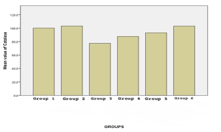
Figure 1: Effect of stone herbal mixture on catalase levels of Male Albino Wister rat

Figure 2: Effect of stone herbal mixture on Glutathione levels of Male Albino Wister rat
Results represented as Mean # SEM n=5 p<0>
Figure 3: Effect of stone herbal mixture on Superoxide Dismutase levels of Male Albino Wister rat

Results represented as Mean # SEM n=5; p<0>
Figure 4: Effect of stone herbal mixture on Malondialdehyde levelsof Male Albino Wister rat
Results represented as Mean # SEM n=5 p<0>
Discussion
Figure 4: Effect of stone herbal mixture on Malondialdehyde levels of Male Albino Wister rat
Results represented as Mean # SEM n=5 p<0>
herbal mixture, and group 5 administered with 650 mg (0.20ml) of stone herbal mixture, in comparison with group
The changes observed in the antioxidant parameters reveal
insights into how the stone herbal mixture can protect against oxidative stress (Usiobeigbeet al., 2024). antioxidant activity was measured by the estimation of serum concentrations of various enzymes by standard biochemical methods. Silymarin (140 mg/kg body weight) was used as a standard drug for assessment of antioxidant status. Silymarin is obtained from the plant silybon marianum, and it has already proven as an antioxidant and antifibrotic.
In figure 1 Serum Catalase level was found to be increased in group 2 administered with (silymarin 140mg/kg body weight) and 1000 mg (0.30ml) of stone herbal mixture (standard group) and decreased in group 3, 4 and 5. This decrease was statistically very significant in the group 3 administered with 1000 mg (0.30ml) of stone herbal mixture, group 4 administered with 850mg (0.25ml) of stone
1 (control group). The increase in group 2 administered with (silymarin 140 mg/kg body weight) and 1000mg (0.30 ml) of stone herbal mixture (standard group) and group 6 administered with 500 mg (0.15ml) of stone herbal mixture, was statistically nonsignificant in comparison with group 1 (control group). In this aspect, group 6 administered with
500 mg (0.15ml) of stone herbal mixture was relatively comparable with the group 2 administered with (Silymarin 140 mg/kg body weight) and 1000mg (0.30ml) of stone herbal mixture.The Group 1,2,3,4,5 and 6 has a percentage of 87%,100%,70%,74%,78% and 99% respectively.
In figure 2 Serum reduced Glutathione level was found to be high in group 2 administered with (silymarin 140 mg/kg body weight) and 1000 mg (0.30ml) of stone herbal mixture (standard group) and decreased in all four administered groups in a graded manner in comparison with group 1
(control group). The administered group 3, group 4 and group 5 showed a very significant decrease as compared with group 1 (control group). Group 6 500 mg (0.15ml) of stone herbal mixture showed a significant decrease (P < 0>
In figure 3 Group 3,4,5 decreased the serum superoxide dismutase level, and the difference was significant when compared with group 1 (control group) indicating potent antioxidant capacity. Oral administration of graded stone herbal mixture also increased serum Superoxide Dismutase levels when compared with group 1 (control group). The highest increase was found in group 2 (standard group) (p>0.05). In group 6, serum superoxide dismutase level was increased but the difference was statistically nonsignificant in comparison with control group. The Group 1,2,3,4,5 and 6 has a percentage of 98%,100%,36%,54%,74%and 99% respectively.
In figure 4 Serum Malondialdehyde level was lowered in the group 2 standard group (110.3 + 12.7) but that was statistically not significant when compared with Group 1 (control group) (113.7 + 7.8). Serum Malondialdehyde levels were also found to be increased in groups 3, group 4, group 5 and group 6, and they were all statistically significant in comparison with group 1 (control group). Malondialdehyde is a stable secondary aldehyde degeneration product of lipid peroxidation and is used as a biological marker for the assessment of lipid peroxidation. serum Malondialdehyde levels in all administered groups proved that stone herbal mixture was an effective antioxidant as standard. The Group 1,2,3,4,5 and 6 has a percentage of 55%,52%,100%,95%,91% and 82% respectively.
Antioxidants are compounds capable of hindering the oxidation of substances. These agents are known as antioxidants and can be classified into two groups according to their mechanism or degree of effectiveness (Somogyi et al., 2017). These categories encompass disruptive antioxidants and preventive antioxidants. The primary distinction between the two lies in preventers slowing the initiation rate, while the disintegrating antioxidants hinder the propagation process. Preventive antioxidants, such as catalase and peroxidase, react with ROOH (hydroperoxide radicals) and metal ion chelators like ethylenediamine tetraacetate (EDTA). These mechanisms block unregulated free radical synthesis or generation and inhibit their interaction with biomolecules. This is how superoxide dismutase (SOD) operates to scavenge superoxide radicals within a living organism (Liang et al., 2018).
The specific action sites of these antioxidants depend on their inherent characteristics within the environment. These antioxidants can also exist in mitochondria and other nuclear compartments. Conversely, hydrophobic antioxidants are situated in membranes, where they counteract lipid
peroxidation (Somogyi et al., 2017). Certain antioxidants are derived from external sources and are called exogenous antioxidants. These antioxidants are acquired through dietary intake. However, acquiring sufficient exogenous antioxidants solely from the contemporary diet is nearly impossible. This is why antioxidant supplementation is frequently advised. These exogenous antioxidants can also be found naturally within the endogenous antioxidant system, including compounds like malondialdehyde, glutathione, superoxide dismutase, and catalase (Asmatet al., 2015).
An antioxidant works by retarding the process of oxidation by free radicals and further damage. Increased levels of measured antioxidant enzymes clearly envisaged the antioxidant potential of this plant. superoxide dismutase is an important endogenous antioxidant enzyme acting as the first line defense system against reactive oxygen species (ROS) which scavenges superoxide radicals to H2O2 and thus provide protection against the deleterious effects of radicals (Mahanteshet al., 2017). H2O2 accumulated by this reaction leads to the formation of hydroxyl radicals which can be harmful too. Glutathione and catalase work as antioxidant enzymes by virtue of scavenging these hydroxyl radicals. Glutathione is a tripeptide and a powerful antioxidant present in the cytosol of cells and is the major intracellular nonprotein thiol compound. SH groups present in glutathione reacts with H2O2 and the hydroxyl radical and prevent tissue damage, and it is also capable of scavenging ROS directly or enzymatically (Olawale et al., 2018). It has been proposed by Kaleet al., (2018) that herbal mixture have the very strong capacity to eliminate free radicals in the blood and promotes the activities of antioxidant enzymes such as SOD, GSH, and CAT. These actions of flavonoids are also dose dependent. Hence, an increase in the serum concentrations of antioxidant enzymes in our study can be explained by this background (Usiobeigbeet al., 2024b). A low dose of stone herbal mixture was unable to increase these enzyme levels, hence failed to exert antioxidant activity.
In this study, we investigated the impact of stone herbal mixture on antioxidant studies in Male Albino Wistar rats. The result shows that stone herbal mixture does not influence the antioxidants in a negative way. It also shows some significant antioxidant properties in experimental animals in this study.
Conclusion
This study demonstrated a significant improvement in the antioxidant status of Male Albino Wistar rats following the administration of the stone herbal mixture. This is indicative of the potential antioxidative properties of the stone herbal mixture. The antioxidant effects observed in this study can be attributed to many phytochemicals in the experimental herbal mixture as they are reported to possess the antioxidant activity. Herbal medicine has been very frequently correlated with the antioxidant potential of any plant extract.
To build on these findings, it is recommended on conducting further research with larger sample sizes and different rat strains. Additionally, exploring the long-term effects and
conducting dose response studies can provide a more comprehensive understanding of the herbal mixture's potential.If the herbal mixture is to be used for therapeutic purposes, establish rigorous quality control measures and standardization to ensure consistency in the composition and potency of the herbal product.Determine the optimal dosage and administration protocols for maximizing the herbal mixture's antioxidant benefits, taking into account variations in age, sex, and health status.
Conflict of Interest:
The authors declare no conflicts of interest. The authors alone are responsible for the content and the writing of the paper.
Funding:
This research did not receive any grant from funding agencies in the public, commercial, or not-for-profit sectors.
Authors’ Contributions:
The entire study procedure was conducted with the involvement of all authors.
Acknowledgements:
The authors would like to acknowledge the management of Lead City University, Ibadan, Oyo State, Nigeria for creating the enabling environment for this study. Thanks to all the technical staff of St Kenny Diagnostic and Research Centre, Ekpoma, Edo State, Nigeria for their excellent assistance and for providing medical writing/editorial support in accordance with Good Publication Practice (GPP3) guidelines.
References
- Asmat, U., Ismail, K. and Abad, K., (2015). Diabetes mellitus and oxidative stress, A concise review. Saudi Pharmaceutical Journal, 3(13), 1-7.
View at Publisher | View at Google Scholar - Deshmukh, P., Unni, S., Krishnappa, G., & Padmanabhan, B. (2016). The Keap1–Nrf2 pathway: promising therapeutic target to counteract ROS-mediated damage in cancers and neurodegenerative diseases. Biophysical Reviews, 9(1), 41–56.
View at Publisher | View at Google Scholar - García-Prat, L., Martínez-Vicente, M., Perdiguero, E., Ortet, L., Rodríguez-Ubreva, J., Rebollo, E., Ruiz-Bonilla, V., Gutarra, S., Ballestar, E., Serrano, A. L., Sandri, M., & Muñoz-Cánoves, P. (2016). Autophagy maintains stemness by preventing senescence. Nature, 529(7584), 37–42. https://doi.org/10.1038/nature16187
View at Publisher | View at Google Scholar - Kale, O. E., Akinpelu, O. B., Bakare, A. A., Yusuf, F. O., Gomba, R., Araka, D. C., ... &Odutola, O. (2018). Five traditional Nigerian Polyherbal remedies protect against high fructose fed, Streptozotocin-induced type 2 diabetes in male Wistar rats. BMC Complementary and Alternative Medicine, 18(1), 1-11.
View at Publisher | View at Google Scholar - Liang, D., Zhou, Q., Gong, W., Wang, Y., Nie, Z., He, H., Li, J., Wu, J., Wu, C. and Zhang, J., (2018). Studies on the antioxidant and hepatoprotective activities of polysaccharides from Talinum triangulare. Journal of Ethnopharmacology, 136(2), 316-321.
View at Publisher | View at Google Scholar - Mahantesh SP, Gangawane AK, Patil CS. (2017) Free radicals, antioxidants, diseases and phytomedicines in human health: Future perspects. World Res J Med Aromat Plants.;1:6–10.
View at Publisher | View at Google Scholar - Mohammadhosseini, M., Sarker, S. D., &Akbarzadeh, A. (2017). Chemical composition of the essential oils and extracts of Achillea species and their biological activities: A review. Journal of Ethnopharmacology, 199, 257–315. https://doi.org/10.1016/j.jep.2017.02.010
View at Publisher | View at Google Scholar - Olawale O, Ikechukwu NE, Grace TO, Chidiebere EU. (2018). Oxidative stress and superoxide dismutase activity in brain of rats fed with diet containing permethrin. Biokemistri.20, 93–98.
View at Publisher | View at Google Scholar - Rastogi, S., Pandey, M. M., & Rawat, A. K. S. (2016). Traditional herbs: a remedy for cardiovascular disorders. Phytomedicine, 23(11),1082–1089. https://doi.org/10.1016/j.phymed.2015.10.012
View at Publisher | View at Google Scholar - Schieber, M., & Chandel, Navdeep S. (2014). ROS Function in Redox Signaling and Oxidative Stress. Current Biology, 24(10), R453–R462. https://doi.org/10.1016/j.cub.2014.03.034
View at Publisher | View at Google Scholar - Somogyi, A., Rosta, K., Pusztai, Peter., Tulassay, Z. and Nagy, G., (2017). Antioxidant measurements. Physiological measurement. 28:41-55.
View at Publisher | View at Google Scholar - Usiobeigbe O.S., Iyevhobu K.O., Obohwemu K.O., Omolumen L.E., Idehen I.C., Asibor E., Udoaka, A., Adeji A.J., Ikede R.E., Omoviye O.E., Lagundoye S.B., Echekwube M.E., and Isiaka S.T. (2024). “Assessment of the Effects of Stone Herbal Mixture Drink on Liver Parameters”. Journal of Advances in Medical and Pharmaceutical Sciences,
View at Publisher | View at Google Scholar - Usiobeigbe O.S., Iyevhobu, K.O., Airhomwanbor, K.O., Omolumen L.E., Asibor, E., Obohwemu K.O., Omoviye, O.E., Omoregie, J., Lagundoye, S.B., Bello, G.O., Adesanya, O.O. (2024b). Evaluation of the Effect of Stone Herbal Mixture Drink on Cardiac Markers (Troponin I, C & T). Sokoto Journal of Medical Laboratory Science, 9 (3): 246 –254.
View at Publisher | View at Google Scholar
