

Review Article | DOI: https://doi.org/10.31579/2835-2971/003
Echocardiographic Markers to Evaluate the Hemodynamic Significance of Patent Ductus Arteriosus in Preterm Infant
*Corresponding Author: Xiaomin GUO. Women and Children's Hospital, Xiaodian, Taiyuan, Shanxi
Citation: Xiaomin GUO (2022). Echocardiographic Markers to Evaluate the Hemodynamic Significance of Patent Ductus Arteriosus in Preterm Infant. Clinical Pediatrics and Mother Health, 1(1); DOI:10.31579/2835-2971/003
Copyright: © 2022 Xiaomin GUO, This is an open access article distributed under the Creative Commons Attribution License, which permits unrestricted use, distribution, and reproduction in any medium, provided the original work is properly cited.
Received: 15 September 2022 | Accepted: 26 September 2022 | Published: 30 September 2022
Keywords: echocardiography; hemodynamic significant patent ductus arteriosus; preterm infant
Abstract
The ductus arteriosus is a vital part of fetal circulation. The poor postnatal transition may lead to persistent patent ductus arteriosus or even hemodynamic significant patent ductus arteriosus (hsPDA), especially in premature infants, because of hypoxia and organ immature. Hemodynamic significance is rested with the volume of shunt from systemic to pulmonary circulation, manifesting in increased pulmonary blood flow and decreased systemic blood flow. For the pathological mechanism above, hemodynamic significant patent ductus arteriosus is associated with severe complications in preterm, such as feeding intolerance, necrotizing enterocolitis (NEC), renal failure, intraventricular hemorrhage (IVH), pulmonary hemorrhage (PH) and chronic lung disease (CLD). It is beneficial to identify hsPDA early for preterm and physician. Echocardiograhpy, as the gold standard for the diagnosis of patent ductus arteriosus, predates clinical markers and biomarkers on evaluating hsPDA. This review aims to summarize echocardiographic parameters overall from three aspects: the characteristics of patent ductus arteriosus, pulmonary overcirculation and systemic hypoperfusion.
Introduction
The ductus arteriosus connect to aortic arch descending section and pulmonary artery, which is an important pathway for fetal circulation. Fetal life depends on placental circulation with less participation of lungs. Umbilical venous blood flows into the right atrium via ductus venosus combined with the inferior vena cava and most oxygenated blood enters the left atrium cross the foramen ovale. Remaining oxygenated blood pumped into the pulmonary artery and a large proportion joins systemic circulation by ductus arteriosus as the high pulmonary vascular resistance and low systemic vascular resistance [1]. The presence of ductus arteriosus is a key factor in fetal organ development. With the establishment of breathing after birth, pulmonary vascular resistance decreases and blood with higher arterial blood oxygen flow through the ductus, coupled with decreased PGE2 secretion, the muscle of catheter contract leading to functional closure of ductus arteriosus in healthy term neonates. Pulmonary immaturity, hypoxia and platelet dysfunction are the crucial factors of the higher incidence of patent ductus arteriosus in preterm infant [2]. The incidence of PDA is inversely proportional to gestational age. About 20% preterm with 32 weeks gestational age had PDA and 80-90% in extremely low birth weight infants with gestation less than 26 weeks [3,4].
However, preterm even with large patent ductus arteriosus have no clinical signs in the first few days resulting from the increasing pulmonary vascular resistance, which may reduce the left-to-right shunt volume. As the application of surfactant and respiratory supporting, the pulmonary vascular resistance decreases leading to a bidirectional shunt or perhaps complete left-to-right flow,combining with organ vulnerability and eventually develop to hemodynamic significant PDA. Symptomatic PDA is closely related to complications and increasing mortality of preterm infants [5,6]. It is crucial to interfere in time. However, there are still controversies over the question of how to treat or when to treat patent ductus arteriosus in preterm infants because of the likelihood of spontaneous closure and impairment of excessive medical treatment. Varied treatment modality mainly owes to the lack consistency of determination of hemodynamic significant patent ductus arteriosus. So far, the published studies discerned hemodynamic significant PDA with echocardiographic evidence and clinical signs. Murmur, bounding pulses, wide pulse pressure, feeding intolerance and pulmonary deterioration are usually used to evaluate hemodynamic significant PDA as clinical signs. Nevertheless, researchers have already found clinical features lag behind echocardiographic indicators. The echocardiographic indexes of hemodynamic significant PDA precede about 1- 2 days than those symptoms [7]. The hsPDA could be monitored by echocardiography even in the first 48 hours after birth [8]. Echocardiography can not only detect the size of the ductus and hemodynamic significance, but also evaluate the myocardial function and cardiac anatomy, which is more reliable and specific than clinical characteristics. Three aspects were involved to evaluate hsPDA: the characteristics of patent ductus arteriosus, pulmonary overcirculation and systemic hypoperfusion (Table 1).
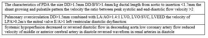
DD ductal diameter, BW body weight, LA left atria, AO aorta, LVO left ventricular output, LVEED left ventricular end diastolic diameter, SVC superior vena cava, LPA left pulmonary artery
The characteristics of patent ductus arteriosus
The size of PDA should be measured at the narrowest site by echocardiography, which is often at the end of the pulmonary [9]. When ductal diameter beyond 1.5mm, end-organ hypoperfusion occurs, the latent risk of hsPDA is decided, [10, 11]. Recently, the ratio of ductal diameter to body weight was also studied in relation to hsPDA. Extremely low birth weight infant with hsPDA can be with a ductal diameter indexed for body weight>1.4mm/kg, which is more sensitive value, because of the strong relationship with echocardiographic indices of increasing shunt volume[12,13]. However, regarding diameter alone as a marker of hemodynamic significance is not valid due to intar- and inter-observer variability[14]. Ductal closure requires ductal contraction and platelet aggregation, short ductus length leads to less constrictive area and has no benefit to platelet aggregation due to deficient blood flow. Researchers found in 2016 that preterm infants with hsPDA had shorter ductal length compared with those no hsPDA and ductal length from aortic to insertion<1>
Shunt is the most critical part of hemodynamic significant PDA. In 1997, researchers from Japan found that premature would undergo four shunt patterns after birth:pulmonary hypertension pattern (right to left shunt mostly), growing pattern (decreased right to left shunt and growing left to right shunt), pulsatile pattern (left to right shunt mostly with highlighted peak velocity in systolic period), closing pattern (continuous left to right shunt with no difference between systolic and diastolic velocity). Pulmonary hypertension pattern usually observed in early postneonatal life in the presence of high pulmonary vascular resistance. Pulsatile pattern was often seen between 2 and 3 days after birth. Premature infants with hsPDA usually experience the above four patterns in order, while infants with asymptomatic PDA changed from pulmonary hypertension pattern to closing pattern. The growing and pulsatile pattern imply the significant left to right shunt and are signals of hsPDA[16].
Velocity of shunt across PDA can be detected by echocardiography. The narrower ductus (restrictive shunt) is often associated with the greater systolic velocity across the ductus and the smaller the difference between the systolic and diastolic peak velocity. While wider ductus (non-restrictive shunt) often has relatively low peak systolic velocity and a bigger systolic to diastolic velocity gradient. When the ratio between peak systolic and end-diastolic flow velocity is beyond 2, the image of ultrasonography is as similar as the pulsatile flow pattern. Therefore, the ratio of >2 is a good predictor of hsPDA[17,18].
Pulmonary overcirculation
Pulmonary overflow caused by significant left-to-right shunt increase the preload of the left atria and largen the left atrial diameter. The aortic root is a relatively fixed structure and it dose not get dilated with left heart overloading. Therefore, the increased ratio of the LA and AO can indirectly reflects the extend of pulmonary venous return. Ductal diameter>1.5mm combining with LA/AO ratio>1.4:1 was used to determine hsPDA by clinicians previously, as beyond these cutoff, peripheral hypoperfusion and higher rate of ductal unsuitable shunt occurs [11,19,20]. Similarly, left ventricular output (LVO) and left ventricular end diastolic diameter (LVEDD) should be surrogate indices of pulmonary venous return. LVO and LVEDD should be considered to determine hsPDA. However, the markers mentioned above may become normal or low due to the inter-atrail left-to-right shunt with the existence of patent foramen ovale or septal defect.
Pulmonary circulation or shunt volume can be estimated by the ratio of left ventricular output(LVO) and superior vena cava(SVC). LVO represents the sum of systemic blood flow and the flow of SVC multiplied by 2.7 is regarded as the real systemic blood flow. Ductal flow can be evaluated by the formula of LVO-2.7×SVC. The higher the left ventricular output is, the bigger the volume of ductal shunt. According the equation above, when LVO/SVC ratio is beyond 4 , the volume of shunt through the patent DA is more than 50% of the total systemic blood flow[21]. Moreover, the LVO:SVC ratio have close relationship with a duct 3 mm in size or larger[22], which is further proved that the ratio of LVO:SVC can evaluate hsPDA well.
The velocity of left pulmonary artery can also be used as an indicator of assessing hsPDA. Normally, the flow through left pulmonary artery is absent during the diastolic period. The end-diastolic velocity of LPA increases as significant ductal shunt exists, with the cut-off value of 0.2m/s[23]. Moreover, comparing with other parameters, the left pulmonary artery end-diastolic velocity at 3 days of age may predict whether the PDA needs surgical closure in the future[24].
The period of ventricular diastole includes the period of isovolumic relaxation, ventricular filling and atrial systole. The mitral valve E/A stands for the ratio of the velocity of the early diastolic phase of ventricular filling and the late diastolic period of atrial contraction. In preterm, the mitral valve E/A is less than 1 due to the poor myocardial diastolic function. However, in the presence of hsPDA, atrial pressure and the velocity passing through mitral valve in phase of ventricular filling increase because of the loaded pulmonary venous return and the ratio E/A may changes, with the E/A ratio >1[25].
The most important but easily overlooked factor to evaluate pulmonary overcirculation is myocardial performance. The myocardium is immature with poor systolic and diastolic function in preterms. It mostly depend on left atrial contraction for ventricular filling due to the stiff myocardium. In preterms with hsPDA, increasing preload of left atrial, with compromised diastolic function will lead to increased left atrial pressure and pulmonary venous congestion in the end. The greater the shunt flow is, the more obvious the characteristics of left ventricle diastolic dysfunction result from large pulmonary venous return exacerbating the load of firm left ventricle. When it is beyond the threshold of LV diastolic capacity, pulmonary hypertension may occur. This may result in some significant clinical complication, such as pulmonary haemorrhage and eventually chronic lung disease. Therefore, left ventricle diastolic function provides basis for assessing hemodynamic significance of PDA[26,27].
Systemic hypoperfusion
Immature myocardium has poor ability to compensate shunt flow from systemic- to-pulmonary through duct by increasing output in the first postnatal life. Limited myocardium ability combined with high shunt volume may lead to systemic hypoperfusion. With the exist of shunt, descending aortic blood flow can be reduced by up to 35%[28,29]. Peripheral arteries blood flow can be assessed by Doppler. In normal physiological conditions, Doppler can detect antegrade blood flow in both systole and diastole. However, the low blood flow may be seen in systole and absent or reversed image in diastole in preterm infant with hsPDA. Researchers found decreased or even reversed diastolic flow in the descending aorta detected by echocardiograph is strongly associated with ductal shunt flow[30].
Diastolic pressure may decrease in infants with hsPDA due to significant left to right shunt. Blood flow supplying to heart is depend on the aortic diastolic pressure. In normal neonates, coronary artery perfusion increase in diastole. While, low coronary artery flow may be found in hsPDA leading to compromised myocardial function. It has been tested 40% coronary artery flow were added after PDA liation. Although this can not reach statistic significance, it infer coronary artery perfusion evaluated by Doppler can assess hsPDA indirectly[31].
Cerebral blood flow is influenced by systemic blood pressure in preterm newborn. Preterm infants with detectable PDA has a alternative cerebral perfusion. Because of the low cerebral artery vascular resistance, the diastolic component monitored by Doppler is evident[32]. While, studies of cerebral blood velocity in preterm with PDA showed a decreasing part in the end diastole. In the presence of PDA, blood velocity reduces or ceases in diastole in the anterior cerebral artery or middle cerebral artery, which will increases after PDA closure[33,34]. This change of cerebral circulation reflects the systemic hemodynamic effects of patent ductus arteriosus. This also explained increasing pulse pressure caused by PDA, coupling with immature autoregulation, preterm infants are vulnerable to get intra-ventricular hemorrhage. In addition to cerebral arteries, the renal arteries blood flow in preterms with hsPDA has also been described[35]. A series of small sample studies showed that symptomatic PDA is usually accompanied by changes in the Doppler waveform in the renal arteries[36,37,38]. Two cases of 25 week gestational age preterm have reported in detail that renal blood flow patterns change in Doppler with the alteration of hemodynamic situation of PDA(reversed end-diastolic wave in the presence of hsPDA and restored end diastolic wave with improvement of hsPDA)[29]. Although large numbers of studies have been executed, the validation to decide significance of PDA remains unknown as well.
Conclusion
In the past fifty years, more and more researchers have studied the predictive parameters of hsPDA to obtain early treatment and control the progression of disease as hsPDA has a strong relationship with many lethal complications in premature infants. This article shows comprehensive markers to evaluate hsPDA in the early postnatal period from three aspects. However, those trails are lack accuracy and specificity, which mostly depend on the diameter of PDA>1.5mm combined with LA/AO>1.4 to define the significance of PDA. In my center, there are large part of preterm using the definition above to diagnose hsPDA having a spontaneous closure, which do not bring cacoethic consequences. Because the hemodynamic significance of PDA assessed by single marker is over-simplified, some scientists have begun to use the scoring method (including multiple color ultrasound parameters mentioned in this article) to determine the severity of PDA and its negative impact on prognosis. We speculate that multi-parameter combination may be more comprehensively and accurately.What kind of combination is better to evaluate hsPDA needs further exploring.
References
- Poorva Deshpande. Patent ductus arteriosus: The physiology of transition. Seminars in Fetal and Neonatal Medicine (2018)
View at Publisher | View at Google Scholar - Joseph J. Vettukattil. Pathophysiology of Patent Ductus Arteriosus in the Preterm Infant. Current Pediatric Reviews, 2016, 12, 120-122
View at Publisher | View at Google Scholar - Bose CL, Laughon MM. Patent ductus arteriosus: lack of evidence for common treatments. Arch Dis Child Fetal Neonatal Ed (2007) 92: F498–502.
View at Publisher | View at Google Scholar - Heuchan AM, Clyman RI. Managing the patent ductus arteriosus: current treatment options. Arch Dis Child Fetal Neonatal Ed (2014) 99: F431–436.
View at Publisher | View at Google Scholar - Lee, J.A., Practice for preterm patent ductus arteriosus; focusing on the hemodynamic significance and the impact on the neonatal outcomes. Korean J Pediatr, 2019. 62(7): p. 245-251.
View at Publisher | View at Google Scholar - Zonnenberg, I. and K. de Waal, The definition of a haemodynamic significant duct in randomized controlled trials: a systematic literature review. Acta Paediatr, 2012. 101(3): p. 247-51.
View at Publisher | View at Google Scholar - W.P. de Boode. Role of neonatologist-performed echocardiography in the assessment and management of patent ductus arteriosus physiology in the newborn. Seminars in Fetal and Neonatal Medicine 23 (2018) 292–297.
View at Publisher | View at Google Scholar - A KHOSITSETH, P NUNTNARUMIT. Echocardiographic Parameters of Patent Ductus Arteriosus in Preterm Infants. INDIAN PEDIATRICS 48(2011)773-778.
View at Publisher | View at Google Scholar - Tschuppert, S., et al., The effect of ductal diameter on surgical and medical closure of patent ductus arteriosus in preterm neonates: size matters. J Thorac Cardiovasc Surg, 2008. 135(1): p. 78-82.
View at Publisher | View at Google Scholar - Evans, N. and P. Iyer, Longitudinal changes in the diameter of the ductus arteriosus in ventilated preterm infants: correlation with respiratory outcomes. Arch Dis Child Fetal Neonatal Ed, 1995. 72(3): p. F156-161.
View at Publisher | View at Google Scholar - Kluckow, M. and N. Evans, Early echocardiographic prediction of symptomatic patent ductus arteriosus in preterm infants undergoing mechanical ventilation. J Pediatr, 1995. 127(5): p. 774-9.
View at Publisher | View at Google Scholar - Gabriele D’Amato. Ductal size indexed to weight and body surface area correlates with morbidities in preterm infants ≤32 weeks.The Journal of Maternal-Fetal & Neonatal Medicine.17 Oct 2019.
View at Publisher | View at Google Scholar - El Hajjar M, Vaksmann G, Rakza T, et al. Severity of the ductal shunt: a comparison of different markers. Arch Dis Child Fetal Neonatal Ed. 2005;90(5): F419–F422.
View at Publisher | View at Google Scholar - Kunal Babla, et al. Repeatability of PDA diameter measurements on echocardiography. European Journal of Pediatrics. 29 June 2021.
View at Publisher | View at Google Scholar - Polat, T.B., I.H. Celik and O. Erdeve, Early predictive echocardiographic features of hemodynamically significant patent ductus arteriosus in preterm VLBW infants. Pediatr Int, 2016. 58(7): p. 589-594.
View at Publisher | View at Google Scholar - Su, B.H., et al., Echocardiographic assessment of patent ductus arteriosus shunt flow pattern in premature infants. Arch Dis Child Fetal Neonatal Ed, 1997. 77(1): p. F36-40.
View at Publisher | View at Google Scholar - Smith, A., et al., Peak systolic to end diastolic flow velocity ratio is associated with ductal patency in infants below 32 weeks of gestation. Arch Dis Child Fetal Neonatal Ed, 2015. 100(2): p. F132-136.
View at Publisher | View at Google Scholar - Singh, Y., et al., Echocardiographic Diagnosis and Hemodynamic Evaluation of Patent Ductus Arteriosus in Extremely Low Gestational Age Newborn (ELGAN) Infants. Front Pediatr, 2020. 8: p. 573627.
View at Publisher | View at Google Scholar - Johnson, G.L., et al., Echocardiographic characteristics of premature infants with patient ductus arteriosus. Pediatrics, 1983. 72(6): p. 864-871.
View at Publisher | View at Google Scholar - El-Saiedi, S., et al., Development of tissue Doppler-derived predictors of hemodynamically significant patent ductus arteriosus and the ability to incorporate it in targeted neonatal echocardiography protocol. J Saudi Heart Assoc, 2019. 31(2): p. 69-74.
View at Publisher | View at Google Scholar - El Hajjar M, Vaksmann G, Rakza T, Kongolo G, Storme L. Severity of the ductal shunt: a comparison of different markers. Arch Dis Child Fetal Neonatal Ed. (2005) 90:F419–422.
View at Publisher | View at Google Scholar - Sehgal A, Menahem S. Interparametric correlation between echocardiographic markers in preterm infants with patent ductus arteriosus. Pediatric cardiology 2013 Jun;34(5):1212-1217.
View at Publisher | View at Google Scholar - Suzmura H, Nitta A, Tanaka O. Diastolic flow velocity of left pulmonary artery of patent ductus arteriosus in preterm infants. Pediatr Int 2001;43:146–151.
View at Publisher | View at Google Scholar - Satoshi Masutani et al. Ductus diameter and left pulmonary artery end-diastolic velocity at 3 days of age predict the future need for surgical closure of patent ductus arteriosus in preterm infants: A post-hoc analysis of a prospective multicenter [25] Singh Y, Fraisse A. Echocardiographic diagnosis and hemodynamic evaluation of patent ductus arteriosus in extremely low gestational age newborn. Front Pedatr. 2020 Nov 8.
View at Publisher | View at Google Scholar - Jennifer L. Shepherd.What is a hemodynamically significant PDA in preterm infants? Congenital Heart Disease. 2019;14:21–26.
View at Publisher | View at Google Scholar - Afif EL-Khuffash. A Patent Ductus Arteriosus Severity Score Predicts Chronic Lung Disease or Death before Discharge.J Pediatr 2015.
View at Publisher | View at Google Scholar - Groves AM, Kuschel CA, Knight DB, Skinner JR. Does retrograde diastolic flow in the descending aorta signify impaired systemic perfusion in preterm infants? Pediatr Res. 2008;63:89–94.
View at Publisher | View at Google Scholar - Arvind Sehgal. Brendan Mulcahy.Pramod Pharande and Suraj Varma.The often forgotten systemic effects of ductus arteriosus: impact on decision-making and future trials.Journal of Perinatology. 22 July 2021.
View at Publisher | View at Google Scholar - Kathryn M. Broadhouse. Assessment of PDA shunt and systemic blood flow in newborns using cardiac MRI.NMR Biomed. 2013; 26: 1135–1141.
View at Publisher | View at Google Scholar - Arvind Sehgal. Coronary artery perfusion and myocardial performance after patent ductus arteriosus ligation. J Thorac Cardiovasc Surg 2012;143:1271-1278.
View at Publisher | View at Google Scholar - Pourcelot L: Diagnostic ultrasound for cerebral vascular disease, in Donald 1, and Levi S, editors: Present and future of diagnostic ultrasound, Rotterdam, 1976, Kooyker Scientific Publications, p 141.
View at Publisher | View at Google Scholar - F. J. Weir A. A patent ductus arteriosus is associated with reduced middle cerebral artery blood flow velocity. Eur J Pediatr(1999)158:484-487.
View at Publisher | View at Google Scholar - Jeffrey M. Perlman. The effect of patent ductus arteriosus on flow velocity in the anterior cerebral arteries: Ductal steal in the premature newborn infant. The Journal of Pediatrics November 1981 VoL 99, No. 5, pp. 767-771.
View at Publisher | View at Google Scholar - Senji Shimada.Effects of patent ductus arteriosus on left ventricular output and organ blood flows in preterm infants with respiratory distress syndrome treated with surfactant.J PEDIATR 1994;125:270-277.
View at Publisher | View at Google Scholar - Bomelburg T, Jorch G. Abnormal blood flow patterns in renal arteries of small preterm infants with patent ductus arteriosus detected by Doppler ultrasonography. Eur J Pediatr 1989; 148:660–664. 10.
View at Publisher | View at Google Scholar - Wong SN, Lo RN, Hui PW. Abnormal renal and splanchnic arterial Doppler pattern in premature babies with symptomatic patent ductus arteriosus. J Ultrasound Med 1990; 9:125–130.
View at Publisher | View at Google Scholar - Damien L. Armstrong et al.The Use of Renal Doppler Ultrasonography to Diagnose Patent Ductus Arteriosus in Preterm Infants.J Ultrasound Med 20:1005–1010, 2001.
View at Publisher | View at Google Scholar
