

Case Report | DOI: https://doi.org/10.31579/2834-8486/017
Double encephalocele in a four-year-old girl: A case report with literature review
MBBS, FCPS Consultant Neurosurgeon & Senior Registrar Department of Neurosurgery, Punjab Institute of Neurosciences, Lahore, Pakistan.
*Corresponding Author: Ahtesham Khizar, MBBS, FCPS Consultant Neurosurgeon & Senior Registrar Department of Neurosurgery, Punjab Institute of Neurosciences, Lahore, Pakistan.
Citation: Ahtesham Khizar, Hassaan Zahid, Manal Khan, Abdul Rahim Farooq, Muhammad Aqeel Nat. (2024), Double encephalocele in a four-year-old girl: A case report with literature review. Biomedical and Clinical Research, 3(1); DOI:10.31579/2834-8486/017
Copyright: © 2024, Ahtesham Khizar. This is an open-access article distributed under the terms of the Creative Commons Attribution License, which permits unrestricted use, distribution, and reproduction in any medium, provided the original author and source are credited.
Received: 11 January 2024 | Accepted: 29 January 2024 | Published: 15 February 2024
Keywords: encephalocele; meningocele;neural tube defects; folic acid deficiency; developing country
Abstract
Encephalocele is a congenital neural tube defect (NTD). The pathophysiology of the NTDs is exceedingly complex. Numerous explanations have been proposed to explain it. Double encephaloceles are highly unusual. There have only been fifteen previously reported cases of double encephalocele in the medical literature, with this index case being the oldest and first from Pakistan. A four-year-old girl presented with two occipital scalp swellings from infancy. The occipital swelling measured about 7x5x3 cm, while the suboccipital swelling measured about 7x9x5 cm. The skin over both the swellings was intact. Following a thorough history, physical examination, and radiological investigations, surgical excision and repair was performed. Postoperative recovery was uneventful. She did not develop hydrocephalus until the three month follow-up.Double encephalocele is a rare entity. The multisite closure theory appears to be the most plausible explanation for the development of multiple NTDs. The management of double encephalocele requires a case based approach.
Introduction
An encephalocele is a congenital neural tube defect (NTD) caused by failure of the cranial part of the developing neural tube to close, resulting in herniation of cranial contents via a defect in the skull.Encephaloceles are uncommon NTDs, affecting one in every 5,000 infants globally, with 70% being occipital.1 The pathophysiology of the NTDs is extremely complicated, including intricate interactions between genes, environment, and nutrition. Multiple hypotheses have been offered to explain neural tube formation using experimental models.2 Based on defect site, encephaloceles are classified as: i) occipital, ii) suboccipital, iii) sincipital (fronto-ethmoidal), iv) basal (trans-sphenoidal, trans-ethmoidal, spheno-ethmoidal, and spheno-orbital), and v) parietal.3 Double encephalocele is extremely rare; the majority of them involve the occipital or suboccipital region.4There are only fifteen previously reported cases of double encephalocele in the medical literature, and this index case is the oldest and the first to be reported from Pakistan. We present the following case in accordance with the CARE-guidelines.
Case Presentation
four years old girl came to us in December 2023 as an outpatient with a history of two occipital scalp swellings since birth. According to her mother, she was born at term in a small local hospital, and they did not seek additional medical care for her occipital swellings. On examination, the occipital swelling measured around 7x5x3 cm, whereas the suboccipital swelling measured about 7x9x5 cm. Overlying skin was intact over both the swellings, and a slight indentation was visible on the bottom edge of the occipital swelling. (Fig.1-A, B, C&D) A bony defect was palpable around the occipital swelling. Transillumination was negative in both the swellings. Computed tomography (CT) brain plain with bone window and magnetic resonance imaging (MRI) brain plain with MR venogram were performed. CT showed both the encephaloceles and the bony defects. (Fig.2-A&B) MRI brain showed soft tissue details (Fig.3-A, B&C) whereas MR venogram showed details of the dural venous sinuses. The patient underwent surgical excision and repair for both the encephaloceles.
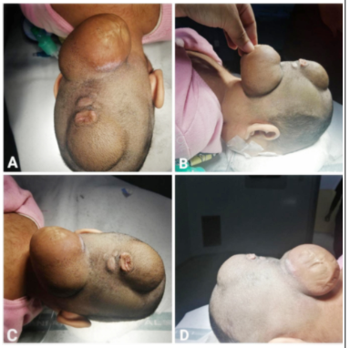
Fig.1: Double encephalocele, A&C: Superior views, B&D: Right lateral and left lateral views.
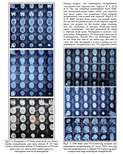
15 ml cystic fluid and applied a tight crepe bandage. There was no recurrent collection after that, and the bandage was removed on her second week of follow-up. She did not develop hydrocephalus until the three month follow-up.
Discussion
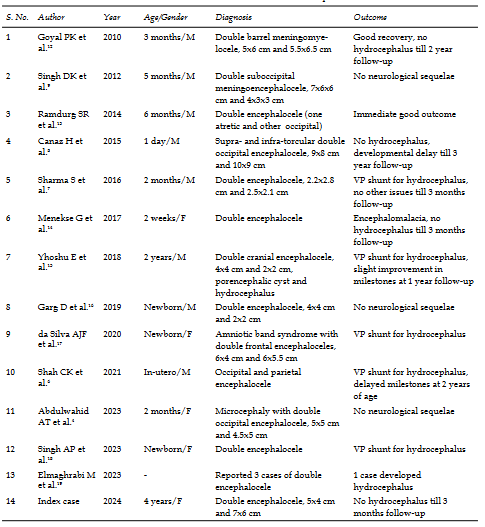
Encephaloceles are uncommon, with an incidence of 0.8 to 5 per 10,000 live births globally.6,7 Male and female carried the same incidence.6 Encephaloceles are typically solitary, with occipital encephaloceles being more common in general.6,8 Double encephaloceles are extremely uncommon, with only a few cases reported in the literature.6 Our literature review, conducted using the PubMed and Google Scholar databases, identified fifteen cases, as indicated in Table-I, with no cases reported on PakMediNet, making this the first case report of double encephalocele from Pakistan.Neural tube genesis and closure need complicated cellular, extracellular, and intracellular processes. There are two basic ideas on neural tube closure. The commonly recognized hypothesis is that neural tube closure occurs in a continuous, bidirectional process that begins in the mid-cervical region and advances in
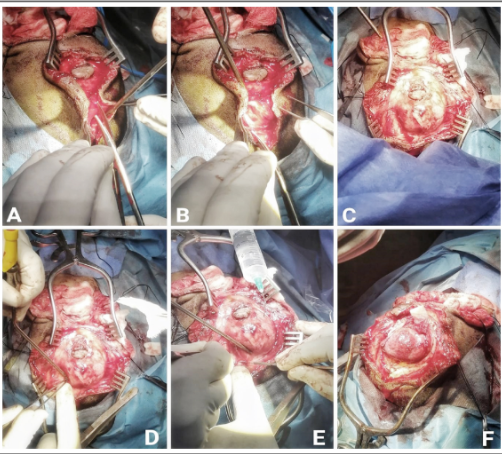
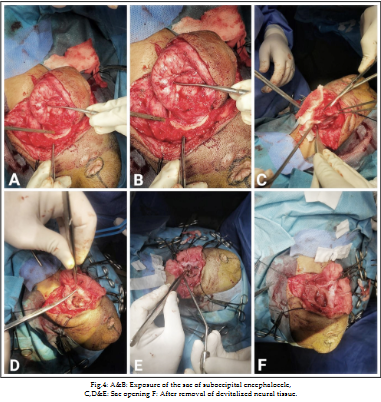
Fig.5: A&B: Dissection of occipital encephalocele, C,D&E: Sac exposure, F: Viable brain tissue inside sac.
a zipper-like pattern both rostrally and caudally, with the cranial and caudal neuropores closing last.
There are several flaws in this relatively rudimentary 'zipper concept'. This idea suggests that meningomyeloceles are more commonly seen at the most cranial or caudal ends, however it does not account for cervical meningomyelocele, multiple NTDs, or double encephalocele.9The findings of multiple meningoceles can be explained at different levels by the multisite closure theory put out by Van Allen et al.10 and Nakatsu et al.11 According to the multisite neural tube closure model, human normal neural tubes have several closure points, or "zippers". Presumably, one or more genes regulate these zippers; mutations in these genes would result in neural tube abnormalities in the vicinity of the affected zipper.9 This could explain why double NTDs develop in embryogenesis at different locations similar to our index case.The mainstay of treatment for encephalocele is surgical. This surgery consists of incising the sac, amputating the extra tissue to the level of the surrounding skull, dural closure, and skin closure. In general, infants born with an occipital encephalocele surrounding the brain have a poor prognosis. In addition to the contents of the sac, the extent of the lesion influences the long-term prognosis.6 In our case, parents of the girl were satisfied with the surgical treatment of their daughter.
Conclusion
Double encephalocele is a rare condition. The multisite closure theory appears to be the most plausible explanation for the development of multiple NTDs. The Double encephalocele in a four-year-old girl
requires unique solutions for each instance.Consent for publication: Consent was obtained from the father of the girl for publication of this case report and the accompanying images.
Conflicts of interest
None.
Grant Support & Financial Disclosures:
None.
References
- Zahid S, Khizar A.(2021). Giant occipital encephalocele: a case report, surgical and anesthetic challenge and review of literature. Egypt J Neurosurg;36(1):1-7. doi: 10.1186/s41984-021-00136-82.
View at Publisher | View at Google Scholar - Yadav JK, Khizar A, Yadav PK, Mustafa G, Bhatti SN.et.al. (2019). A case report of triple neural tube defect: revisiting the multisite closure theory. BMC Surg;19(1):164. doi:10.1186/s12893-019-0633-23.
View at Publisher | View at Google Scholar - Suwanwela C, Suwanwela N.(1972). A morphological classification of sincipital encephalomeningoceles. J Neurosurg. ;36(2):201-211. doi: 10.3171/jns.1972.36.2.0201Pak J Med Sci December Part-II 2024 Vol. 40 No. 12 PINS Supplement www.pjms.org.pk S864.
View at Publisher | View at Google Scholar - Abdulwahid AT, Al-Obaidi AD, Al-Obaidi MN, Hashim HT. (2023). Double encephalocele with an excellent outcome postoperatively: A case report from Iraq. eNeurologicalSci. 2023; 30:100449. doi: 10.1016/j.ensci.1004495.
View at Publisher | View at Google Scholar - Riley DS, Barber MS, Kienle GS, Aronson JK, von Schoen-Angerer T,et al. (2017). CARE guidelines for case reports: explanation and elaboration document. J Clin Epidemiol. 2017;89:218-235. doi: 10.1016/j.jclinepi.04.0266.
View at Publisher | View at Google Scholar - Shah CK, Lee RY, Jeph S. (2021). In-utero Diagnosis of Double Encephalocele - Imaging Features and Review of Literature. J Radiol Case Rep;15(12):1-9. doi: 10.3941/jrcr.v15i12.42307.
View at Publisher | View at Google Scholar - Sharma S, Ojha BK, Chandra A, Singh SK, Srivastava C.et.al.(2015). Parietal and occipital encephalocele in same child: A rarest variety of double encephalocele. Eur J Paediatr Neurol. 2016;20(3):493-496. doi: 10.1016/j.ejpn.2015.12.0088.
View at Publisher | View at Google Scholar - Canaz H, Ayçiçek E, Akçetin MA, Akdemir O, Alataş I. (2015). Supra- and infra-torcular double occipital encephalocele. Neurocirugia (Astur). 2015;26(1):43-47. doi: 10.1016/j.neucir.2014.09.0029.
View at Publisher | View at Google Scholar - Singh DK, Singh N, Kumar P.(2012). Double suboccipital meningoencephalocele: a unique case report. Pediatr Neurosurg. 2012;48(5):331-332. doi: 10.1159/00034888610.
View at Publisher | View at Google Scholar - Van Allen MI, Kalousek DK, Chernoff GF, et al.(1993). Evidence for multi-site closure of the neural tube in humans. Am J Med Genet;47(5):723-743. doi:10.1002/ajmg.132047052811.
View at Publisher | View at Google Scholar - Nakatsu T, Uwabe C, Shiota K. (2000). Neural tube closure in humans initiates at multiple sites: evidence from human embryos and implications for the pathogenesis of neural tube defects. Anat Embryol (Berl);201(6):455-466. doi: 10.1007/s00429005033212.
View at Publisher | View at Google Scholar - Goyal PK, Singh D, Singh H, Tandon M.(2010). Suboccipital double barrel twin meningocoele: Another new theory?. J Pediatr Neurosci;5(2):126-128. doi:10.4103/1817-1745.7610913.
View at Publisher | View at Google Scholar - Ramdurg SR, Gubbi S, Odugoudar A, Kadeli V.(2014). A rare case of split pons with double encephalocoele, dermal sinus tract, and lipomeningomyelocele: a case report and review of literature. Childs Nerv Syst;30(1):173-176. doi: 10.1007/s00381-013-2207-414.
View at Publisher | View at Google Scholar - Menekse G, Celik H, Bayar MA.(2017). Giant Parietal Encephalocele with Massive Brain Herniation and Suboccipital Encephalocele in a Neonate: An Unusual Form of Double Encephalocele. World Neurosurg;98:867.e9-867.e11. doi: 10.1016/j.wneu.2016.11.03015.
View at Publisher | View at Google Scholar - Yhoshu E, Dash V, Bawa M. (2018). Double Encephalocele: An Unusual Presentation. J Pediatr Neurosci;13(2):264-266. doi: 10.4103/jpn.JPN_22_1816.
View at Publisher | View at Google Scholar - Garg D, Singh AP, Tanger R, Gupta AK.(2019). Double encephalocele arising from single bone defect: A rare case. Journal of Clinical Neonatology;8(3):176-177. doi:10.4103/jcn.JCN_10_1917.
View at Publisher | View at Google Scholar - da Silva AJF, Silva CSME, Mariano SCR. (2020). Amniotic band syndrome with double encephalocele: A case report. Surg Neurol Int. 2020;11:448. doi: 10.25259/SNI_454_202018.
View at Publisher | View at Google Scholar - Singh AP, Kumar A, Barolia DK, Solanki N, Bathia HV. Double encephalocele: A rare neural tube defect. J Pediat Neurosci. 2023:10-4103. doi: 10.4103/jpn.JPN_116_2119.
View at Publisher | View at Google Scholar - Elmaghrabi M, Arab A, El Awady M, Mourad M.(2023). Management of encephalocele in infants: a 5-years retrospective study in Benha, Egypt. Benha Medical Journal;40(Special issue (Surgery)):235-245. doi: 10.21608/bmfj.2022.162432.
View at Publisher | View at Google Scholar
