

Review Article | DOI: https://doi.org/10.31579/2835-2882/058
Development of silk scaffolds for newer areas of applications in tissue engineering
- N. Gokarneshan *
- A. Jothimanikandan
- P. Periyasamy
- M. Ponmaniselvam
- K. Sridhar
Department of Textile Chemistry, SSM College of Engineering, Komarapalayam, Tamil Nadu, India.
*Corresponding Author: N.Gokarneshan, Department of Textile Chemistry, SSM College of Engineering, Komarapalayam, Tamil Nadu, India.
Citation: N.Gokarneshan, A.Jothimanikandan, P.Periyasamy, M.Ponmaniselvam and K.Sridhar, (2024), Development of silk scaffolds for newer areas of applications in tissue engineering, Clinical Research and Studies, 3(4); DOI:10.31579/2835-2882/058
Copyright: © 2024, N.Gokarneshan. This is an open access article distributed under the Creative Commons Attribution License, which permits unrestricted use, distribution, and reproduction in any medium, provided the original work is properly cited.
Received: 12 July 2024 | Accepted: 18 July 2024 | Published: 25 July 2024
Keywords: Silk, scaffold; musculoskeletal system; tissue engineering; regeneration
Abstract
During the past decade, various novel tissue engineering (TE) strategies have been developed to maintain, repair, and restore the biomechanical functions of the musculoskeletal system. Silk fibroins are natural polymers with numerous advantageous properties such as good biocompatibility, high mechanical strength, and low degradation rate and are increasingly being recognized as a scaffolding material of choice in musculoskeletal TE applications. This current systematic review examines and summarizes the latest research on silk scaffolds in musculoskeletal TE applications within the past decade. Scientific databases searched include PubMed, Web of Science, Medline, Cochrane library, and Embase. The following keywords and search terms were used: musculoskeletal, tendon, ligament, intervertebral disc, muscle, cartilage, bone, silk, and tissue engineering. Our Review was limited to articles on musculoskeletal TE, which were published in English from 2010 to September 2019. The eligibility of the articles was assessed by two reviewers according to prespecified inclusion and exclusion criteria, after which an independent reviewer performed data extraction and a second independent reviewer validated the data obtained. A total of 1120 articles were reviewed from the databases. According to inclusion and exclusion criteria, 480 articles were considered as relevant for the purpose of this systematic review. Tissue engineering is an effective modality for repairing or replacing injured or damaged tissues and organs with artificial materials. This Review is intended to reveal the research status of silk-based scaffolds in the musculoskeletal system within the recent decade. In addition, a comprehensive translational research route for silk biomaterial from bench to bedside is described in this Review.
1.Introduction
Musculoskeletal tissues, which include bone, cartilage, ligament/tendon, skeletal muscle, and intervertebral disc, are highly susceptible to injury or damage arising from degenerative changes, external force impingement, or sports-related activities. Within the clinic, growing concerns over the complications of autografting (e.g., donor site morbidity, infection increased surgery time) and allografting (e.g., graft rejection, limited quantity) as well as the limited availability and efficacy of these tissue repair options have prompted the development of various tissue engineering (TE) strategies [1-3]. TE is an interdisciplinary field that combines the principles of engineering and biomedical sciences for the development of biological substitutes that restore, maintain, or improve tissue function. Through the effective integration of biological scaffold materials, cells, and bioactive factors, the goal of replacing or supporting the function of defective or injured body parts is expected to be realized [4].
Silk fibroin (SF), a natural protein material that has been clinically used as a suture for decades, is now widely lucubrated and utilized in a variety of new biomedical applications including TE [5]. Silk fibers possess advantageous properties over most synthetic and natural fibers with a unique combination of toughness, biocompatibility, biodegradability, low immunogenicity, and thermal stability, which may better meet the requirements of musculoskeletal TE [6]. Silk from silkworms can be broadly categorized into mulberry and non-mulberry silk, depending on the food source of the worm [7]. The domesticated mulberry silk worm Bombyx mori is commonly cultivated through large-scale sericulture [8]. The non-mulberry silkworms, including Antheraea mylitta (A. mylitta), muga silkworm Antheraea assamensis (A. assamensis), oak silkworm Antheraea pernyi (A. pernyi), Philosamia ricini (P. ricini), and Samia cynthia ricini (S. cynthia ricini), produce silk that have a particular peptide recognition sequence: Arg-Gly-Asp (RGD) [8,9]. This is a cell attachment domain in extracellular matrix proteins recognized by integrins, which promote cell adhesion of various different cell types. Hence, non-mulberry silk biomaterials are superior over mulberry silks in TE applications [10]. Silk fibers with a triangular cross section are primarily composed of two proteins: The central protein known as silk fibroin is covered by a glue-like coating composed of another protein called sericin [11]. The earliest use of silk for suture materials was found to have significant biocompatibility issues, which provoke immunological reactions ranging from delayed hypersensitivity to acute and chronic inflammatory processes [12]. However, sericin-free fiber was later found to exhibit only a weak immunoreactivity, which greatly increases its application potential in the medical field [13,14]. In the past few years, SF materials and their derivatives have been the target of intensive research in the biomedical field.
Silk materials have numerous advantages in TE applications that are unmatched by other materials, including (1) increased biocompatibility suitable for cell adhesion and proliferation with less inflammatory responses in vivo; (2) enhanceable and modifiable mechanical properties with different silk fibroin solution concentrations and porosities to better meet target tissue requirements; (3) nontoxic degradation products and controllable biodegradability achieved through modification of the β-sheet structure; (4) excellent structural adjustability enabling the fabrication of a scaffold with desirable features for specific applications [15]. At present, the most widely used biodegradable implantable polymer materials include poly(lactic acid) (PLA), polyglycolic acid (PGA), and their copolymers, which can basically meet the application of scaffold materials in terms of degradability but are not as good as silk fibroin materials in terms of biocompatibility and cell adhesion [16]. In addition, silk materials also have unique advantages over other biopolymer materials. Collagens exhibit a variety of characteristics making them highly biocompatible and nontoxic, but their poor mechanical properties (Young’s modulus: 0.0018–0.046 GPa) make them play a minor role in the process of musculoskeletal regeneration [17]. Chitosan exhibits structural similarity to the extracellular matrix and has a hydrophilic surface that promotes cell adhesion, proliferation, and differentiation [18]. Chitosan alone lacks sufficient mechanical strength, which limits its application as a three-dimensional scaffold in musculoskeletal tissue engineering (MTE). Natural biopolymers have shown superiority in biomedical applications since they have proven to be most compatible with the native extracellular matrix (ECM). Despite the great compatibility with native ECM, all biopolymers were insufficient in delivering the desired performances in one or more aspects [19]. Relatively speaking, silk material is more balanced in properties and more suitable for MTE. By modifying the molecular structure and morphology of silk proteins through the use of certain organic solvents for processing or surface modification, we can further improve various aspects of the scaffold properties and function, thus expanding their applications in drug delivery and TE.
The following aspects have been considered
a) Improved mechanical properties
b) Increased biocompatibility
c) Controllable biocompatibility
d) Excellent structural processability
2. Morphollogical classification
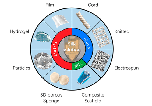
In recent years, many reports have been published on the application of silk materials in tissue engineering and drug delivery. Among them, the degummed silk fiber is usually reconstructed into different morphological types for increasing use in various applications. The mechanical properties as well as various primary features of silk can be modified through different processing methods. In general, varying conditions, such as silk concentration, methanol/salt treatment, pore size and porosity, processing temperature, etc., are capable of modulating the properties of the silk scaffold. Additionally, silk fibroin can also be physically blended or chemically cross-linked with various other complementary materials for the reinforcement of scaffolds and enhance their mechanical properties. The control of different morphological types of scaffolds therefore delivers the control of their specific characteristics, providing a pathway for the regeneration application in MTE. To obtain different types of scaffolds, degummed silk is dissolved into fresh SF solution, which can be used to fabricate into films, knitted scaffold, cords, 3D porous/sponge, hydrogels, electrospun fibers, particles, and composite scaffolds (Figure 1). The different characteristics of various morphological types of silk scaffolds are thus individually discussed.
The following types have been considered
a) Films [20,21]
b) Knitted scaffold [22]
c) 3D porous or spongs [23]
d) Hydrogel [24]
e) Electrospinning [25]
f) Composite scaffolds [26]
3. Areas of applications
a) Silk scaffolds in muscoskeletal tissue engineering [27]
The following aspects have been considered
i) Progress in the Utilization of Silk Scaffolds in Musculoskeletal TE over the Years
ii) Classification of Studies
b) Silk scaffolds in bone tissue engineering [28]
The following have been considered
i) Silk source
ii) Morphological Type of Scaffolds
iii) Cells
iv) Animal Models
v) Mechanical Stimuli
vi) Bioactive Factors
c) Silk Scaffolds in Ligament/Tendon Tissue Engineering [29]
The following have been considered
i) Silk Source
ii) Morphological Type of Scaffolds
iii) Cells
iv) Animal Models
v) Mechanical Stimuli
vi) Biological Factors
d) Silk Scaffolds in Cartilage Tissue Engineering [30]
The following aspects have been considered
i) Silk Source
ii) Morphological Type of Scaffolds
iii) Cells
iv) Animal Models
v) Mechanical Stimuli
vi) Biological Factors
e) Silk Scaffolds in Osteochondral Tissue Engineering [31]
f) Silk Scaffolds in Skeletal Muscle Tissue Engineering [32]
g) Silk Scaffolds in IVD Tissue Engineering [33]
4. Future perspectives
The following aspects have been considered [34 -45]
a) Controllable Parameters in Musculoskeletal Tissue Engineering
b) Limitations of Silk Scaffolds in Clinical Applications
c) How to Promote the Clinical Applications of Silk Scaffold
5. Conclusions
Musculoskeletal tissues, including bone, cartilage, tendon, ligament, and skeletal muscles, are easily damaged, which severely threatens human health. Tissue engineering is an effective modality to repair or replace damaged or injured tissues and organs with man-made material, and the disadvantages and problems of conventional surgical methods have further promoted the development and progress of tissue engineering. This Review intends to reveal the current research status of silk-based scaffolds for application in the musculoskeletal system within the past decade. The successful application of silk scaffolds in the musculoskeletal system proves the value of silk fibroin in tissue engineering, thus providing strong evidence to support the fabrication of silk scaffolds with superior performance and integrated designs. In addition, a comprehensive translational research route for silk biomaterials from bench to bedside is described in this study, providing a feasible route for researchers interested in or intending to take further steps for clinical translation of their own research work.
References
- Ma, P. X. (2004). Scaffolds for tissue fabrication. Mater. Today 7, 30– 40.
View at Publisher | View at Google Scholar - Langer, R; Vacanti, J. (1993). Tissue ngineering. Science 260, 920– 926.
View at Publisher | View at Google Scholar - Laurencin, C. T.; Ambrosio, A. M.; Borden, M. D.; Cooper, J. A. Jr. (1999). Tissue engineering: orthopedic applications. Annu. Rev. Biomed. Eng. 1, 19– 46.
View at Publisher | View at Google Scholar - Atala, A. (2012). Regenerative medicine strategies. J. Pediatr. Surg. 2012, 47, 17– 28.
View at Publisher | View at Google Scholar - Kundu, S. C.; Kundu, B.; Talukdar, S.; Bano, S.; Nayak, S.; et al. (2012). Invited review nonmulberry silk biopolymers. Biopolymers 97, 455– 467.
View at Publisher | View at Google Scholar - Ho, M. P.; Wang, H.; Lau, K. T. (2012). Effect of degumming time on silkworm silk fibre for biodegradable polymer composites. Appl. Surf. Sci., 258, 3948– 3955.
View at Publisher | View at Google Scholar - Kundu, B.; Kurland, N. E.; Bano, S.; Patra, C.; Engel, F. B.; et al. (2014). Silk proteins for biomedical applications: Bioengineering perspectives. Prog. Polym. Sci. 39, 251– 267.
View at Publisher | View at Google Scholar - Hardy, J. G.; Scheibel, T. R. (2009). Silk-inspired polymers and proteins. Biochem. Soc. Trans. 37, 677– 681.
View at Publisher | View at Google Scholar - Patra, C.; Talukdar, S.; Novoyatleva, T.; Velagala, S. R.; Mühlfeld, C.; et al. (2012). Silk protein fibroin from Antheraea mylitta for cardiac tissue engineering. Biomaterials 33, 2673– 2680.
View at Publisher | View at Google Scholar - Chen, J.; Altman, G. H.; Karageorgiou, V.; Horan, R.; Collette, A.; et al. (2003). Human bone marrow stromal cell and ligament fibroblast responses on RGD-modified silk fibers. J. Biomed. Mater. Res. 67a, 559– 570.
View at Publisher | View at Google Scholar - Kasoju, N.; Bora, U. (2012). Silk fibroin in tissue engineering. Adv. Healthcare Mater. 1, 393– 412.
View at Publisher | View at Google Scholar - Gong, X.; Liu, H.; Ding, X.; Liu, M.; Li, X.; et al. (2014). J. Physiological pulsatile flow culture conditions to generate functional endothelium on a sulfated silk fibroin nanofibrous scaffold. Biomaterials 35, 4782– 4791.
View at Publisher | View at Google Scholar - Kundu, B.; Rajkhowa, R.; Kundu, S. C.; Wang, X. (2013). Silk fibroin biomaterials for tissue regenerations. Adv. Drug Delivery Rev. 65, 457– 470.
View at Publisher | View at Google Scholar - Altman, G. H.; Diaz, F.; Jakuba, C.; Calabro, T.; Horan, R. L.; et al. Silk-based biomaterials. Biomaterials 24, 401– 416.
View at Publisher | View at Google Scholar - Wang, Y.; Kim, H. J.; Vunjaknovakovic, G.; Kaplan, D. L. (2006). Stem cell-based tissue engineering with silk biomaterials. Biomaterials 27, 6064– 6082.
View at Publisher | View at Google Scholar - Wenk, E.; Merkle, H. P.; Meinel, L. (2011). Silk fibroin as a vehicle for drug delivery applications. J. Controlled Release 150, 128– 141.
View at Publisher | View at Google Scholar - Koh, L. D.; Cheng, Y.; Teng, C. P.; Khin, Y. W.; Loh, X. J.; et al. (2015). Structures, mechanical properties and applications of silk fibroin materials. Prog. Polym. Sci. 46, 86– 110.
View at Publisher | View at Google Scholar - Wang, F.; Pang, Y.; Chen, G.; Wang, W.; Chen, Z. (2020). Enhanced physical and biological properties of chitosan scaffold by silk proteins cross-linking. Carbohydr. Polym. 229, 115529,
View at Publisher | View at Google Scholar - Wang, Y.; Wang, X.; Shi, J.; Zhu, R.; Zhang, J.; et al. (2016). M. A Biomimetic Silk Fibroin/Sodium Alginate Composite Scaffold for Soft Tissue Engineering. Sci. Rep. 6, 39477,
View at Publisher | View at Google Scholar - Jiang, C.; Wang, X.; Gunawidjaja, R.; Lin, Y. H.; Gupta, M. K.; et al. (2007). V. V. Mechanical Properties of Robust Ultrathin Silk Fibroin Films. Adv. Funct. Mater. 2229– 2237.
View at Publisher | View at Google Scholar - Lawrence, B. D.; Wharram, S.; Kluge, J. A.; Leisk, G. G.; Omenetto, F. G.; et al. (2010). Effect of hydration on silk film material properties. Macromol. Biosci. 10, 393– 403.
View at Publisher | View at Google Scholar - Wang, X.; Han, C.; Hu, X.; Sun, H.; You, C.; et al. (2011). Applications of knitted mesh fabrication techniques to scaffolds for tissue engineering and regenerative medicine. J. Mech Behav Biomed Mater. 4, 922– 932.
View at Publisher | View at Google Scholar - Wang, X.; Han, C.; Hu, X.; Sun, H.; You, C.; et al. (2011). Applications of knitted mesh fabrication techniques to scaffolds for tissue engineering and regenerative medicine. J. Mech Behav Biomed Mater. 4, 922– 932.
View at Publisher | View at Google Scholar - Kluge, J. A.; Rosiello, N. C.; Leisk, G. G.; Kaplan, D. L.; Dorfmann, A. L. (2010). The consolidation behavior of silk hydrogels. J. Mech Behav Biomed Mater. 3, 278– 289.
View at Publisher | View at Google Scholar - Shan, Y. H.; Peng, L. H.; Liu, X.; Chen, X.; Xiong, J.; et al. (2015). Silk fibroin/gelatin electrospun nanofibrous dressing functionalized with astragaloside IV induces healing and anti-scar effects on burn wound. Int. J. Pharm. 479, 291– 301.
View at Publisher | View at Google Scholar - Sahoo, S.; Toh, S. L.; Goh, J. C.(2010). PLGA nanofiber-coated silk microfibrous scaffold for connective tissue engineering. J. Biomed. Mater. Res., Part B 95B, 19– 28.
View at Publisher | View at Google Scholar - Yang, M.; Zhou, G.; Castano-Izquierdo, H.; Zhu, Y.; Mao, C. (2015). Biomineralization of Natural Collagenous Nanofibrous Membranes and Their Potential Use in Bone Tissue Engineering. J. Biomed. Nanotechnol. 11, 447– 456.
View at Publisher | View at Google Scholar - Gupta, P.; Adhikary, M.; Joseph, C. M.; Kumar, M.; Bhardwaj, N.; et al. (2016). Biomimetic, Osteoconductive Non-mulberry Silk Fiber Reinforced Tricomposite Scaffolds for Bone Tissue Engineering. ACS Appl. Mater. Interfaces 8, 30797,
View at Publisher | View at Google Scholar - Goh, J. C.; Ouyang, H. W.; Teoh, S. H.; Chan, C. K.; Lee, E. H.(2003). Tissue-engineering approach to the repair and regeneration of tendons and ligaments. Tissue Eng. 9 (Suppl 1), S31– S44
View at Publisher | View at Google Scholar - Makris, E. A.; Gomoll, A. H.; Malizos, K. N.; Hu, J. C.; Athanasiou, K. A.(2015). Repair and tissue engineering techniques for articular cartilage. Nat. Rev. Rheumatol. 11, 21– 34,
View at Publisher | View at Google Scholar - Ding, X.; Zhu, M.; Xu, B.; Zhang, J.; Zhao, Y.; et al. (2014). Integrated trilayered silk fibroin scaffold for osteochondral differentiation of adipose-derived stem cells. ACS Appl. Mater. Interfaces 6, 16696– 16705,
View at Publisher | View at Google Scholar - Manchineella, S.; Thrivikraman, G.; Khanum, K. K.; Ramamurthy, P. C.; Basu, B.; et al. (2016). Pigmented Silk Nanofibrous Composite for Skeletal Muscle Tissue Engineering. Adv. Healthcare Mater. 5, 1222– 1232,
View at Publisher | View at Google Scholar - Ghorbani, M.; Ai, J.; Nourani, M. R.; Azami, M.; Bordbar, S.(2017). Injectable natural polymer compound for tissue engineering of intervertebral disc: In vitro study. Mater. Sci. Eng., C 2017, 80, 502,
View at Publisher | View at Google Scholar - (2021). ACS Biomater. Sci. Eng. 7, 3, 817–840.
View at Publisher | View at Google Scholar - N.Gokarneshan,(2017). Adances in silk research, Woodhead Publishing India,
View at Publisher | View at Google Scholar - N.Gokarneshan,(2023). Development of Medical Textiles from Sericin Coated Polyester Materials
View at Publisher | View at Google Scholar - N.Gokarneshan and S.Mrudhulla,(2024). Nanomaterials in functional textiles, Handbook of Nanomaterials.
View at Publisher | View at Google Scholar - N.Gokarneshan and D.Maria Jenita,(2023). Development of Artificial Skin Scaffold Using Electrospun Nano Fibres for Wound Healing, Mathew’s journal of case reports.
View at Publisher | View at Google Scholar - N.Gokarneshan et.al.,(2021). Mechanical properties of textile sutures for orthopaedic surgery, GSC Advanced research and reviews, 07(03), 072–074.
View at Publisher | View at Google Scholar - Gokarneshan N,Ratna U.(2021). Development of a Textile Suturewith Improved Properties. Invest GynecolRes Women’s Health. 4(2). IGRWH.000582.
View at Publisher | View at Google Scholar - N. Gokarneshan1 and V. Krishna kumar,(2020). Some Significant Advances in Antibacterial Treatment of Textile Materials, Applied Medical Research, NO. 2, PAGE 1 – 6.
View at Publisher | View at Google Scholar - N Gokarneshan, Anish khan. (2020). Antimicrobial Properties of Bamboo Fibres. Biomed J Sci & Tech Res 32(5)- BJSTR. MS.ID.005309.
View at Publisher | View at Google Scholar - N Gokarneshan, U Dhatchayani. (2018). Performance Evaluation of Newer Types of Silk Surgical Sutures. OAJ Gerontol & Geriatric Med. 4(2): 555631.
View at Publisher | View at Google Scholar - N.Gokarneshan, (2019). Advances in jute research, Woodhead publishing India, 2019.
View at Publisher | View at Google Scholar - N.Gokarenshan and N.Jegadeesan,(2009). Synthesising of spider silk – A new era, Asian textile journal.
View at Publisher | View at Google Scholar
