

Research | DOI: https://doi.org/10.31579/2835-835X/088
Comparative Data on the Effect of the Venom of the Viper (Macrovipera Lebetina Obtusa Linnaeus, 1758) On the Composition of Blood Elements in Experimental Animals
1Institute of Zoology of the Ministry of Science and Education of the Republic of Azerbaijan.
2Research Center of Azerbaijan Medical University.
3Tokat Gaziosmasha University, Department of basic medical sciences/Physiology, Turkey.
4School of Education, general Education Program, ADA University, Azerbaijan.
5Azerbaijan Medical University, Baku.
*Corresponding Author: Sh.A. Topchiyeva, Institute of Zoology of the Ministry of Science and Education of the Republic of Azerbaijan
Citation: Sh.A. Topchiyeva, R.E. Jafarova, l.Z. Yildiz, A. Gojayev, R.Z. Karimova, (2025), Comparative Data on the Effect of the Venom of the Viper (Macrovipera Lebetina Obtusa Linnaeus, 1758) On the Composition of Blood Elements in Experimental Animals, Clinical Trials and Case Studies, 3(5); DOI:10.31579/2835-835X/088
Copyright: © 2025, Sh.A. Topchiyeva. This is an open-access artic le distributed under the terms of the Creative Commons Attribution License, which permits unrestricted use, distribution, and reproduction in any medium, provided the original author and source are credited.
Received: 24 November 2025 | Accepted: 01 December 2025 | Published: 08 December 2025
Keywords: viper venom; Macrovipera lebetina obtusa Linnaeus; leukocytes; lymphocytes; erythrocytes; platelets.
Abstract
The purpose of the presented work is to study the effect of microdoses of viper venom on the composition of blood cells.
Materials and methods. Experiments were carried out on 48 white outbred laboratory rats, weighing in the range of 200-220 g, which were intraperitoneally injected with viper poison (Macrovipera lebetina obtusa Linnaeus, 1758) at a dose of 0.01 mg/kg body weight.
Research results and discussion. As a result, after the introduction of the poison, the content of leukocytes in the blood of animals exceeded intact values after 30 minutes by 2.7%. Further, its content in the blood continued to increase and already exceeded IZ by 5.9%. Analysis of daily blood showed that the content of leukocytes in the blood began to decrease, tending to intact values, exceeding them slightly by 1.6%. The content of lymphocytes in the blood increased in the first 30 minutes and an hour after the administration of the poison by 1.0% and 2.5%, respectively, and then gradually decreased, exceeding the intact values by only 1.0%. The content of erythrocytes in the blood, on the contrary, during the entire period of research decreased by 2.7%, 4.1% and 7.8%, respectively. The platelet content in the blood in the first 30 minutes decreased by 1.1%, then, after an hour, the balance was practically restored, falling short of intact values by only 0.4%, and in a daily blood test it was 1.3% higher than intact values.
Conclusion. The results of laboratory studies showed that there is a slight fluctuation in the content of leukocytes, lymphocytes, erythrocytes, and platelets in the blood, which do not pose a threat to the animal’s body. Intraperitoneal administration of microdoses of viper venom leads to quantitative changes in the composition of blood cells. These changes are insignificant and are not statistically confirmed. Intraperitoneal administration of microdoses of viper venom 0.01 mg viper venom per kg body weight to white laboratory rats does not pose a serious threat to life and health.
Introduction
The study of poisonous biosynthesis products of snakes, which are a unique group of biologically active substances in their chemical composition and physiological action, is one of the important problems of modern medical and biological science. The study of the effect of the venom of the viper (Macrovipera lebetina obtusa Linnaeus, 1758) on the composition of blood cells is an important area of scientific research that has medical significance in connection with the understanding of the mechanisms of poisoning and the development of treatment methods. In this review, we will consider the main aspects of this topic, including the composition of the poison, the mechanisms of action on the body and its effect on the formed elements of the blood.
The viper (Macrovipera lebetina) is a venomous snake found in various regions of Eurasia and Africa. Its subspecies, Macrovipera lebetina obtusa, is common in a number of countries in Central Asia and the Middle East [1-3]. It belongs to the viper family (Viperidae) and is characterized by the presence of venom, which is used for hunting prey and in self-defense. Viper's venom consists of a complex of biologically active substances, including enzymes, toxins, anticoagulants and other components. The main components of the poison are phospholipases, as well as neurotoxins and hemotoxins, which affect the nervous system and circulatory system [3-5].
When a viper is bitten, the poison enters the human or animal body through the circulatory system. The components of the venom interact with tissue structures and biological molecules, causing a variety of pathological effects. Proteases and phospholipases destroy tissue, which leads to the development of necrosis at the bite site. Neurotoxins can cause paralysis and breathing problems, and hemotoxins can cause blood clotting and bleeding disorders. In cases of viper poisoning, a disturbance in the composition and functions of blood cells is observed. Hemotoxins can cause hemolysis (destruction of red blood cells), leading to anemia and an increased risk of bleeding. Neurotoxins and other venom components can affect platelets and their functions, which can lead to bleeding disorders and thromboembolic complications [6-8].
In medicine, the venom of the Caucasian viper, also known as Vipera kaznakovi, is usually not used directly due to its toxicity and danger. However, the composition and properties of the venom of other snakes can be the subject of research for the development of drugs. Some components of snake venoms may have potential in medical science, for example in the development of antivenoms, as well as in studying the mechanisms of action on the nervous system and muscles. In some cases, snake venom extracts are used for research in the fields of biochemistry, pharmacology and neurobiology. However, the use of snake venom in medicine requires a very careful approach due to its toxicity.
The development and use of drugs based on snake venoms requires extensive research and clinical trials to ensure their effectiveness and safety for patients. Therefore, it is necessary to identify the balance between toxicity and pharmacological effect of this natural biologically active substrate. That is, find a dose at which the pronounced pharmacological effect will not be offset by the toxic effect of the poison.
Based on the above, we considered it appropriate to study the effect of microdoses of poison on the composition of blood cells.
Materials and Methods
The experiments were carried out on 48 white outbred laboratory rats. Criteria for inclusion of animals in the experiment: sexually mature age, weight in the range of 200-220 g, coat without damage, gender - males. All animals used in the experiments were kept under the same conditions of care and diet. All animal experiments were carried out in accordance with the European Convention for the Protection of Vertebrate Animals Used for Experimental and Other Scientific Purposes, Strasbourg, 1986.
The animals were divided into 4 groups of 12 animals each; Group 1 consisted of animals in an intact state. In the remaining groups, animals received viper poison at a dose of 0.01 milligrams per kilogram of body weight of the test animal. Experimental animals were decapitated after 30 minutes, 1 hour, 24 hours, followed by blood sampling for laboratory studies.
Laboratory blood tests were carried out using an Auto Hematology Analyzen Ratyo RT -7600 device, made in China, 2019. 20 indicators were determined in the blood of animals, but in these studies the content of erythrocytes, leukocytes, lymphocytes and platelets in the blood was assessed as the most informative indicators.
The research material was whole venom of the Transcaucasian viper (Macrovipera lebetina obtusa), dried in a desiccator over calcium chloride vapor and samples of venom dried in 2015. The poisonous secretion of the Transcaucasian viper was dried under standard conditions in a desiccator over calcium chloride at room temperature for at least 10-12 days. Next, the crystalline poison was collected and analyzed. Samples of viper venom were stored in glass containers in the refrigerator at a temperature of +5-60 C. With this method of drying and storage, the poison retained its biological activity for at least 3 years. The poison used in this work was prepared in 2015. According to experts, storage and harvesting conditions guarantee the safety of biological activity. Dose – 0.01 milligrams of poison per kilogram of body weight of the test animal.
Statistical analysis of quantitative data was carried out using nonparametric methods - the Wilcoxon-Mann-Whitney test. When studying the dependencies between indicators, the Spearman formula for the rank correlation coefficient was applied:
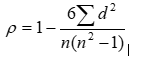
where: d – rank difference, n – number of ranks.
When applying the above methods, MS EXCEL and S-PLUS programs were used for statistical processing.
Research Results and Discussion
Integral indicators, including data on changes in weight, food and water consumption, assessment of the general condition of the animals’ motor activity and respiratory rate showed that after the administration of poison, the animals showed restless behavior in the first 10 minutes, then calmed down and behaved according to the species characteristics. The results of laboratory studies (Table 1) showed that there is a slight fluctuation in the content of leukocytes, lymphocytes, erythrocytes, and platelets in the blood, which do not pose a threat to the body.
Groups
Indicators | Intact animals | Against the background of poison administration | ||
After 30 minutes | After 1 hour | After 24 hours | ||
1st group | 2nd group | 3rd group | 4th group | |
Leukocytes 109/L | 15,87(12,0-22,6) | 16,3(12,0-22,7) | 16,8(12,0-20,9) | 16,13(11,6-20,6) |
% |
| 2,7 | 5,9 | 1,6 |
P |
| 0,87 | 0,344 | 0,531 |
Lymphocytes 109/L | 8,39(6,3-11,3) | 8,47(6,4-11,2) | 8,6(6,3-11,6) | 8,47(5,9-12,0) |
% |
| 1,0 | 2,5 | 1,0 |
P |
| 0,89 | 0,832 | 0,736 |
Red blood cells 109/L | 7,06(6,4-8,3) | 6,87(6,0-8,4) | 6,77(6,0-8,0) | 6,51(5,7-7,8) |
% |
| -2,7 | -4,1 | -7,8 |
P |
| 1,000 | 0,221 | 0,069 |
Platelets 109/L | 833,8(710-988) | 825,0(730-980) | 837,0(739-980) | 845,0(743-991) |
% |
| -1,1 | 0,4 | 1,3 |
P |
| 0,903 | 0,912 | 1,000 |
Table 1: Changes in the blood picture after intraperitoneal administration of viper venom at a dose of 0.01 mg per 1 kg of animal weight
From the data presented in the table it is clear that in the second group the leukocyte content increased to 16.3x10⁹/L with minimum values (Min) of 12.0x10⁹/L and maximum values (Max) of 22.7x10⁹/L from 15.877x10⁹/L at Min =12.0x10⁹/L and Max =22.6x10⁹/L in animals in the intact state (IS). The number of lymphocytes in this group also increased very slightly to 8.47x10⁹/L with Min =6.4x10⁹/L and Max =11.2x10⁹/L from 8.39x10⁹/L with Min =6.3x10⁹/L and Max =11 ,3 x109/L in IZ. Red blood cells decreased to 6.87 x 10⁹/L with Min = 6.0 x 10⁹/L and Max = 8.4 x 10⁹/L from 7.06 x 10⁹/L with Min = 6.4 x 10⁹/L and Max = 8.3 x 10⁹/L in IZ. Platelets in the blood of animals of this group decreased to 825.0 x 10⁹/L with Min = 730.0 x 10⁹/L and Max = 980.0 x 10⁹/L from 833.8 x 10⁹/L with Min = 710.0 x 10⁹/L and Max = 988 .0 x109/L in IS.
In the third group, the leukocyte content continued to increase, reaching 16.8 x10⁹/L at Min = 12.0 x10⁹/L and Max = 20.9 x10⁹/L, the lymphocyte content also increased, reaching 8.6 x10⁹/L at Min =6, 3x10⁹/L and Max =11.6 x10⁹/L, the content of erythrocytes decreased even more and reached 6.77x10⁹/L with Min =6.0x10⁹/L and Max =8.0 x10⁹/L, platelets increased to 837, 0x109/L at Min =739.0x10⁹/L and Max =980.0x10⁹/L.
In the fourth group, the content of all studied indicators with the exception of platelets decreased, reaching leukocytes - to 16.13 x10⁹/L at Min = 11.6 x10⁹/L and Max = 20.6 x10⁹/L, lymphocytes - to 8.47 x10⁹/L at Min = 5.9 x 10⁹/L and Max = 12.0 x 10⁹/L, red blood cells – up to 6.51 x 10⁹/L with Min = 5.7 x 10⁹/L and Max = 7.8 x 10⁹/L, platelets – increased to 845 .0x109/L at Min =743.0x10⁹/L and Max =991.0x10⁹/L.
Analysis of the obtained data in dynamics (graph) showed that after the introduction of poison, the content of leukocytes in the blood of animals exceeded the IS after 30 minutes by 2.7%, P = 0.87. Further, its content in the blood continued to increase and already exceeded IS by 5.9% at P = 0.344. Analysis of 24-hour blood showed that the content of leukocytes in the blood began to decrease, tending to IS, exceeding them slightly by 1.6% at P = 0.531.
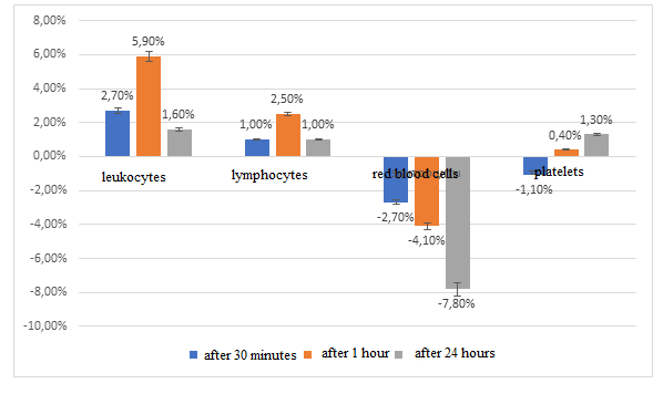
Figure: Changes in the content of formed elements in the blood after intraperitoneal injection of 0.01 mg of viper venom per kg of body weight into rats
The content of lymphocytes in the blood increased in the first half and hour of administration, respectively, by 1.0% (P = 0.89) and 2.5% (P = 0.832), and then gradually decreased, exceeding IS by only 1.0% (P =0.736). The content of erythrocytes in the blood, on the contrary, during the entire study period decreased by 2.7% (P = 1.00), 4.1% (P = 0.221) and 7.8% (P = 0.069), respectively.
The platelet content in the blood in the first 30 minutes decreased by 1.1% at P = 0.903, then, after an hour, the balance was practically restored, falling short of the IS by only 0.4% at P = 0.912, and in the daily blood test it was higher IS by 1.3% at P=1.00.
As can be seen from the results, the content of leukocytes in the blood - indicators of the inflammatory process - increases with the introduction of microdoses of viper venom, which can be explained by general intoxication of the body, as a result of which inflammatory mediators are activated. And the blood, as one of the most reactive systems of the body, responds to this by increasing the activity of immunocompetent systems, which include blood leukocytes and lymphocytes [9]. The synthesis of erythrocytes against the background of increased synthesis of other blood cells is somewhat reduced [9]. And the indicator of a hidden inflammatory process and tissue damage - the content of platelets in the blood [9], starting from a period of 1 hour after the administration of the poison, increases and the process continues throughout the entire observation period, which indicates the protracted nature of intoxication. However, all identified changes in the content of formed elements in the blood in percentage terms are insignificant and are not statistically confirmed.
Thus, intraperitoneal administration of microdoses of viper venom leads to quantitative changes in the composition of blood cells. These changes are insignificant and are not statistically confirmed. Intraperitoneal administration of viper venom at a dose of 0.01 mg per kg of body weight to white laboratory rats does not pose a serious threat to life and health.
Conclusion
1. It was experimentally revealed that after intravenous administration of viper venom at a dose of 0.01 mg/kg body weight after 30 minutes, 1 hour and 24 hours, there was a change in the percentage of leukocytes in the blood of experimental animals from 2.1%, 4.77% to 1.5%, respectively.
2. It was found that after intravenous administration of viper venom at a dose of 0.01 mg/kg body weight after 30 minutes, 1 hour and 24 hours, there was a change in the percentage of lymphocytes from 0.22%, 2.61% to 0.21%, respectively.
3. It was revealed that after intravenous administration of viper venom at a dose of 0.01 mg/kg body weight after 30 minutes, 1 hour and 24 hours, there was a change in the percentage of erythrocytes in the blood of rats from 1.15%, 5.27% to 7.63%, respectively.
4. It was established that the change in the percentage of platelets after i.p. administration of zootoxin at a dose of 0.01 mg/kg body weight after 30 minutes, 1 hour and 24 hours corresponded to 0.45%, 0.02%, 0.71%.
The study of the effect of viper venom on the composition of blood cells is an important aspect of research in the field of toxicology and medicine. Understanding the mechanisms of venom action helps develop effective treatments for poisonous snake envenomations and provide medical care for snakebite victims.
References
- Kasheverov I., Kudryavtsev D., Shelukhina I., Nikolaev G., Utkin Y., Tsetlin V.. Marine Origin Ligands of Nicotinic Receptors: Low Molecular Compounds, Peptides and Proteins for Fundamental Research and Practical Applications. в журнале Biomolecules, издательство MDPI (Basel, Switzerland), 2022,vol. 12, № 2, p. 189
View at Publisher | View at Google Scholar - Topciyeva Sh.A (2000). Influence of venom transcaucasian adder (vipera lebetina obtusa) on metabolic conversion of Hepatic and renal proteins of white mice / Билги, Баку, 2000, No 1, с. 38-40.
View at Publisher | View at Google Scholar - Pereira Teixeira C. D F., Fernandes Cristina Maria, Zuliani Juliana Pavan, Zamuner Silvia Fernanda Inflammatory effects of snake venom metalloproteinases. Mem Inst Oswaldo Cruz, Rio de Janeiro, 2005, Vol. 100, p.181-184
View at Publisher | View at Google Scholar - Kukushkin O.V., Bakiev A.G., Malenev A.L (2012). Some biochemical characteristics of the venom of the steppe viper from the Feodosia steppes (Crimea) // News of the Samara Scientific Center of the Russian Academy of Sciences. 2012, v. 14, no. 1, p. 158-161
View at Publisher | View at Google Scholar - Tan N.H.,Fung S.E. Snake venom L-aminoacid axidases. Handbook of venoms and toxins of reptiles.Ch. 10. Boca Raton; London; New York., CRC Press, Taylor, Francis Group, 2010,p.221-237.
View at Publisher | View at Google Scholar - Dubovskii P.V., Ignatova A. A., Alekseeva A. Set al (2022). Membrane-Disrupting Activity of Cobra Cytotoxins Is Determined by Configuration of the N-Terminal Loop. в журнале Toxins, издательство MDPI (Basel, Switzerland), 2022, vol. 15, №
View at Publisher | View at Google Scholar - Dubovskii P. V., Dubova K. M., Bourenkov G.et al (2022). Variability in the Spatial Structure of the Central Loop in Cobra Cytotoxins Revealed by X-ray Analysis and Molecular Modeling. Toxins, MDPI (Basel, Switzerland). vol. 14, № 2 DOI
View at Publisher | View at Google Scholar - Dyachenko I.A., Palikova Yu A., Palikov V.A., et al (2022). Alpha-Conotoxin RgIA and oligoarginine R8 in the mice model alleviate long-term oxaliplatin induced neuropathy в журнале Biochimie, Elsevier BV (Netherlands)vol. 194, p. 127-136 DOI
View at Publisher | View at Google Scholar - Dmitrenko M.M., Osyko Y.A., Kuligin A.V (2013). The role of the ratio of blood cells in the syndrome of endogenous intoxication // Bulletin of modern clinical medicine, 2013, Volume 6, issue. 5, p145-149
View at Publisher | View at Google Scholar
