

Case Report | DOI: https://doi.org/Bılateral Renal-Perırenal Lymphangıomatosıs: Ct an
Bılateral Renal-Perırenal Lymphangıomatosıs: Ct and Mrı Fındıngs
- Hasan Aydin *
- Yasin Ozdemir
Hasan Aydin, SB. Ankara Oncology Res. Hospital Radiology Department.
*Corresponding Author: Hasan Aydin, SB. Ankara Oncology Res. Hospital Radiology Department.
Citation: Yasin Ozdemir, Hasan Aydin, (2024), Bılateral Renal-Perırenal Lymphangıomatosıs: Ct and Mrı Fındıngs, International Journal of clinical and Medical Case Reports, 3(1); Doi:10.31579/2834-8664/038
Copyright: © 2024, Hasan Aydin. This is an open-access article distributed under the terms of the Creative Commons Attribution License, which permits unrestricted use, distribution, and reproduction in any medium, provided the original author and source are credited.
Received: 03 January 2024 | Accepted: 18 January 2024 | Published: 26 January 2024
Keywords: renal-lymphangioma-ct-mrı
Abstract
Renal lymphangiectasia is a rare, benign developmental disease of the kidney characterized by abnormal enlargement of the intrarenal and/or perirenal lymphatics.
Diagnosis of Lymphangiomatosis can be done by CT and MRI.
Here, We report a case of bilateral intrarenal and perirenal lymphantiectasia with CT and MRI findings.
Introduction
Renal lymphangiectasia is a rare, benign developmental disease of the kidney characterized by abnormal enlargement of the intrarenal and/or perirenal lymphatics. This condition, often asymptomatic, can be congenital or acquired, it may be due to renal failure and/or hypertension in undiagnosed patients[1] Diagnosis of Lymphangiomatosis can be done by cross-sectional imaging methods, Computerized Tomography(CT) and Magnetic Resonance Imaging (MRI) without any need for an invasive procedure[2]. In this case, we presented the CT and MRI findings of the patient who was diagnosed with bilateral intrarenal and perirenal lymphantiectasia.
Case Report
57-year-old male patient was under follow-up due to a previous colon tumor and surgery. In his routine Contrast-enhanced Abdominopelvic CT: Multiple bilateral perirenal and intramedullary cysts were observed. The cysts did not compress the renal cortex, surrounded the cortex and perirenal fatty tissue, were in conglomerated appereance and showed a hypodense surrounding feature with an average density of 8.5 HU. Bilateral kidney sizes and parenchymal thicknesses were in normal range, bilateral corticomedullary separations in both kidneys were clearly performed (Figure.1)
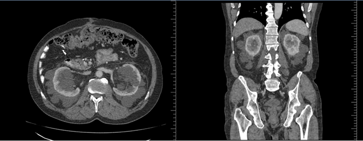
Figure 1: a-b: Renal and perirenal Lymphangiomas in the axial and coronal contrast enhanced CT
Dynamic abdominal MRI was requested from the patient in order to exclude possible cystic metastases of colon tumor. Lesions were diagnosed as "renal lymphangiomatosis in MRI those were hyperintense on T2W images, without any diffusion restrictions on DW images, and without any enhancement in the postcontrast acquisitions. There was a discrete connection between the intrarenal and perirenal lymphangiomas which was thought to be a lymphatic drainage and leakage (Figure .2)
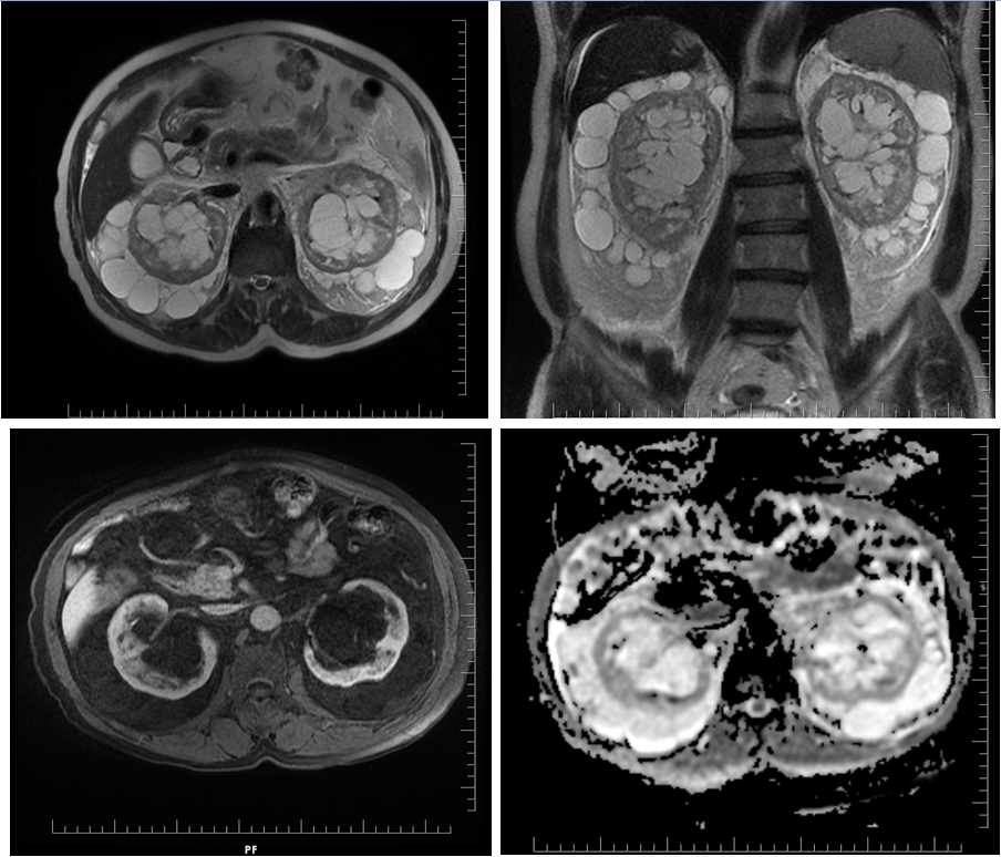
Figure 2a-d: Bilateral hyperintense Lymphangiomas in axial and Coronal T2W images.
These renal and perirenal Lymphangiomas do not enhance in contrast enhanced axial MRI and do not show any diffusion restrictions on ADC mapping.
In the routine follow-ups: There was no increase in the size, amount and dimensions of the renal-perirenal cysts.
Discussion
Renal and perirenal lymphangiectasias are rare diseases of kidneys with dilatation of the renal lymphatics, seen in both children and adults. They can be unilateral and/or bilateral, occur equally in both sexes, are also known as renal lymphangiomatosis, renal lymphangiomas and renal hygromas [1-3]. Since these lympangiectasias were manifested by perirenal, intrarenal and parapelvic cystic structures due to the dilatation of lymphatic vessels, it should be considered in the differential diagnosis of renal pathologies such as hydronephrosis, medullary lipomatosis and other cystic renal diseases particularly with the autosomal recessive polycystic kidney diseases of childhood [1,4,5].
Renal lymphangiectasias are known to be congenital and benign, hereditary cases have also been reported although they were mostly sporadic[4-6]. USG, CT and MRI have typical imaging appearances and have a great role in the diagnosis of Lympangiomatosis. On CT, typical findings are: The cysts located in perirenal and/or parapelvic region with fluid density, with or without any septa, without any compression towards the renal parenchyma, with prominent conglomerating features and without any contrast enhancement. On MRI, the appearance of cysts that appear hypointense on T1W and hyperintense on T2W sequences, are typical[4,6,7].
In most of the cases, no treatment is required, follow-up of lymphangiomas is sufficient. Percutaneous aspiration and nephrectomy can be applied in complicated patients[3,8].
In our case, there was no familial history without any clinical complaints. The diagnosis was made by contrast-enhanced
abdominal CT and with dynamic abdominal MRI. Any invasive procedures like biopsy, fluid aspiration and cytology were not required in the diagnosis due to the typical imaging findings which were mainly included in the diagnosis of renal lymphangiectasias.
References
- Mookadam F, Thota VR, Garcia-Lopez AM, Zamorano J. (2010). et al. Unicuspid aortic valve in adults: a systematic review. J Heart Valve Dis; 19:79-85
View at Publisher | View at Google Scholar - Edwards JE. (1958). Pathologic aspects of cardiac valvular insufficiencies. AMA Arch Surg ;77: 634–649
View at Publisher | View at Google Scholar - Novaro GM, Mishra M, Griffin BP.(2003).Incidence and echocardiographic features of congenital unicuspid aortic valve in an adult population. J Heart Valve Dis;12: 674–678
View at Publisher | View at Google Scholar - Roberts WC, Ko JM. (2005). Frequency by decades of unicuspid, bicuspid, and tricuspid aortic valves in adults having isolated aortic valve replacement for aortic stenosis, with or without associated aortic regurgitation. Circulation;111: 920–925
View at Publisher | View at Google Scholar - Brantley HP, Nekkanti R, Anderson CA, Kypson AP. (2012).Three-dimensional echocardiographic features of unicuspid aortic valve stenosis correlate with surgical findings. Echocardiography; 29: E 204–207
View at Publisher | View at Google Scholar - Yamanaka T, Fukatsu T, Ichinohe Y. (2021). et al. An adult case of unicommissural unicuspid aortic valve diagnosed based on the intraoperative findings. Intern Med 2016; 55: 2643–2648
View at Publisher | View at Google Scholar - von Stumm M, Gross TS, Petersen J, et al. Narrative review of the contemporary surgical treatment of unicuspid aortic valve disease. Cardiovasc Diagn Ther; 11:503-517.
View at Publisher | View at Google Scholar
