

Research Article | DOI: https://doi.org/10.31579/2834-8761/032
Acute Rheumatic Fever: The Rare Association with Vitamin B12 Deficiency Induced Neutropenia
- Aamir Al-Mosawi *
The National Center of Training and Development and Baghdad Medical City Baghdad, Iraq
*Corresponding Author: Aamir Al-Mosawi, Advisor doctor and expert trainer the National Center of Training and Development and Baghdad Medical City Baghdad, Iraq.
Citation: Aamir Al-Mosawi (2023), Acute Rheumatic Fever: The Rare Association with Vitamin B12 Deficiency Induced Neutropenia, Clinical Endocrinology and Metabolism, 2(5) DOI:10.31579/2834-8761/032
Copyright: © 2023, Aamir Al-Mosawi. This is an open access article distributed under the Creative Commons Attribution License, which permits unrestricted use, distribution, and reproduction in any medium, provided the original work is properly cited.
Received: 14 November 2023 | Accepted: 27 November 2023 | Published: 30 November 2023
Keywords: acute rheumatic fever; vitamin B12 deficiency-induced neutropenia
Abstract
Background: Acute rheumatic fever is an autoimmune disorder that develops commonly few weeks following a group A beta hemolytic Streptococcal tonsillopharyngitis. The autoimmune response can affect joints, skin, heart, and the brain. One of the important serious consequences of acute rheumatic fever that have to be prevented is the the long-term heart disease resulting from valves damage which may occur after one severe illness or following multiple recurrence of the illness. The diagnosis of acute rheumatic fever is generally based on the presence of either two Major diagnostic criteria (multiple joint arthritis, carditis, erythema marginatum, chorea, and subcutaneous nodules) or one Major diagnostic criteria and two Minor criteria (Fever, arthralgia, first degree heart block, elevated inflammatory markers [ESR and C-reactive protein]) plus appropriate evidence of preceding streptococcal infection.
The association of acute rheumatic fever with vitamin B12 deficiency-induced neutropenia has not been reported previously in the medical literature.
Patients and methods: The case of a ten-year old girl who developed acute febrile illness associated with arthralgia, arthritis, skin rash and neutropenia is described.
Results: The patient developed fever and arthritis of one elbow (Painful and swollen) followed shortly by the development of macular rash consisting of pink rings mostly on the extensor surfaces and arthritis of the second elbow. The patient had a previous febrile illness associated with sore throat which occurred during the previous 2 to 3 weeks. ASO titer was 150 iu/ml and CRP was positive 300 mg/dL. The patient had a high ESR, leukopenia, and blood film showed severe neutropenia, poikilocytosis and, frequent elliptocytosis. Serum vitamin B12 level was low, 152 pg/mL. Echocardiography showed low mitral valve prolapse.
Conclusion: The novel association of acute rheumatic fever with vitamin B12 deficiency-induced neutropenia is reported.
Introduction
Acute rheumatic fever is an autoimmune disorder that develops commonly few weeks following a group A beta hemolytic Streptococcal tonsillopharyngitis. The autoimmune response can affect joints, skin, heart, and the brain. One of the important serious consequences of acute rheumatic fever that have to be prevented is the the long-term heart disease resulting from valves damage which may occur after one severe illness or following multiple recurrence of the illness.
Some of the important features of acute rheumatic fever have been known as early as the 1700s.
In 1797, Matthew Baillie quoted David Pitcairn (Figure-1A) who suggested that rheumatism is one of the causes of a morbid growth of the heart.
Matthew Baillie (Figure-1B) reported the presence of a thickening of some valves of heart in autopsies of patients who had acute rheumatism [1].
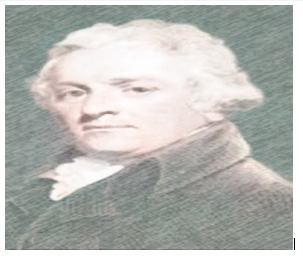
Figure 1A: David Pitcairn (1749-1809), a Scottish physician
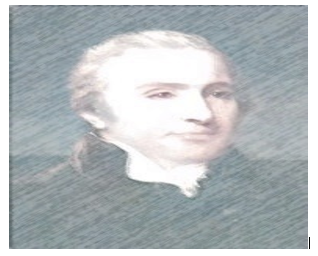
Figure 1B: Matthew Baillie (1761-1823), a British physician and pathologist
In 1803, Wagstaffe of Southwark reported a fatal case of rheumatic pericarditis with enlargement of the heart [2], and Crowfoot of Beccles reported two cases of rheumatic carditis in 1809 [3].
In 1809, David Dundas reported nine cases of heart disease associated with rheumatism that had been observed since 1770. The cases reported by Dundas were considered typical cases of acute rheumatic carditis. The heart disease started after, or sometimes during the same time of rheumatism. The cases of Dundas were mostly young patients who developed chest pain cough, dyspnea, and palpitations. There was a high death rate with most patients died because of congestive cardiac failure [4].
In 1812, William Charles Wells was still thinking that the association of rheumatism with heart disease was a concurrence rather than a specific association as he quoted Morgagni and Ferrier who had regarded the association as apparently an interesting coincidence [5, 6].
Richard Bright (Figure-1C) was probably the first to recognize the association of neurological manifestations of rheumatic fever (Sydenham’s chorea) with joint manifestations (Rheumatism) in 1831 [7].
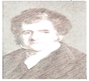
Figure-1C: Richard Bright (1789-1858), an English physician
Walter Butler Cheadle (Figure-1D) was most probably the first to link rheumatism and heart disease with tonsillitis in 1889. He reported his observations during twenty years on rheumatic fever at the Hospital for Sick Children, Great Ormond Street [8].
Frederic John Poynton (Figure-1E) and his colleague Paine were most probably the first to identify the diplococcus which they isolated from patients with acute rheumatic fever as the causative bacteria for the disease [9].
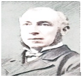
Figure-1D: Walter Butler Cheadle (October 1836-March 1910), an English pediatrician
Figure-1E: Frederic John Poynton (June 26, 1869-October, 29 1943), an English physician
In 1944, Thomas Duckett Jones (Figure-1F) suggested diagnostic criteria for its diagnosis [10].

Figure-1F: Thomas Duckett Jones (February 2, 1899-November 22, 1954) was an American physician, cardiologist
Jones diagnostic criteria have been revised and modified in 1965, 1984, 1992, and 2002. The diagnosis of acute rheumatic fever is generally based on the presence of either two Major criteria (multiple joint arthritis, carditis, erythema marginatum, chorea, and subcutaneous nodules) or one Major criterion and two Minor criteria (Fever, arthralgia, first degree heart block, elevated inflammatory markers [ESR and C-reactive protein]) plus appropriate evidence of preceding streptococcal infection [11-14].
The association of acute rheumatic fever with vitamin B12 deficiency-induced neutropenia has not been reported previously in the medical literature.
Patients and methods
The case of a ten-year old girl who developed acute febrile illness associated with arthralgia, arthritis, skin rash and neutropenia is described.
Results
The patient developed fever and arthritis of one elbow (Painful and swollen) on the 22nd of August, 2023 followed shortly by the development of macular rash consisting of pink rings mostly on the extensor surfaces and arthritis of the second elbow. The face was not involved by the rash. She was treated with oral antibiotics and analgesics without obvious improvement. The patient was brought to us on the 26th of August, and she still had painful swollen elbows and rash (Figure-2). The patient had a previous febrile illness associated with sore throat which occurred during the previous 2 to 3 week
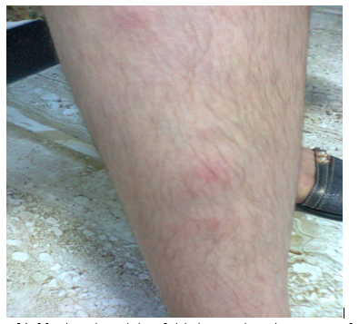
Figure-2A: Macular rash consisting of pink rings mostly on the extensor surfaces
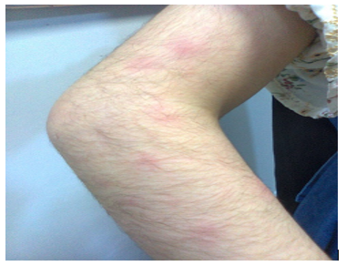
Figure-2B: Macular rash consisting of pink rings mostly on the extensor surfaces
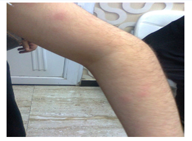
Figure-2C: Macular rash consisting of pink rings mostly on the extensor surfaces
ASO titer was 150 iu/ml and CRP was positive 300 mg/dL.
Erythrocyte Sedimentation Rate (ESR) was 100 cubic mm/hour.
Hemoglobin was 8.9 g/dL.
White blood cell count was 1.58 × 10⁹/L (Normal: 4.0 to 11.0 × 10⁹/L).
Granulocyte (Including neutrophils) count was 0.9 × 10⁹/L (Normal: 2.0 to 7.0 × 10⁹/L).
Blood film showed:
White blood cells: Severe neutropenia, no immature or abnormal cells.
Red blood cells: Poikilocytosis (10% or more of blood cells have abnormal shape), frequent eliptocytosis, occasional polychromasia (Excessive immature blood cells).
Platelets: Normal.
Serum folic acid was within normal range.
Serum vitamin B12 level was 152 pg/mL (Normal: 200-900 pg/mL).
Radiographs of the left elbow showed no significant abnormalities (Figure-3).
Ultrasound of the elbows showed fluid collection suggesting joint effusion (Figure-4).
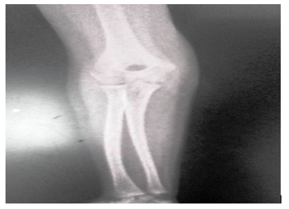
Figure-3A: Radiographs of the left elbow
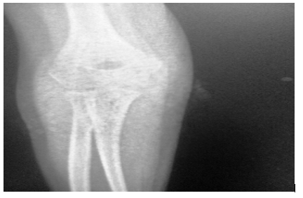
Figure-3B: Radiographs of the left elbow
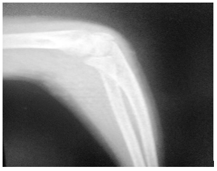
Figure-3C: Radiographs of the left elbow
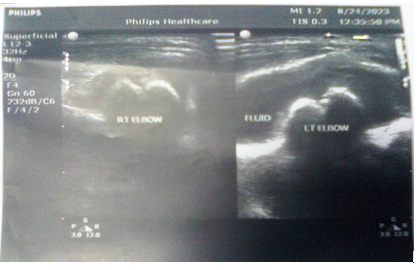
Figure-4: Ultrasound of the elbows showed fluid collection suggesting joint effusion
Echocardiography showed low mitral valve prolapse.
The patient was treated with oral ibuprofen 800 mg in two divided doses after meals instead of aspirin based on the evidence provided by Morino et al (1973), Altay et al (2021), Yılmaz and colleagues (2022) [15,16,17].
The girl was also treated with single injections of benzathine penicillin given every month based on the evidence provided by Cham Ovitz et al (1954), Stoller man (1954), Markowitz and Hemphill (1955), Stoller man and colleagues (1955), Wilson (2013), and Ralph and Currie (2022) [18-23].
She also received three doses of vitamin B12 1000 mcg given every other day.
On the seventh of October, 2023, oral ibuprofen has already been stopped before few days and she was asymptomatic. Laboratory tests showed:
Erythrocyte Sedimentation Rate (ESR) was 19 cubic mm/hour.
Hemoglobin was 10.1 g/dL.
White blood cell count was 2.7 × 10⁹/L (Normal: 4.0 to 11.0 × 10⁹/L).
Granulocyte (Including neutrophils) was 29.6 %.
Serum vitamin B12 level was 210 pg/mL (Normal: 200-900 pg/mL).
Discussion
Zeynep Canan Ozdemir from Turkey and her research group reported the etiological causes of neutropenia in 94 children. 55 children (58.5%) had post-infectious neutropenia with 38.4% occurring following upper respiratory airway. 23 (24.6%) children had idiopathic neutropenia. Six children (6.3%) had drug-induced neutropenia. Five children (5.3%) had vitamin B12 deficiency-induced. Two children (2%) had chronic benign neutropenia. One child (1.1%) had immune deficiency, one child (1.1%) had autoimmune lymphoproliferative syndrome, and one child (1.1%) had post-infection hemophagocytic lymph histiocytosis.
Neutropenia improved in ninety-one children (96.8%).
Neutropenia persisted in 3 children (3.2%).
One died because of infection [24].
Although, the patient in this report had lab-confirmed vitamin B12 deficiency, the blood film showed no macrocytosis, but showed poikilocytosis and frequent elliptocytosis.
As early as 1982, Spivak emphasized that megaloblastic anemia can be masked by the absence of macrocytosis. Spivak also emphasized that poikilocytosis may indicates a vitamin deficiency [25].
As early as 1982, Schoomaker and colleagues, emphasized that vitamin B deficiency may result in an abnormal red blood cells’ membrane causing elliptocytosis [26].
In this patient, echocardiography showed low mitral valve prolapse.
As early as 1980, Storozhakov et al suggested the potential of echocardiographic diagnosis of rheumatic valvulitis and mitral valve prolapse in children [27].
In 1988, Lembo et al emphasized that acute rheumatic fever commonly results in mitral regurgitation without associated mitral valve stenosis. They reported that the surgical findings have showed a high frequency of mitral prolapse in rheumatic valvular heart disease suggesting that acute rheumatic fever can cause mitral valve prolapse.
Lembo et al reported thirty patients who had previously acute rheumatic fever. 25 patients (84%) were found to have mitral regurgitation on doppler examination. Echocardiography showed mitral prolapse in 24 patients (80%). Only one patient had mitral stenosis when examined by echocardiography.
Therefore, Lembo et al suggested that acute rheumatic fever can cause mitral valve prolapse [28].
The use of ibuprofen in acute rheumatic fever was reported as early as the 1970s [15].
In 2021, Derya Altay from Turkey and her research team reported 286 patients with acute rheumatic fever. 53 patients (18.5%) [Mean age: 10.7 ± 2.5 years] were treated initially with aspirin for arthritis.
Aspirin-associated hepatotoxicity occurred in 9 (17%) of the 53 patients, and therefore ibuprofen or naproxen was given as an alternative to aspirin. Both ibuprofen and naproxen were not associated with side effects.
30% of 53 patients had anemia, and aspirin was used for longer period in the hepatotoxic anemic patients than non-anemic patients (p=0.02) [16].
Münevver Yılmaz and her colleagues emphasized that the use of Aspirin (Acetylsalicylic acid) in acute rheumatic fever can be associated with severe side effects [17].
The use of single injections of benzathine penicillin given every month in the treatment of acute rheumatic fever was reported as early as the 1950s [18, 19, 20, 21].
In 2013, Nigel Wilson from New Zealand reviewed the literature and emphasized that randomized controlled studied showed four weekly intramuscular long-acting benzathine penicillin G was found to be more effective than penicillin given orally to prevent recurrence of rheumatic fever [22].
In 2022, Anna P Ralph and Bart J Currie from Australia emphasized that the aims of the treatment of acute rheumatic fever include relieving symptoms, lessening heart valve damage and eliminating streptococcal infection. They recommended the use of monthly intramuscular injections benzathine benzyl penicillin G for the prevention of recurrences [23].
Conclusion
The novel association of acute rheumatic fever with vitamin B12 deficiency-induced neutropenia is reported.
Acknowledgement
The author has the copy right of all figures and sketches included in this paper.
Conflict of interest: None.
References
- Baillie M. (1797). The Morbid Anatomy of Some of the Most Important Parts of the Human Body, 2nd ed. J. Johnson and G. Nicol, London,
View at Publisher | View at Google Scholar - 2-Wagstaffe MF. (1803). A Case of Thoracic Adhesions. Med Phys J Apr 1; 9(50):359-362.
View at Publisher | View at Google Scholar - Crowfoot WH. (1809). Two cases of carditis. Edinburgh Medical and Surgical Journal; 5: 298-300.
View at Publisher | View at Google Scholar - Dundas D. (1809). An account of a peculiar Disease of the Heart. Med Chir Trans ;1:37-46. Doi: 10.1177/095952870900100105.
View at Publisher | View at Google Scholar - Wells WC. (1812). On rheumatism of the heart. Transactions of the Society for Improving Medical and Chirurgicat Knowledge.3:373-424.
View at Publisher | View at Google Scholar - Morgagni GB. De sedibus, et (1761). causis morborum Translated by B Alexander. A. Millar and T. Cadell, London, 1769.
View at Publisher | View at Google Scholar - Bright R. Reports of medical cases. Vol 2 Disease of the brain and nervous system: selected with a view of illustrating the symptoms and cure of diseases by reference to morbid anatomy. London: Longman, 1831:493.
View at Publisher | View at Google Scholar - Cheadle WB. (1889). Harveian lectures on the various manifestations of the rheumatic state as exemplified in childhood and early life. Lancet 133:821-827.
View at Publisher | View at Google Scholar - Poynton FJ, Paine A. The etiology of rheumatic fever. Lancet 1900; 156:861-869.
View at Publisher | View at Google Scholar - 10-Jones TD. (1944). Diagnosis of rheumatic fever. JAMA 126(8):481-484.
View at Publisher | View at Google Scholar - Jones criteria (revised) (1965). for guidance in the diagnosis of rheumatic fever. Circulation Oct; 32(4):664-668.
View at Publisher | View at Google Scholar - Jones (1984). Criteria (revised) for guidance in the diagnosis of rheumatic fever. Circulation Jan; 69(1):204A-208A.
View at Publisher | View at Google Scholar - Guidelines for the diagnosis of rheumatic fever. Jones Criteria, (1992) update. Special Writing Group of the Committee on Rheumatic Fever, Endocarditis, and Kawasaki Disease of the Council on Cardiovascular Disease in the Young of the American Heart Association. JAMA 1992 Oct 21; 268(15):2069-2073.
View at Publisher | View at Google Scholar - Ferrieri P. (2002). Jones Criteria Working Group. Proceedings of the Jones Criteria workshop. Circulation. Nov 5; 106(19):2521-2523.
View at Publisher | View at Google Scholar - Morino F, Possati F, Santarelli P, Paolini E, Calafiore AM. (1973). L'impiego dell'ibuprofen nel controllo dell'attività reumatica [Use of ibuprofen in the control of rheumatic activity]. Minerva Med Jun 23; 64(47):2475-2478 [Article in Italian].
View at Publisher | View at Google Scholar - Altay D, Pamukçu Ö, Baykan A, Üzüm K, Arslan D. (2021). Aspirin-induced hepatotoxicity and anemia in children with acute rheumatic fever. Turk J Pediatr. 63(2):193-199.
View at Publisher | View at Google Scholar - Yılmaz M, Gürses D, Tükenmez G. The effectiveness and safety of ibuprofen and acetylsalicylic acid in acute rheumatic fever. Pediatr Int 2022 Jan; 64(1):e15133.
View at Publisher | View at Google Scholar - Chamovitz R, Catanzaro FJ, Stetson CA, Rammelkamp CH (1954). Jr. Prevention of rheumatic fever by treatment of previous streptococcal infections. I. Evaluation of benzathine penicillin G. N Engl J Med Sep 16; 251(12):466-471. Doi: 10.1056/NEJM 195409162511203.
View at Publisher | View at Google Scholar - Stollerman GH. (1954). Repository benzathine penicillin for the control of rheumatic fever. Bull Rheum Dis Dec; 5(4):79-80.
View at Publisher | View at Google Scholar - Markowitz M, Hemphill W. (1955). A comparison of oral benzathine penicillin G and sulfonamides for the prevention of streptococcal infections and recurrences of rheumatic fever. Pediatrics May; 15(5):509-515.
View at Publisher | View at Google Scholar - Stollerman GH, Rusoff JH, Hirschfeld I. (1955). Prophylaxis against group A streptococci in rheumatic fever; the use of single monthly injections of benzathine penicillin G. N Engl J Med May 12;252(19):787-92.
View at Publisher | View at Google Scholar - Wilson N. (2013). Secondary prophylaxis for rheumatic fever: simple concepts, difficult delivery. World J Pediatr Congenit Heart Surg Oct; 4(4):380-384.
View at Publisher | View at Google Scholar - Ralph AP, Currie BJ. (2022). Therapeutics for rheumatic fever and rheumatic heart disease. Aust Prescr Aug; 45(4):104-112.
View at Publisher | View at Google Scholar - Ozdemir ZC, Kar YD, Kasaci B, Bor O. (2021). Etiological causes and prognosis in children with neutropenia. North Clin Istanb May 24; 8(3):236-242.
View at Publisher | View at Google Scholar - Spivak JL. Masked megaloblastic anemia. Arch Intern Med 1982 Nov; 142(12):2111-2114.
View at Publisher | View at Google Scholar - Schoomaker EB, Butler WM, Diehl LF. (1982). Increased heat sensitivity of red blood cells in hereditary elliptocytosis with acquired cobalamin (vitamin B12) deficiency. Blood Jun; 59(6):1213-1219.
View at Publisher | View at Google Scholar - Storozhakov GI, Kuz'mina NN, Shaikov AV, Voronina NM, Malysheva NV.(1980). Vozmozhnosti ékhokardiograficheskoĭ diagnostiki revmaticheskogo val'vulita i prolapsa mitral'nogo klapana u deteĭ [Potentials of echocardiographic diagnosis of rheumatic valvulitis and mitral valve prolapse in children]. Vopr Revm Jan-Mar ;(1):18-22 [Article in Russian].
View at Publisher | View at Google Scholar - Lembo NJ, Dell'Italia LJ, Crawford MH, Miller JF, Richards KL, et all., (1988). Mitral valve prolapse in patients with prior rheumatic fever. Circulation Apr; 77(4):830-836.
View at Publisher | View at Google Scholar
