

Review Article | DOI: https://doi.org/10.31579/2835-9291/026
Single Coronary Presenting with Acute Coronary Syndrome-Ptca through Trans-Radial Route
- Rajeev Bhardwaj *
- Sivaji Patibandla
- Gaurav Aggrawal
- Mehroj Mirza
Department of cardiology, MM Institute of Medical Sciences and Research, Mullana, Ambala, India.
*Corresponding Author: Rajeev Bhardwaj, Department of cardiology, MM Institute of Medical Sciences and Research, Mullana, Ambala, India.
Citation: R Bhardwaj, S Patibandla, G Aggrawal, M Mirza. (2024). Single Coronary Presenting with Acute Coronary Syndrome- Ptca through Trans-Radial Route, International Journal of Clinical case Studies 3(5); DOI: 10.31579/2835-9291/026
Copyright: © 2024 Rajeev Bhardwaj, this is an open-access article distributed under the terms of the Creative Commons Attribution License, which permits unrestricted use, distribution, and reproduction in any medium, provided the original author and source are credited.
Received: 25 September 2024 | Accepted: 10 October 2024 | Published: 22 October 2024
Keywords: coronary single coronary anomalies; artery; PTCA
Abstract
Isolated single coronary artery is a rare congenital anomaly occurring in approximately 0.024% of the population. It is usually diagnosed incidentally during coronary artery angiograms or on postmortem evaluations. The single coronary artery anomaly is usually asymptomatic, but may present as myocardial ischemia, syncope or sudden cardiac death depending on its course and the presence and severity of atherosclerosis.
Introduction
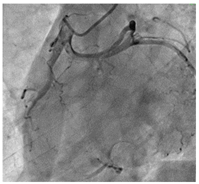
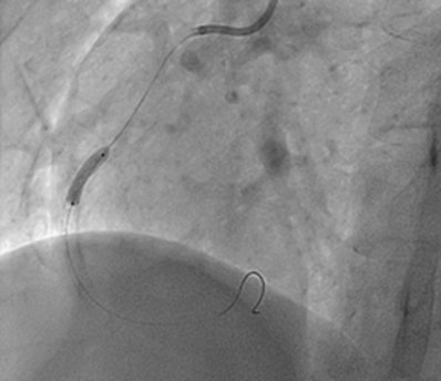
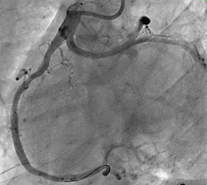
Case: 62 years male patient was admitted in internal medicine department with acute inferior wall myocardial infarction (MI) around one month back. He was treated on medical therapy. He remained admitted for five days. There was no complication. He was not offered invasive therapy. He was discharged on 6th day with advice to follow up in cardiology out patient department (OPD) after one month. After two weeks, he started having angina on routine activities. Severity of pain increased over next two days, when he had episodes of rest pain, lasting few minutes. He was admitted in Cardiology. His ECG showed T wave inversion in leads II, III and AVF. Echocardiography showed hypokinetic inferior with ejection fraction of 55%. He was taken up for coronary angiography through trans radial route. We were not able to hook left coronary artery (LCA). After struggling for around 2-3 minutes, we gave sinus injection, but still LCA was not visible. We then did right coronary angiography. We found single coronary artery arising from right coronary sinus, which showed total occlusion after giving rise to left main coronary artery, which then devided into left anterior descending (LAD) and left circumflex (LCx) arteries (Fig 1). There was retrograde filling of distal right coronary artery (RCA) form LCA. Patient was taken up for percutaneous transluminal coronary angioplasty (PTCA).
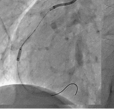
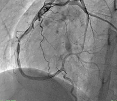
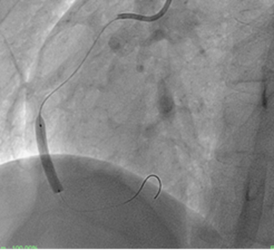
Judkinn’s right could not properly hook the coronary artery, and so Amplatzer right (AR) guiding catheter was used. Lesion crossed with balanced middle weight miracle 3 wire (Fig. 2). Tandom dilatations were done with 2.0X10 mm balloon (Fig 3). There was good flow (Fig 3,4) 3.0X15 mm stent was deployed. there was some narrowing proximal to the stent. 3.0X12 mm stent was deployed proximal to the first stent. (Fig 5). There was no residual stenosis, with TIMI 3 flow (Fig. 6).
Discussion
Most coronary artery anomalies are asymptomatic and are usually encountered as coincidental findings during coronary angiography or autopsy [1]. Coronary arteries originating from a single coronary ostium in the aorta are rare, occurring in less than 0.03% of the general population [2]. The first report of a single coronary artery was by Hyrtl in 1841 [3]. Anomalies are classified according to the relation of the coronary anomaly to the aorta and pulmonary artery-that is, anterior, between, septal, posterior, and combined. Previous investigation of coronary anomalies and analysis of the causes of sudden cardiac death has identified the course of the left main coronary artery between the aorta and pulmonary artery as a potential cause of significant coronary ischaemia [1,2]. This anomaly can present with exertional angina, dyspnoea, and palpitations, as well as sudden death. There are several proposed potential mechanisms for the clinical manifestation of this anomaly, such as compression of the left coronary artery between the aorta and pulmonary artery during exercise when the vessels become enlarged; and compromise of the lumen of the left coronary artery due to the acute angle formed at its origin from the right aortic sinus resulting in a slit-like orifice. Horan et al reported SCA in father and daughter, raising the possibility of genetic link [4].
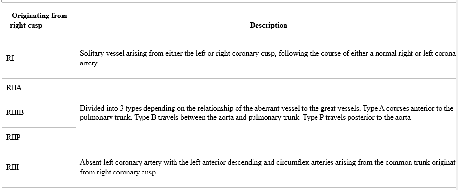
Lipton et al. [5] originally proposed the angiographic classification of SCA, which was later modified by Yamanaka et al. [6]. This classification takes into account the origin of the ostium from the sinus of Valsalva, anatomical course of the vessel, and the course of the transverse trunk. Alphabets R or L are used to identify the ostial origin of the vessel, roman numerals I, II, or III are used to represent the anatomical distribution of the vessel, and letters A, B, P, S, and C are used to delineate the course of the vessel with respect to the pulmonary artery and the aorta [6].
References
- Yamanaka O, Hobbs RE. (1990) Coronary artery anomalies in 126,595 patients undergoing coronary arteriography. Cathet Cardiovasc Diagn. 21:28-40.
View at Publisher | View at Google Scholar - Roberts WC. Major anomalies of coronary arterial origin seen in adulthood. Am Heart J. 111:941-963.
View at Publisher | View at Google Scholar - Hyrtl J. Einige in chirurgischer Hinsicht wichtige Gefässvarietäten. Med Jahrb Österr Staats. 33:17-38.
View at Publisher | View at Google Scholar - Horan PG, Murtagh G, Mckeown PP. Single coronary artery-a familial clustering. Heart;12: 27.
View at Publisher | View at Google Scholar - Lipton MJ, Barry WH, Obrez I, Silverman JF, Wexler L. (1979) Isolated single coronary artery: diagnosis, angiographic classification, and clinical significance. Radiology. 130:39-47.
View at Publisher | View at Google Scholar - Yamanaka O, Hobbs RE. (1990) Coronary artery anomalies in 126,595 patients undergoing coronary arteriography. Catheter Cardiovasc Interv. 21:28-40
View at Publisher | View at Google Scholar
