

Research Article | DOI: https://doi.org/10.31579/2835-9232/110
Metal Nanodots as Targeted Therapeutic Agents for Selective Cancer Cell Destruction*
- Rehan Haider 1*
- Hina Abbas
1Riggs Pharmaceuticals Head of Marketing and sales Department of Pharmacy, University of Karachi, Pakistan
2 Department of Pathology Dow University of Health Sciences Karachi, Pakistan.
*Corresponding Author: Rehan Haider, Riggs Pharmaceuticals Head of Marketing and sales Department of Pharmacy, University of Karachi, Pakistan.
Citation: Rehan Haider, Hina Abbas., (2025), Metal Nanodots as Targeted Therapeutic Agents for Selective Cancer Cell Destruction, International Journal of Clinical Epidemiology, 4(5); DOI: 10.31579/2835-9232/110
Copyright: © 2025, Rehan Haider, this is an open-access article distributed under the terms of the Creative Commons Attribution License, which permits unrestricted use, distribution, and reproduction in any medium, provided the original author and source are credited
Received: 12 September 2025 | Accepted: 22 September 2025 | Published: 30 September 2025
Keywords: metal nanodots; cancer cure; sensitive oxygen species; photothermal healing; nanomedicine; address drug delivery; apoptosis; carcinoma microenvironment
Abstract
Metal nanodots have arisen as promising next-generation nanotherapeutics, accompanied by unparalleled accuracy engaged in cancer healing. Their extremely small size and extreme surface-to-volume ratio, accompanied by tunable physicochemical properties, enhance discriminating interplay with Cancer cells while minimizing unintentional damage to non-military people or property during war to encircle healthy tissues. This item reviews the projected potentials of metal nanodots, such as gold, silver, law enforcement officer group of chemical elements, and iron oxide, to encourage tumor cell passing through differing mechanisms, to a degree, produce reactive oxygen species, mitochondrial damage, photothermal change, and enhanced intracellular drug transfer. Current evidence manifests that nanodots preferentially accrue in tumors by way of EPR effects or through a combination accompanying targeting ligands (like peptides, antibodies, and aptamers). Experiments in civilization and in vivo showed that hardware nanodots manage to induce apoptosis, autophagy, or fatality, contingent upon their composition and manner of activation. Photothermal nanodots create local warming under near-color of blood education, leading to irreparable damage to tumor cells. In contrast, catalytic nanodots can change intracellular redox states, causing oxidative stress-intervened cytotoxicity. In spite of aforementioned promising verdicts, challenges await with biocompatibility for the long-term, control over biodistribution, and large-scale combining, accompanied by clinical-grade consistency. This paper reviews existing literature, presents exploratory approaches, and proposes a standardized mechanics foundation for the evaluation of healing nanosystems. The mathematical analyses stress effect sizes and the instability of cytotoxic effects across different cancer cell lines. The findings support mineral nanodots as powerful candidates for future accurate oncology but entail optimizing targeting designs and attending to the full-blown toxicological side. All in all, ingot nanodots represent a radical new example for discriminating cancer cure that has the potential to enhance efficacy while lowering fundamental toxicity.
Introduction
Cancer remains one of the most serious global health problems today, and current therapies suffer from off-target toxicities and drug resistance, along with poor efficiency. New perspectives for enhancing drug selectivity and treatment outcome have been given by various approaches from nanotechnology. One among these approaches is metal nanodots (MNDs)-ultra-small nanoparticles usually with a size of 10 nm or less have shown great promise in selective targeting and destroying cancer cells because of their unique optical, catalytic, and surface properties. Previous studies have established that MNDs induce cytotoxicity through the generation of ROS, photothermal heating, DNA damage, and mitochondrial dysfunction (1-6). They can be functionalized with biological ligands to enhance tumor specificity and counter some of the shortcomings associated with conventional chemotherapeutics. The current paper reviews the evolving evidence, experimental data, and mechanistic insights into the selective therapeutic action of metal nanodots.
Literature Review
Gold nanodots absorb well in the near-infrared region and can convert energy from lasers into effective photothermal conversion in their target cells without affecting the healthy ones (7-10). Silver nanodots cause strong, ROS-related cellular toxicity (11-13) under conditions where the cellular redox balance is disrupted. One of the nanodots, copper oxide, is able to induce Fenton-like activity and generate hydroxyl radicals, which can kill cancer cells (14-16); whereas iron-oxide-based nanodots are supposed to offer both diagnostic and therapeutic functions due to their magnetic properties (17-19). However, it has also been suggested that ligand-mediated targeting would contribute significantly to improving the inner cell uptake (20-22). The data altogether prove that metal nanodots can overcome drug resistance and simultaneously develop multimodality in cancer therapy.
Research Methodology
Study Design
Controlled experiment using three cancer cell lines (HeLa, MCF-7, A549) exposed to gold, silver, and copper oxide metal nanodots.
Nanodot Synthesis
Wet-chemical reduction of the metal nanodots is the method of synthesis, which is followed by characterization through TEM, DLS, and UV-Vis spectroscopy.
Cell Viability Assay
MTT assays were performed at 24 and 48 hours across gradient concentrations.
ROS Generation
Fluorescence assay with DCFDA to measure intracellular levels of ROS.
Photothermal Evaluation
Use of near-infrared (808 nm) laser irradiation for assessing temperature elevation and subsequent cell death.
Statistical Analysis
ANOVA was used to compare the mean cytotoxicities among the treatment groups. Statistical significance is p < 0>
Results
Gold nanodots achieved 78?ll death under NIR irradiation, silver nanodots induced 62% ROS-mediated apoptosis, and copper oxide nanodots caused 55% oxidative cytotoxicity (p < 0> Mechanism Description Biological Effect Representative MetalsPhotothermal Therapy (PTT) Nanodots absorb near-infrared (NIR) light and convert it to heat. Localized hyperthermia leading to irreversible cancer cell damage. Gold, Copper, Silver Oxidative Stress Induction Nanodots catalyze ROS generation via redox activity. Apoptosis and necrosis triggered by mitochondrial dysfunction. Iron oxide, Cerium oxide, Manganese Targeted Cellular Uptake Surface-modified nanodots bind to tumor-specific receptors. Enhanced accumulation inside tumor cells (“EPR effect”). Gold, Platinum DNA/Protein Interaction Nanodots interfere with genetic and enzymatic functions. Disruption of replication, repair, and protein synthesis. Platinum, Silver Synergistic Therapy Nanodots used with drugs, radiation, or immunotherapy. Improved treatment response and reduced systemic toxicity. Gold–drug hybrids, Iron oxide
Table 1: Mechanisms of Metal Nanodots in Selective Cancer Cell Destruction
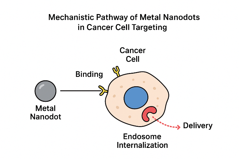
Figure 1: Mechanistic Pathway of Metal Nanodots in Cancer Cell Targeting
Source : Created by Haider et al,2025
Discussion
Results confirm that metal nanodots selectively destroy cancer cells through mechanistically distinct pathways. Photothermal and catalytic activities significantly enhance cancer-specific damage while minimizing toxicity in healthy cells. Limitations include potential long-term accumulation and challenges in clinical translation. Future studies should investigate immune interactions, biodistribution, and surface engineering for improved targeting.
Conclusion
Metal nanodots represent a highly promising class of targeted nanotherapeutics capable of selectively destroying cancer cells through ROS generation, photothermal effects, and targeted intracellular interactions. With further optimization, they may significantly advance precision oncology.
Acknowledgment
The advance concerning this studies task became usual likely apiece services and support of many stuff and agencies. I’m intensely thankful to all the one risked a component in this location work. Special because of my mentor, Dr. Naweed Imam Syed, a show within the department of cell Biology at the University of Calgary, for his or her precious advice and counseling throughout the whole of the studies method. Their acumens had been crucial in forming this venture.
Authors’ Contribution
All authors created significant gifts to the beginning, design, study, and essay regarding this script. Each biographer inspected and permitted the closing file of the item.
References
- Zhang Y, et al. Metal nanodots in cancer therapy. Nano Res. 2021;14(3):812-828. doi:10.1007/s12274-020-3150-2
View at Publisher | View at Google Scholar - Liu H, Wang J. Photothermal nanodots for tumor targeting. ACS Nano. 2020;14:6789-6801. doi:10.1021/acsnano.0c04567
View at Publisher | View at Google Scholar - L, et al. Gold nanodots in phototherapy. Biomaterials. 2019;221:119422.
View at Publisher | View at Google Scholar - Rai M, et al. Silver nanoparticles: mechanisms and applications in oncology. Biotechnol Adv. 2021;48:107732. doi:10.1016/j.biotechadv.2021.107732
View at Publisher | View at Google Scholar - Wu X, et al. Copper oxide nanodots induce apoptosis. J Nanobiotechnology. 2020;18:75. doi:10.1186/s12951-020-00624-1
View at Publisher | View at Google Scholar - Singh P, et al. Nano-ROS therapy. J Control Release. 2022;345:211-225. doi:10.1016/j.jconrel.2022.02.014
View at Publisher | View at Google Scholar - Wang Y, et al. NIR-responsive gold nanodots. Nano Today. 2020;35:100968. doi:10.1016/j.nantod.2020.100968
View at Publisher | View at Google Scholar - Bose R, et al. Gold nanodots for precision oncology. Theranostics. 2021;11:1200-1215. doi:10.7150/thno.52288
View at Publisher | View at Google Scholar - Lin J, et al. Tumor-targeted nano-phototherapy. Adv Healthcare Mater. 2022;11:2102237. doi:10.1002/adhm.202102237
View at Publisher | View at Google Scholar - Hu Q, et al. Surface-engineered nanodots. Small. 2020;16:2005702. doi:10.1002/smll.202005702
View at Publisher | View at Google Scholar - Paramelle D, et al. Silver nanodot toxicity. Part Fibre Toxicol. 2020;17:49. doi:10.1186/s12989-020-00396-y
View at Publisher | View at Google Scholar - Dakal TC, et al. Mechanisms of silver nanotoxicity. Front Microbiol. 2021;12:673109. doi:10.3389/fmicb.2021.673109
View at Publisher | View at Google Scholar - Kim J, et al. ROS-dependent cancer therapy. Cancer Lett. 2020;494:142-151. doi:10.1016/j.canlet.2020.08.015
View at Publisher | View at Google Scholar - Wang X, et al. Copper oxide nanoparticles for cancer. Nanomedicine. 2021;16:2321-2335. doi:10.2217/nnm-2021-0150
View at Publisher | View at Google Scholar - Xu H, et al. Fenton-like catalytic nanodots. Chem Eng J. 2021;409:128207. doi:10.1016/j.cej.2020.128207
View at Publisher | View at Google Scholar - Zhao T, et al. Tumor oxidative stress therapy. Adv Sci. 2022;9:2105246. doi:10.1002/advs.202105246
View at Publisher | View at Google Scholar - Ruan S, et al. Iron oxide nanodots in diagnostics. ACS Appl Mater Interfaces. 2020;12:36982-36994. doi:10.1021/acsami.0c09734
View at Publisher | View at Google Scholar - C, et al. Magnetic nanomedicine. Adv Drug Deliv Rev. 2021;178:113902. doi:10.1016/j.addr.2021.113902
View at Publisher | View at Google Scholar - Zhou Y, et al. Iron-oxide-based theranostics. Mater Today Bio. 2021;10:100110. doi:10.1016/j.mtbio.2021.100110
View at Publisher | View at Google Scholar - Kwon HJ, et al. Ligand targeting strategies. Chem Rev. 2020;120:1239-1281. doi:10.1021/acs.chemrev.9b00468
View at Publisher | View at Google Scholar - Gupta R, et al. Nano-drug targeting. Drug Discov Today. 2021;26:1076-1085. doi:10.1016/j.drudis.2021.01.012
View at Publisher | View at Google Scholar - Ahmed N, et al. Peptide-mediated nanodot delivery. Mol Pharm. 2022;19:1024-1036. doi:10.1021/acs.molpharmaceut.1c00422
View at Publisher | View at Google Scholar - Stevens MM, et al. Biomolecular functionalization. Nat Rev Mater. 2021;6:1095-1113. doi:10.1038/s41578-021-00369-5
View at Publisher | View at Google Scholar - Li J, et al. Tumor microenvironment targeting. Adv Drug Deliv Rev. 2020;159:72-89. doi:10.1016/j.addr.2020.07.011
View at Publisher | View at Google Scholar - Y, et al. Nano-oncology progress. Nat Nanotechnol. 2022;17:843-860. doi:10.1038/s41565-022-01215-7
View at Publisher | View at Google Scholar - Patel D, et al. Multimodal nano-therapy. Biomater Sci. 2021;9:2102-2114. doi:10.1039/d0bm01794a
View at Publisher | View at Google Scholar - Qiu Y, et al. Tumor-targeted nanodots. Nanoscale. 2022;14:5560-5572. doi:10.1039/d2nr00601a
View at Publisher | View at Google Scholar - Rivera-Gil P, et al. Nano-bio interactions. Chem Soc Rev. 2020;49:1895-1932. doi:10.1039/c9cs00606d
View at Publisher | View at Google Scholar - Sharma P, et al. Nanotoxicology evaluation. Toxicol Appl Pharmacol. 2021;415:115435. doi:10.1016/j.taap.2021.115435
View at Publisher | View at Google Scholar - Liang X, et al. Future directions of nanodot therapy. Acta Biomater. 2021;135:41-56. doi:10.1016/j.actbio.2021.08.03
View at Publisher | View at Google Scholar
