

Research Article | DOI: https://doi.org/10.31579/2835-7957/001
Sex Difference in The Occurrence of Supernumerary Teeth Among People Living in Gombe State, Nigeria
1 Department of Human Anatomy, Gombe State University, Nigeria
2 Department of Dental Surgery, Lamido School of Hygiene and Health Sciences, Nigeria.
3 Department of Human Anatomy, College of Health Sciences, Bauchi State University Itas-Gadau, Nigeria
*Corresponding Author: Rayyanu Z.S, Department of Human Anatomy, Gombe State University.
Citation: Rayyanu Z. S, Muhammad H. B, Abdullahi N, Murtala M.J(2022). Sex Difference in The Occurrence of Supernumerary Teeth Among People Living in Gombe State, Nigeria. Clinical Reviews and Case Reports.1(1); DOI:10.31579/2835-7957/001
Copyright: © 2022 Rayyanu Z. S, This is an open-access article distributed under the terms of the Creative Commons Attribution License, which permits unrestricted use, distribution, and reproduction in any medium, provided the original author and source are credited.
Received: 12 September 2022 | Accepted: 22 September 2022 | Published: 29 September 2022
Keywords: sex; occurrence; supernumerary; teeth, hospital; gombe
Abstract
The present study is aimed to evaluate sex difference in the occurrence of supernumerary teeth among people living in Gombe State Nigeria. The total number of 300 individuals with equal number of males (n=150) and females (n=150) with ages ranges from 5-78 years that are attending dental clinic, specialist hospital Gombe for different reasons of dental complain and routine dental check-up were randomly selected for this research. The data was collected with the subject sited on a chair, under a sun light, the oral examination was carried out using a mouth mirror, hand gloves and a blunt probe. The teeth were cleaned of food debris with cotton wool for proper visibility. The observed supernumerary teeth were classified under different classifications. The obtained data were subjected Chi-squared test in order to obtain sex differences in supernumerary teeth distribution using SPSS version 20.0 software (IBM Corporation, USA). The result shows that there is 5.2% prevalence of supernumerary teeth in which is more in the incisor presented 3.39 %. The location was more in the maxillary arch 90 % (n = 311), about 35.8% (n = 124) of the supernumerary teeth were erupted. This study find that the frequency of supernumerary teeth was higher in children (5-10 years), which is more around the incisor of the maxillary region with the females having the highest frequency than males, in which most of them are singly erupted and asymptomic, although some of them are accompanied by some symptoms like impaction, crowding and displacement.
Introduction
The supernumerary teeth are any extra tooth that developed away from normal dentition, this condition is also known as “hyperdontia.” The occurrence of supernumerary teeth in the permanent dentition is between 0.5 and 5.3 Percentage and in primary dentition is between 0.2 and 0.8 Percentage in different populations (Sasaki et al., 2007; Ferrés-Padró et al., 2009; Diaz et al., 2009; Kaya et al., 2011; Demiriz et al., 2015; Rayyanu et al., 2020). The prevalence of supernumerary teeth or hyperdontia is more frequent in males than in females, which may be associated with several complications like cleidocranial dysplasia, Gardner’s syndrome, Ehlers–Danlos syndrome, and Fabry–Anderson syndrome (Fernandez et al., 2006; Leco Berrocal et al., 2007; Ferrés-Padró et al., 2009; Çelikoğlu et al., 2010; Demiriz et al., 2015; Rayyanu et al., 2020; Rayyanu et al., 2020). In some cases, the supernumerary teeth may appear in different forms such as single, double or multiple, which will be unilaterally or bilaterally located and may be associated with complications (Moore et al., 2002; Rajab and Hamdan, 2002; Ferrés-Padró et al., 2009; Demiriz et al., 2015; Rayyanu et al., 2020).
Although the main causes of supernumerary teeth are not well known, but many researches proposed that it developed due to hyperactivity or horizontal proliferation of the dental lamina (Rajab and Hamdan, 2002; De Oliveira et al., 2008; Ferrés-Padró et al., 2009; Demiriz et al., 2015; Rayyanu et al., 2020). The supernumerary teeth are located in different region of the oral arch, but they are mostly appeared between two central teeth followed by molar, lateral incisor teeth of the maxillary region, in the mandibular region it mostly appeared around the premolar and molar teeth (De Oliveira et al., 2008; Kara et al., 2012; Demiriz et al., 2015; Rayyanu et al., 2020).
The supernumerary teeth may be morphologically classified according to their shape into conical, tuberculate, supplemental, and odontomatous, which may be either erupted or impacted and they causes some complications such as failure of eruption, displacement, crowding, diastemas, development of odontogenic cyst, and resorption of neighboring teeth (De Oliveira et al., 2008; Kara et al., 2012; Demiriz et al., 2015; Rayyanu et al., 2020). The positions of supernumerary teeth were located using radiological examinations. The treatment options of supernumerary teeth include clinical follow-up for a particular period, surgical removal, and orthodontic intervention were used to treat supernumerary teeth complications (De Oliveira et al., 2008; Esenlik et al., 2009; Kara et al., 2012; Martínez-González et al., 2012; Demiriz et al., 2015; Rayyanu et al., 2020).
The supernumerary teeth lead to different complications such as un eruption, delayed eruption, ectopic eruption, displacement, diastemas, occlusal problems, rotated neighboring teeth, radicular resorption and cyst formation. Although, sometimes the supernumerary teeth are asymptomatic and cannot be diagnosed without examination if their location is not in the oral and maxillofacial region (Zilberman et al., 1992; De Oliveira et al., 2008; Mevlut et al., 2010; Kara et al., 2012; Mali et al., 2012; Fidele et al., 2016; Rayyanu et al., 2020).
The occurrence of supernumerary teeth varies according to race sex, ethnicity and geographical location. To the best of our knowledge no study has been carried in order to assess sex difference in the occurrence of supernumerary teeth in this region.
The present study is aimed to evaluate the sex difference in occurrence of supernumerary teeth among people living in Gombe State Nigeria.
Materials and Methods
Sampling
The total number of 300 children and adults, consist of equal number of males (n-150) and females (n=150) that are attending dental clinic specialist hospital Gombe for different reasons of dental complications and a routine dental check-up. With the ages ranges from 5 – 76 years were randomly selected for this research, after been informed about the research.
Procedure of Data Collection
Initially, the basic information which includes: Age, Gender, Address and Date of birth were recorded. The subject were allowed to sit on a chair under sun light and open their mouth, the dental examination was carried out using a mouth mirror, hand gloves and blunt probe. The teeth were cleaned of food debris with cotton wool for proper visibility.
All the observed supernumerary teeth were classified in to location (anterior or posterior part of maxilla or mandible), position (vertical, horizontal, angled or inverted), morphology (conical, tuberculated, supplemental or odontoma), and eruption (erupted or unerupted). The clinical complications and treatment protocols were also observed.
Data Analysis
The obtained data were subjected to Chi-squared test in order to determine sex differences in supernumerary teeth distribution. The data analysis was carried out using SPSS software version 18.0. The confidence interval of 95 Percentage (P ≤ 0.05) was considered statistically significance.
Result
The total number of subjects used for this study was 300 patients that are diagnosed with supernumerary teeth, among which 150 were males (50 Percentage) and 150 were females (50 Percentage). The age of the subjects ranges from 5 - 76 years with the mean age of (18 ± 4) years. Out of which 346 supernumerary were discovered. 171 (49.6 Percentage) were discovered from males and 175 (50.4 Percentage) were from females Table 1.
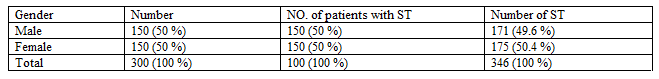
Key: ST = Supernumerary teeth
The prevalence of supernumerary teeth was found to 5.2 Percentage among which the incisor was the most prevalent with 3.39 Percentage (n = 194), then premolars with (0.75 Percentage; n = 43), then canines with (0.46 Percentage; n = 27), then molars with (0.42 Percentage; n = 24). The deciduous teeth was observed with (0.20 Percentage; n = 12) Table 2.
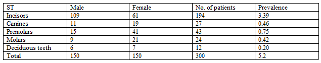
Key: ST= Supernumerary teeth, No.= Number,
Table 3 shows that the supernumerary teeth frequency was high in maxilla (89.9 Percentage; n = 311) than mandible (10.1 Percentage; n = 35) and more in female (50.6 Percentage; n = 175) than male (49.4 Percentage; n = 171). The prevalence of supernumerary teeth was significantly higher in female (P = 0.03). A significant difference was also found between the maxilla and mandible (P = 0.01).
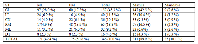
Key: ST= supernumerary teeth, ML= male, FM= female, CI= central incisors, LI= lateral incisosr, CN= canine, PM= premolars, ML= molars, DT= deciduous teeth.
Table 4 shows that the frequency of supernumerary teeth is higher in children between 5 - 10 years (61.4 Percentage; n = 212), then young adolescent between 11 - 16 years (25.6 Percentage; n = 88).
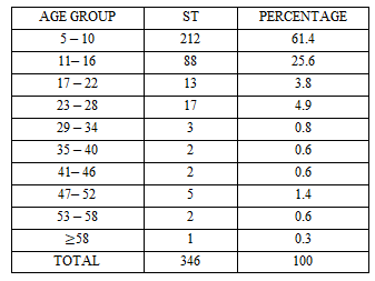
Key: ST= supernumerary teeth
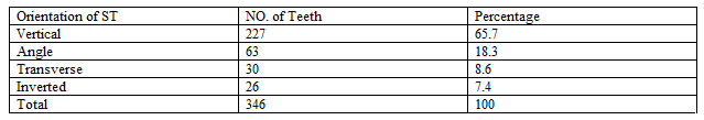
Table 5 shows the orientation of supernumerary teeth, in which 65.7 Percentage (n= 227) are vertically oriented, 18.3 Percentage (n=63) are angular oriented, 8.6 Percentage (n= 30) are transverse oriented and 7.4 Percentage (n= 26) are inverted.
Key: ST= supernumerary teeth, NO. = number
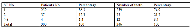
Table 6 shows the number of supernumerary teeth in each patient, in which 86.3 Percentage 9 (n= 259) of the patients were observed with one supernumerary tooth, 12.3 Percentage (n=37) of the patients were observed with two supernumerary teeth and 1.3 Percentage (n=4) of the patients were observed with multiple supernumerary teeth.
Key: ST= supernumerary teeth, NO= number
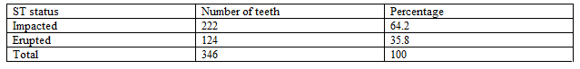
Table 7 shows the state of supernumerary eruption within the arch, in which 124 of the supernumerary teeth (35.8 Percentage) had erupted and 222 supernumerary teeth (64.2 Percentage) were impacted.
Key: ST= supernumerary teeth.
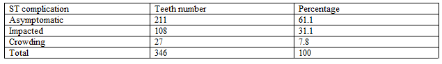
Table 8 shows the supernumerary teeth complications, in which 211 (61.1 Percentage) did not cause any complication, while 108 teeth (31.1 Percentage) caused teeth impaction and 27 teeth (7.8 Percentage) caused adjacent teeth displacement.
Key: ST= supernumerary teeth.
Discussion
The prevalence of supernumerary teeth was reported by different researchers among different racial and ethnic groups. The prevalence of supernumerary teeth was reported to be about 1 Percentage - 3 Percentage in Caucasian population, it was found to be greater than 3 Percentage in Asians and about 0.42 Percentage to 5.6 Percentage in Africa (Tay et al., 1984; Celikoglu et al., 2010).
The present study find the incidence of supernumerary teeth to be significantly higher in females than in males (p less than 0.001), which disagreed with the previous studies this may be due to difference in the of male to female, in which in previous studies the ratio male to female was between 1.18:1 to 4.5:1, where as in the present study the ratio was 1:1. This study ratio was found to be diverged from other studies, such as study of Liu et al. (2007) for Chinese population with ratio of 2.64:1 (male/female), the study of Esenlik et al. (2009) for Turkish population with ration of (1.13:1), study of Rajab and Hamdan (2002) the ratio was (2.2:1) and the study made by Çelikoğlu (2010) whose ratio was 1.8:1 for male to female respectively. The male to female ratio of 6.5:1 was used in different study of Chinese children by Davis (1987).
This study discovered the supernumerary teeth prevalence among Gombe state population to be 5.2 Percentage in which the incisor presented 3.39 Percentage. This finding was in disagreement with previous findings due to the differences in demographic and environmental factors and different sample size which may have impact on the reported prevalence rate (Patil and Maheshwari, 2014; Ferres-Padro et al., 2019). Also the included population in the previous studies was only the children and young population but this study included different ages which ranges from 5 to 76 years old.
In the present study the supernumerary teeth were found to be more frequent in age group between 5 - 10 years (61.4 Percentage; n = 212), followed by age group between 11 - 16 years (25.6 Percentage; n = 88). This result was supported previous research who reported that the supernumerary teeth were mostly observed between the age group of 7 and 10 (Rajab and Hamdan. 2002; Mukhopadhyay, 2011). Esenlik et al. (2009) also reported in his study that most cases of supernumerary teeth were found between the ages of 7-9.
Many studies reported that the most common location of supernumerary teeth is the premaxilla (Esenlik et al., 2009; Mukhopadhyay, 2011). This agreed with our study, which find the premaxillary regions as the predominant location of supernumerary teeth and 50.9 Percentage of this teeth were mesiodens, this is supported by studies of Montenegro et al. and Bäckman and Wahlin (Backman and Wahlin, 2001; Montenegro et al., 2006). This situation usually leads to complications of mesiodens which can be easily diagnosed by the parents.
The present study find the location of the supernumerary teeth to be 90 Percentage (n = 311) in the maxillary arch. These results agreed with that of De Oliveira et al. who reported that 91.3 Percentage of the supernumerary teeth were found in the maxillary arch (De Oliveira et al., 2008). Our results was also in agreement with that of Hattab et al. (1994) and Zhu et al. (1996) who reported that 90 Percentage of supernumerary teeth were found in the maxillary bone. The incisor (56.8 Percentage; n = 197) was the most commonly appearing supernumerary teeth with high frequency in the central incisor (45.3 Percentage; n = 157). This is in agreement with the studies by Hyun et al. (2009) and Dermiriz et al. (2015).
The present study discovered the provalence of supernumerary teeth in the deciduous teeth to be 4.6 Percentage , which varies from findings by others authors who had showed that, the prevalence of supernumerary teeth ranges from 0.2 Percentage to 0.8 Percentage in the deciduous dentition (Rajab and Hamdan, 2002; Gábris et al., 2006).
According to our finding, 74.9 Percentage (n = 259) of the supernumerary teeth were single, 21.7 Percentage (n = 75) were double and 3.4 Percentage (n=12) were multiples supernumerary teeth. Our findings coincide with previous studies who reported that the supernumerary teeth are more frequently single, while multiple supernumerary teeth are normally two in number (Rajab and Hamdan, 2002; De Oliveira Gomes et al., 2008; Çelikoğlu et al., 2010). This is due to the fact that the supernumerary teeth may occur in either single or multiples number in any region, but it is well known that, multiple supernumerary teeth co-exist rarely without any diseases or syndromes.
Our study find that 35.8 Percentage (n = 124) of the supernumerary teeth were erupted. Our results were close to other studies by (Rajab and Hamdan, 2002; Esenlik et al., 2009; Mukhopadhyay, 2011; Demiriz et al., 2015) who reported that all supernumerary teeth were mostly erupted. We also verified that erupted supernumerary teeth were vertically oriented 65.7 Percentage (n = 227). This is supported by studies of (Rajab and Hamdan, 2002; Esenlik et al., 2009; Mukhopadhyay, 2011; Demiriz et al., 2015) who reported that all the supernumerary teeth were normally vertically orientated while none of the transverse or inverted supernumerary teeth were erupted.
Our study find that the displacement (38.9 Percentage) as the most frequent clinical complication of the supernumerary teeth. This was supported by report of (Rajab and Hamdan, 2002; Esenlik et al., 2009; Mukhopadhyay, 2011; Anthonappa et al., 2012).
Conclusion
The occurrence of supernumerary teeth in the Gombe region was higher and it is more frequent in children with ages ranges between 5 to 10 years. This is followed young adolescent population between 11 to 16 years old. Its frequency is more in the permanent incisor of the maxillary region with the females having the highest frequency than males. Most of the supernumerary teeth are singly erupted and asymptomic, even though some of them are accompanied by some complications such as impaction, crowding and displacement. The detection of supernumerary teeth is very necessary. The early diagnosis helps to prevent or minimize possible complications.
References
- Anthonappa R. P., King N. M, and Rabie A. B. (2013). “Aetiology of supernumerary teeth: a literature review,” European Archives of Paediatrc Dentistry. 14; 279–288.
View at Publisher | View at Google Scholar - Anthonappa R. P., King N. M, and Rabie A. B. M. (2012) “Diagnostic tools used to predict the prevalence of supernumerary teeth: a meta-analysis,” Dentomaxillofacial Radiology. 41 (6) 444–449.
View at Publisher | View at Google Scholar - Anthonappa R. P., King N. M, and Rabie A. B. M. (2013). “Prevalence of supernumerary teeth based on panoramic radiographs revisited,” Pediatric Dentistry. 35 (3) 257–261.
View at Publisher | View at Google Scholar - Anthonappa R. P., King N. M., Rabie A. B. M, and Mallineni S. K. (2012). “Reliability of panoramic radiographs for identifying supernumerary teeth in children,” International Journal of Paediatric Dentistry. 22 (1) 37–43.
View at Publisher | View at Google Scholar - Anthonappa R. P., Omer R. S. M, and King N. M. (2008). “Characteristics of 283 supernumerary teeth in Southern Chinese children,” Oral Surgery, OralMedicine, Oral Pathology, Oral Radiology and Endodontology. 105;6;48–54.
View at Publisher | View at Google Scholar - Anthonappa, R.P., King, N.M., Rabie, A.B. and Mallineni, S.K. (2012) Reliability of Panoramic Radiographs for Identifying Supernumerary Teeth in Children. International Journal of Paediatric Dentistry, 22, 37-43.
View at Publisher | View at Google Scholar - Backman B, and Wahlin Y. B. (2001). “Variations in number and morphology of permanent teeth in 7-year-old Swedish children,” International Journal of Paediatric Dentistry. 11 (1) 11–17.
View at Publisher | View at Google Scholar - Çelikoğlu, M., Kamak, H. and Oktay, H. (2010) Prevalence and Characteristics of Supernumerary Teeth in a Non- Syndrome Turkish Population: Associated Pathologies and Proposed Treatment. Medicina Oral Patologia Oral y Cirugia Bucal, 15, 575-578.
View at Publisher | View at Google Scholar - Davis, P.J. (1987) Hypodontia and Hyperdontia of Permanent Teeth in Hong Kong School Children. Community Dentistry and Oral Epidemiology, 15, 218-220.
View at Publisher | View at Google Scholar - Mukhopadhyay, S. (2011) Mesiodens: A Clinical and Radiographic Study in Children. Journal of Indian Society of Pedodontics and Preventive Dentistry, 29, 34-38.
View at Publisher | View at Google Scholar - De Oliveira Gomes, C., Drummond, S. N., Jham, B.C., Abdo, E. N. and Mesquita, R. A. (2008). A Survey of 460 Supernumerary Teeth in Brazilian Children and Adolescents. International Journal of Paediatric Dentistry, 18, 98-106.
View at Publisher | View at Google Scholar - Demiriz, L., Mısır, A.F. and Durmuşlar, M.C. (2015). The Prevalence and the Characteristics of Supernumerary Teeth of Children and Young Adolescents from North-Western Region of Turkey. BJMMR, 7, 369-377.
View at Publisher | View at Google Scholar - De-Oliveira G. C., Drummond S. N., Jham B. C., Abdo E. N, and Mesquita R. A. (2008). A survey of 460 supernumerary teeth in Brazilian children and adolescents. Int J Paediatr Dent. 18: 98-106.
View at Publisher | View at Google Scholar - Diaz A., Orozco J, and Fonseca M. (2009). Multiple hyperodontia: Report of a case with 17 supernumerary teeth with non-syndromic association. Med Oral Patol Oral Cir Bucal. 14: 229-231.
View at Publisher | View at Google Scholar - Esenlik, E., Sayın, M.Ö., Atilla, A.O., Özen, T., Altun, C. et al. (2009) Supernumerary Teeth in a Turkish Population. American Journal of Orthodontics and Dentofacial Orthopedics, 136, 848-852.
View at Publisher | View at Google Scholar - Fernández M. P., Valmaseda C. E., Berini A. L, and Gay E. C. (2006). Retrospective study of 145 supernumerary teeth. Med Oral Patol Oral Cir Bucal. 11: 339- 344.
View at Publisher | View at Google Scholar - Ferrés-Padró E., Prats-Armengol J, and Ferrés-Amat E. (2009). A descriptive study of 113 unerupted supernumerary teeth in 79 pediatric patients in Barcelona. Medicina Oral, Patología Oral y Cirugía Bucal. 14: 146- 152.
View at Publisher | View at Google Scholar - Fidele N. B., Sekele I. B., Em K. K., Mantshumba M. A., Rubina S., Muyembi M., et al. (2016). Prevalence and Pattern Occurrence of Supernumerary Teeth in the North-East Heilongjiang Population of China. Open Journal of Stomatology. 6: 47-53.
View at Publisher | View at Google Scholar - Hattab, E.N., Yassin, O.M. and Rawashdeh, M.A. (1994) Supernumerary Teeth: Report of Three Cases and Review of the Literature. ASDC Journal of Dentistry for Children, 61, 382-393.
View at Publisher | View at Google Scholar - Hyun, H.K., Lee, S.J., Lee, S.H., Hahn, S.H. and Kim, J.W. (2009) Clinical Characteristics and Complications Associated with Mesiodentes. Journal of Oral and Maxillofacial Surgery, 67, 2639-2643.
View at Publisher | View at Google Scholar - Kara M. I., Aktan A. M., Ay S., Bereket C., Şener İ., et al. (2012). Characteristics of 351 Supernumerary Molar Teeth in Turkish Population. Medicina Oral, Patología Oral y Cirugía Bucal. 17: 395- 400.
View at Publisher | View at Google Scholar - Kaya G. Ş., Yapıcı G., Ömezli M. M, and Dayı E. (2011). Non-syndromic supernumerary premolars. Med Oral Patol Cir Bucal. 16: 522-525.
View at Publisher | View at Google Scholar - Leco B. M., Martin - Morales J. F, and Martinez González J. M. (2007). An observational study of the frequency of supernumerary teeth in a population of 2000 patients. Med Oral Patol Oral Cir Bucal. 12: 134- 138.
View at Publisher | View at Google Scholar - Mali S., Karjodkar F. R., Sontakke S, and Sansare K. (2012) Supernumerary Teeth in Non-Syndromic Patients. Imaging Science in Dentistry. 42: 41-45.
View at Publisher | View at Google Scholar - Martínez-González J. M., Cortés-Bretón B. J., Calvo-Guirado J. L., Arias I. O, and Barona-Dorado C. (2012). Clinical epidemiological analysis of 173 supernumerary molars. Acta Odontol Scand. 70: 398-404.
View at Publisher | View at Google Scholar - Mevlut C., Hasan K, and Hüsamettin O. (2010). Prevalence and characteristics of supernumerary teeth in a non-syndrome Turkish population: Associated pathologies and proposed treatment Journal section. Oral Medicine and Pathology. 15 (4) 575-578.
View at Publisher | View at Google Scholar - Montenegro, F. P., Valmaseda Castellón, E., Berini Aytés, L. and Gay Escoda, C. (2006) Retrospective Study of 145 Supernumerary Teeth. Medicina Oral Patologia Oral y Cirugia Bucal, 11, 339-344.
View at Publisher | View at Google Scholar - Moore S. R., Wilson D. F, and Kibble J. (2002). Sequential development of multiple supernumerary teeth in the mandibular premolar region-a radiographic case report. Int J Paediatr Dent. 12: 143-145.
View at Publisher | View at Google Scholar - Mukhopadhyay, S. (2011) Mesiodens: A Clinical and Radiographic Study in Children. Journal of Indian Society of Pedodontics and Preventive Dentistry, 29, 34-38.
View at Publisher | View at Google Scholar - Patil. S. and Maheshwari, S. (2014) Prevalence of Impacted and Supernumerary Teeth in the North Indian Population. Journal of Clinical and Experimental Dentistry, 6, e116-e120.
View at Publisher | View at Google Scholar - Rajab L. D, and Hamdan M. A. M. (2002) “Supernumerary teeth: review of the literature and a survey of 152 cases,” International Journal of Paediatric Dentistry. 12 (4) 244–254.
View at Publisher | View at Google Scholar - Rayyanu Z. S., Muhammad H. B. and Abdullahi N. (2020) Occurrence and Complications of Supernumerary Teeth Among People Living in Gombe State, Nigeria. Bima Journal of Science and Technology, 4(2) 2536-6041.
View at Publisher | View at Google Scholar - Sasaki H., Funao J., Morinaga H., Nakano K, and Ooshima T. (2007). Multiple supernumerary teeth in the maxillary canine and mandibular premolar regions: A case in the postpermanent dentition. Int J Paediatr Dent. 17: 304- 308.
View at Publisher | View at Google Scholar - Tay, F., Pang, A. and Yuen, S. (1984) Unerupted Maxillary Anterior Supernumerary Teeth: Report of 204 Cases. Journal of Dentistry for Children, 51, 289-294.
View at Publisher | View at Google Scholar - Zhu, J., Marcushamer, M., King, D.L. and Henry, R.J. (1996) Supernumerary and Congenitally Absent Teeth: A Literature Review. The Journal of Clinical Pediatric Dentistry, 20, 87-95.
View at Publisher | View at Google Scholar - Zilberman Y., Malron M, and Shteyer A. (1992). Assessment of 100 children in Jerusalem with supernumerary teeth in the premaxillary region. ASDC J Dent Child. 59: 44 - 47.
View at Publisher | View at Google Scholar
